www.australiandoctor.com.au 6 August 2010 | Australian Doctor |
29
Background
Diagnostic and
classification
criteria
Differential
diagnoses
Investigation
Complications
Treatment
Prognosis and
outcome
inside
DAVINDER SINGH-GREWAL, paediatric rheumatologist; staff specialist, the Children’s Hospital at Westmead, Sydney Children’s Hospital, Randwick, and John Hunter Children’s Hospital, Newcastle; senior clinical lecturer, discipline of paediatrics and child health, University of Sydney; conjoint senior lecturer, school of women’s and children’s health, University of NSW; and in private practice at the Children’s Hospital Medical Centre, Westmead, and Prince of Wales Private Hospital, Randwick, NSW.The author
COMPLETE HOW TO TREAT QUIZZES ONLINE (www.australiandoctor.com.au/cpd) to earn CPD or PDP points.
PULL-OUT SECTION
How
to
Treat
www.australiandoctor.com.au
JUVENILE idiopathic arthritis (JIA) is a chronic, autoimmune, inflammatory joint disease. It is the most common rheumatic disease in children and ado-lescents and is defined as persistent arthritis of unknown aetiology that begins before age 16 and persists for at least six weeks.
At least 5000 children in Australia have JIA and more than 20,000 Aus-tralian adults were diagnosed with arthritis in childhood. Delayed diag-nosis and delayed definitive therapy are common in patients with JIA. Such delays are known to be detri-mental to the long-term outcomes of children with JIA.
Arthritis in childhood has been plagued by many misconceptions, including the belief that arthritis does not occur in children and all muscu-loskeletal complaints of childhood
are due to trauma or ‘growing pains’. Furthermore it has incorrectly been felt that children who have arthritis will grow out of the condi-tion without any long-term conse-quences and not require any specific interventions.
JIA is a very different condition from adult arthritis and impacts sig-nificantly on a child’s growth and development. It requires very specific expertise and approaches to treat-ment to ensure effective manage-ment.
The GP has an essential role in the recognition of symptoms, initial workup, exclusion of differential diagnoses, referral and ongoing care of children with JIA in partnership with a paediatric rheumatologist.
This article describes the presen-tations of JIA, the differential
diag-noses, classification, investigation and management of the condition. It also discusses complications and outcomes of the disease along with recent advances in management.
Epidemiology
Juvenile idiopathic arthritis is a term used to describe a group of conditions that share the common clinical mani-festation of chronic joint inflamma-tion. There are several subtypes of JIA, which are discussed below and proba-bly represent a heterogenous group with differing aetiologies.
Juvenile arthritis affects at least one in 1000 children under 16 and as such is one of the most common chronic inflammatory diseases of childhood. Overall it is more common in girls than boys and the age of onset varies with subtype.
Aetiology and pathogenesis
As with most autoimmune diseases, the aetiology and pathogenesis of JIA is not well understood. It is hypothesised that a genetic predisposition exists in some individuals that is most likely inherited.Several HLA associations have been described for some of the subtypes of JIA and are thought to represent sus-ceptibility markers. However, genetic studies of twins, siblings and families suggest that susceptibility is most likely polygenic.
This predisposition in conjunction with an external trigger such as a viral infection, environmental exposure or trauma initiates an autoimmune phe-nomenon that manifests in the symp-toms of the disease.
Abnormalities in both the cellular
cont’d next page
JUVENILE idiopathic arthritis is defined as persistent arthritis of unknown aetiology that begins before age 16 and persists for at least six weeks. It is diagnosed only after all other causes of arthritis are excluded. The duration of six weeks is to exclude arthritis due to causes such as reactive arthritis that are not persistent but can closely mimic rheumatic causes. The important and common differen-tial diagnoses are discussed below. In the past, childhood arthritis has been classified by a number of different systems. More recently a unifying classification system has been developed by the International League Against Rheumatism (ILAR) using standardised nomen-clature. The ILAR criteria for JIA divides patients into seven distinct groups based predominantly on the pattern of joint involvement at onset and through the course of the disease, along with the presence of other clinical and laboratory fea-tures.
The criteria for six of these sub-types are shown in table 1 (page 32) and are designed more for use in clinical trials than as diagnostic criteria, yet they provide a useful framework for considering the dis-ease. The seventh subtype — ‘undifferentiated’ — is JIA that does not fit into the other cate-gories.
Oligoarticular
This is the most common subtype of JIA, affecting 50-60% of patients. It affects four or fewer joints at onset and predominantly affects large joints of the lower limbs, especially the knees and ankles (figure 1), but rarely the hips. Elbows and wrists are also often affected.
Presentation is usually in the pre-school years and this subtype is more common in girls than boys. Stiffness and limp are often the pre-dominant features rather than com-plaints of pain, meaning that the condition can go unrecognised for some time before diagnosis.
One-half of children who pres-ent with an oligoarticular pattern will ‘extend’ to involve more than four joints six months or more after onset, while the remaining 50% will display what is described as a persistent oligoar-ticular pattern. Eighty per cent of those with oligoarticular JIA will have a positive antinuclear anti-body (ANA) test but, other than mildly elevated inflammatory markers (ESR and CRP), the
remaining blood counts and chem-istry are usually normal.
Patients with persistent oligoar-ticular disease have a fairly good prognosis from an articular point of view. However, 20-30% will have uveitis as a complication of their arthritis. This uveitis is asymptomatic and vision-threaten-ing. Most patients with JIA there-fore require regular ophthalmolog-ical review even after their articular disease is in remission. Uveitis is more common in patients with JIA who are ANA positive.
Polyarticular
Polyarticular JIA (figure 2) affects five or more joints at onset and comprises 25-35% of all JIA. It is further subdi-vided into two groups based on rheumatoid factor (RF) status. At about 5-10% of all JIA, RF-positive polyarticular arthritis constitutes a small proportion of this group.
Polyarticular disease is more common in girls than boys and onset shows a bimodal pattern, with RF-negative disease peaking in the pre-school age and RF-positive disease in adolescence. ANA is frequently posi-tive in the RF-negaposi-tive group, and patients in this group are prone to uveitis.
The pattern of joint involvement is often asymmetric in RF-negative polyarticular JIA, and symmetric in RF-positive disease, in a pattern simi-lar to that of adult-onset RA. Small and large joints are commonly involved in both types.
RF-positive polyarticular JIA is an aggressive disease that can cause sig-nificant joint destruction and may be associated with nodules and vasculi-tis, as seen in RA. Inflammatory markers and white cell count may be elevated in this form of JIA, and anti-cyclic citrullinated peptide (anti-CCP) antibodies are also elevated, as in adult RA.
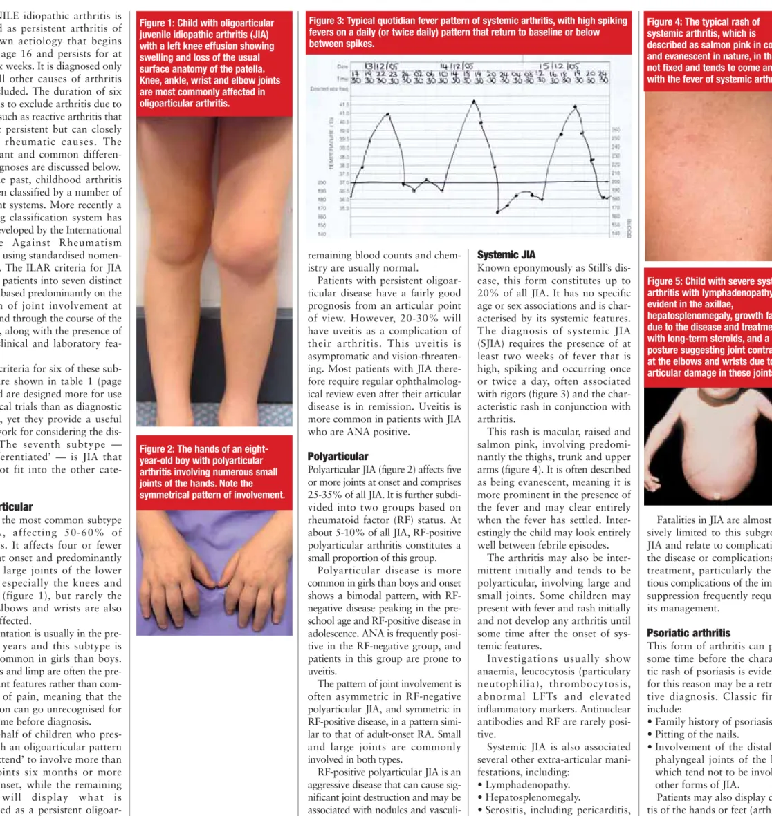
Systemic JIA
Known eponymously as Still’s dis-ease, this form constitutes up to 20% of all JIA. It has no specific age or sex associations and is char-acterised by its systemic features. The diagnosis of systemic JIA (SJIA) requires the presence of at least two weeks of fever that is high, spiking and occurring once or twice a day, often associated with rigors (figure 3) and the char-acteristic rash in conjunction with arthritis.
This rash is macular, raised and salmon pink, involving predomi-nantly the thighs, trunk and upper arms (figure 4). It is often described as being evanescent, meaning it is more prominent in the presence of the fever and may clear entirely when the fever has settled. Inter-estingly the child may look entirely well between febrile episodes.
The arthritis may also be inter-mittent initially and tends to be polyarticular, involving large and small joints. Some children may present with fever and rash initially and not develop any arthritis until some time after the onset of sys-temic features.
Investigations usually show anaemia, leucocytosis (particulary neutophilia), thrombocytosis, abnormal LFTs and elevated inflammatory markers. Antinuclear antibodies and RF are rarely posi-tive.
Systemic JIA is also associated several other extra-articular mani-festations, including:
• Lymphadenopathy. • Hepatosplenomegaly.
• Serositis, including pericarditis, pleuritis and peritonitis (figure 5). These features may precede the joint manifestation, and, in the case of pericarditis, may be life-threaten-ing if the patient develops tampon-ade.
Fatalities in JIA are almost exclu-sively limited to this subgroup of JIA and relate to complications of the disease or complications of its treatment, particularly the infec-tious complications of the immuno-suppression frequently required in its management.
Psoriatic arthritis
This form of arthritis can present some time before the characteris-tic rash of psoriasis is evident and for this reason may be a retrospec-tive diagnosis. Classic findings include:
• Family history of psoriasis. • Pitting of the nails.
• Involvement of the distal inter-phalyngeal joints of the hands, which tend not to be involved in other forms of JIA.
Patients may also display dactyli-tis of the hands or feet (arthridactyli-tis of the interphalyngeal joints in con-junction with tenosynovitis of the associated tendons, resulting in the appearance of swelling of the entire digit, sometimes called a ‘sausage-and humoral immune
response have been demon-strated in JIA. It is felt that T cells have a central role in the expression of pro-inflamma-tory cytokines such as TNF-α, interleukin 1 (IL-1) and IL-6, which are known to mediate much of the damage seen in this disease. Genes that influ-ence cytokine production and T-lymphocyte function are likely to be heavily implicated in the pathogenesis of these diseases.
Blocking cytokines using
biological therapies has proven to be an effective means of treatment of many of these diseases and will be discussed in detail later in this article.
The basic pathological process involves inflammation of the synovial lining of the joint, which results in increased production of syn-ovial fluid and degradation of the articular cartilage of the joint through a combination of enzymes such as metallo-proteinases and inflammatory molecules. The destructive
pannus seen in adult rheuma-toid arthritis (RA) with promi-nent erosions may be seen in JIA but is less common.
Symptoms and signs
Arthritis is literally defined as inflammation of the joint. Like headache, abdominal pain or haematuria it is a symptom and sign of disease rather than a specific diagno-sis. There are many possible causes of arthritis ranging from trauma through to sys-temic inflammatory disease and the main task facing theclinician is to determine the cause of the signs and symp-toms. This is particularly important, as JIA is a diagno-sis of exclusion.
The cardinal features of the rheumatic causes of arthritis include the symptoms of pain, swelling and stiffness, which are classically worse in the morning and after prolonged periods of inactivity, for exam-ple, after naps or car trips. The onset may be either acute or insidious.
Complaints of pain are often not as prominent as those seen
in adult arthritis due to under-reporting, but limp and decreased function or school performance may be keys to the presence of arthritis in chil-dren. Toddlers with regression of motor milestones such as crawling, cruising or walking must also be considered for a diagnosis of arthritis.
On examination, arthritis is defined by the presence of joint swelling or effusion or the coexistence of two of the following three findings: • Tenderness or pain on
motion.
• Limited range of motion. • Increased warmth of the
skin overlying the joint. The systemic form of JIA may also present with char-acteristic systemic symp-toms and signs, including rash, fever, organomegaly, lymphadenopathy and serositis. These clinical fea-tures may precede the onset of arthritis, making the diagnosis difficult and the exclusion of the differential diagnoses of these features, especially malignancy, even more critical.
from previous page
Diagnostic and classification criteria
Figure 1: Child with oligoarticular juvenile idiopathic arthritis (JIA) with a left knee effusion showing swelling and loss of the usual surface anatomy of the patella. Knee, ankle, wrist and elbow joints are most commonly affected in oligoarticular arthritis.
Figure 4: The typical rash of systemic arthritis, which is described as salmon pink in colour and evanescent in nature, in that it is not fixed and tends to come and go with the fever of systemic arthritis.
Figure 2: The hands of an eight-year-old boy with polyarticular arthritis involving numerous small joints of the hands. Note the symmetrical pattern of involvement.
Figure 5: Child with severe systemic arthritis with lymphadenopathy evident in the axillae,
hepatosplenomegaly, growth failure due to the disease and treatment with long-term steroids, and a posture suggesting joint contracture at the elbows and wrists due to articular damage in these joints. Figure 3: Typical quotidian fever pattern of systemic arthritis, with high spiking
fevers on a daily (or twice daily) pattern that return to baseline or below between spikes.
32
| Australian Doctor | 6 August 2010 www.australiandoctor.com.auJuvenile idiopathic arthritis
HOW TO TREAT
AS mentioned above, the most important aspect of the diagnosis of JIA is the exclusion of other causes of arthritis in childhood. As the primary care practitioner is usually the first point of contact for a child with mus-culoskeletal symptoms, it is impor-tant that the physician be familiar not only with the presentation of JIA but also the important differential diag-noses.
As we will see, some of these con-ditions require specific therapy and others require prompt diagnosis and management to prevent complica-tions. Table 2 presents a non-exhaus-tive list, and some of the more impor-tant conditions are described below.
Infection
Septic arthritis (SA) and osteomyelitis (OM) are caused by direct bacterial infection of the bone or joint. They are usually characterised by a rela-tively rapid onset of symptoms, while JIA is ordinarily more gradual in onset.
Both SA and OM can cause joint effusions (in the case of OM due to a sympathetic effusion in an adjacent joint) and are also frequently associ-ated with fevers. Children with SA or OM are much more likely to refuse to bear weight on an affected limb than those with JIA.
A patient with a painful monoarthritis of rapid onset associ-ated with fever requires immediate investigation and treatment for sus-pected bone or joint infection. It is important to remember that rarer types of bone and joint infections
such as those due to tuberculosis or fungi can cause sub-acute infections that may be difficult to distinguish from JIA.
Post-infectious arthritis
This condition can be impossible to distinguish from JIA at initial presen-tation. Like JIA, it is a true autoim-mune arthritis usually triggered by a viral infection. The diagnostic criteria of six weeks’ duration before diag-nosis of JIA excludes the misdiagdiag-nosis of JIA in these cases.
Post-infectious arthritis usually involves the large joints and includes conditions such as ‘irritable hip’ or ‘transient synovitis’, which is a rela-tively common but short-lived phe-nomenon in preschool-age children
that usually follows an URTI. Viruses such as rubella and parvovirus are known to cause polyarticular small-joint arthritis, and enteric bacteria are a well-known cause of severe reac-tive arthritis that may have a pro-longed course.
Malignancy
Some malignant conditions of child-hood can present with joint and limb pain or arthritis. Primary bone tumours and haematological malig-nancies can cause these symptoms. Key features include constitutional symptoms such as fever, weight loss and general malaise, which are not usually seen in JIA except in the SJIA subtype.
Thus SJIA may be difficult to
dis-tinguish from malignancy at initial presentation. Keys to the possibility of an underlying malignancy include the presence of severe night pain that wakes the child from sleep, or the presence of abnormalities on the blood count, including thrombocy-topenia, lymphocytosis or neutrope-nia.
Rheumatic fever
Rheumatic fever is a form of reac-tive arthritis and is an immunolog-ical reaction to Streptococcus species. It is uncommon before age five and may present with fever and migratory large-joint arthritis, along with carditis, erythema mar-ginatum, nodules and, rarely, chorea. This diagnosis needs to be
considered in children presenting with arthritis, as a delay in antibi-otic treatment may result in long-term cardiac complications.
Slipped capital femoral
epiphysis
This condition predominantly affects boys in early adolescence and may have an acute or subacute pres-entation. Symptoms usually include hip and knee pain associated with limp. Examination will often reveal some reduction in the range of hip movement.
Radiographs are diagnostic and will usually be abnormal at presenta-tion. Frog-leg or abduction/external rotation views are the most sensitive image for detecting the condition early (figure 6).
Management requires immediate consultation with an orthopaedic sur-geon, and no weight bearing until the slip stabilises. Surgical intervention is sometimes required and the outcome depends on the degree of slip.
Avascular necrosis of the femoral
head (Legg–Calve–Perthes
disease)
This condition affects boys pre-dominantly aged 5–10. It presents with varying degrees of hip and/or knee pain and, while radiographs are diagnostic, early images may fail to show the characteristic abnormalities (figure 7). Examina-tion will show a progressive loss of hip range of motion. Manage-ment is usually conservative, with a good outcome in most cases. digit’).Psoriatic arthritis can
be associated with ANA pos-itivity in about 50% of patients and can be associ-ated with asymptomatic uveitis.
Enthesitis-related
arthritis
Entheses are sites of the insertion of tendons, liga-ments and muscles to bone. These sites are prone to inflammation in a similar way to synovium, and this process is the clinical hall-mark of this class of JIA, which can be seen as homol-ogous with seronegative arthritis and ankylosing spondylitis in adults.
Enthesitis-related arthritis (ERA) is more common in boys over age eight and has a strong association to the pres-ence of HLA-B27. In adults, HLA-B27-associated disease is characterised by the pres-ence of symptoms of back pain and by findings of peripheral and axial arthritis, but enthesitis is less frequent.
In children, back pain is far less prevalent, and enthesitis along with peripheral arthritis is the usual pattern seen in the HLA-B27-associated arthri-tides. Joint involvement is typ-ically asymmetric, with knees, hips and ankles commonly affected. Sacroiliitis is also seen but again with a lower frequency than in adult patients.
This group of conditions is closely related to the arthritis seen in inflamma-tory bowel disease (IBD) and thus signs and symp-toms of IBD should be sought in patients with
ERA. Other extra-articular manifestations include iritis, which, in contrast to that of ANA-positive JIA, is acute and painful and thus does not often escape detection. Other manifestation such as
aortitis may also be seen but are rare and do not usually develop in patients until they reach adulthood.
Undifferentiated
This final group is used to
describe patients who have clearly had inflammatory arthritis lasting six weeks but who do not fit into one of the above categories, and will not be dealt with in any further detail.
Differential diagnoses
Table 1: Features of juvenile idiopathic arthritis by subtype
Oligoarticular Polyarticular Systemic Psoriatic Enthesitis-related arthritis
RF negative RF positive
% of JIA 50-60 20-30 5-10 10-20 5-15 15%
Gender (F:M) 4:1 9:1 4:1 1:1 3:2 1:9
Usual onset
age 2-12 (peak 1-2) 2-12 (peak 1-3) Adolescence Any Mid-childhood Adolescence Joint pattern Asymmetric
Large joints (knee, ankle, wrist, elbow)
Often asymmetric Multiple Small and large joints
Symmetric Multiple
Small and large joints
Small and large joints
Small and large joints, including hips and especially the DIPs
Asymmetric
Large joints, including hips; axial skeleton, including sacroiliitis Extra-articular Involvement Asymptomatic uveitis 20% (especially if ANA positive) Asymptomatic uveitis (especially if ANA positive)
Rheumatoid nodules Fever Rash Lymphadenopathy Hepatosplenomegal y Serositis Psoriasis Nail pitting Dactylitis Symptomatic uveitis in 10% Enthesitis Enthesitis Symptomatic uveitis in 20% IBD Aortitis FBC N N N ↑WCC/Plt ↓Hb N N ESR/CRP N N or ↑ N or↑ ↑ ↑ N N or ↑ ANA 80% 60-80% 50% <10% 50% negative Rheumatoid
factor negative negative negative negative negative negative HLA-B27 Absent Absent Absent Absent 30% 80%
Remission (%) 52% 24% 0% 35% Low Low
JIA = Juvenile idiopathic arthritis ANA= antinuclear antibody RF = rheumatoid factor N = normal
Adapted from McKay D, Singh-Grewal D. Rheumatology. In: Kilham H, et al. (editors). Paediatrics Manual, The Children’s Hospital at Westmead Handbook, 2ndedition. McGraw Hill, Sydney, 2009.
Figure 7: Avascular necrosis of the right femoral head of the right hip, showing loss of height and sclerosis of the right femoral epiphysis along with loss of the normal rounded contour seen in the normal left hip. Figure 6: Patient with a high-grade slipped capital
femoral epiphysis of the right hip. It is important to remember that very mild grades may not be seen easily on antero-posterior films, making the ‘frog’s leg’ view with the knees apart and the ankles together a more useful film in these cases.
AS there is no diagnostic test for JIA, diagnosis relies on clinicians main-taining an index of suspicion, careful clinical history-taking and examina-tion. This said, investigations can be valuable for excluding other
differen-tial diagnoses and surveying for com-plications of the disease.
Plain X-rays of affected joints should be obtained at diagnosis and in the follow-up of patients with per-sistently active disease. Joint
aspira-tion, synovial fluid cell count and culture are warranted if septic arthri-tis is suspected.
Blood tests, including FBC and inflammatory markers, are fre-quently done at diagnosis and can
be useful, particularly the ANA, which we have seen is a strong marker of uveitis risk. RF and anti-CCP, which are markers of aggres-sive disease, are also useful prog-nostically. Other radiological
investigations such as bone scans, ultrasound, CT and MRI are proba-bly not warranted as tools to diag-nose arthritis but are often useful when excluding the differential diag-noses of JIA.
Benign nocturnal limb
pains or growing pains
Growing pains are a rela-tively common phenomenon in preschool or early school-age children. Children wake in the late evening or early night complaining of often severe bilateral leg or shin pains. These pains settle with massage or simple analgesia and the child sleeps soundly for the rest of the night. Growing pains are never associated with limp or stiff-ness in the morning or day, which is important in distin-guishing this condition from JIA.Hypermobility
Many children have hyper-mobility of their joints that is not associated with any other form of connective tissue disease (eg, Marfans or Elhers–Danlos syndrome). These children will often complain of pain that is
exacerbated by activity or that occurs after exercise, and will display hypermobil-ity on physical examination.
Overuse injury
Overuse injuries, including Osgood–Schlatter’s disease, are seen in adolescence and are also characterised by pain, especially that related to exercise.
Pain syndromes
It is important to remember that pain conditions such as complex regional pain syn-drome and fibromyalgia are seen in children and adoles-cents.
Other inflammatory or
rheumatic diseases
Conditions such as SLE, IBD and juvenile dermatomyosi-tis may also present with arthritis and should be con-sidered in the differential diagnosis.Investigation
AS the aetiology of JIA is unknown, no preventive measures exist. Thus the aim of therapy is to improve qual-ity of life and prevent cations. Preventable compli-cations in most cases are a result of delayed diagnosis and treatment, poor compliance with treatment, or inadequacy of therapy. While complica-tions were formerly common, in this era only a small number of children should be left with sequelae as a result of uncontrollable disease.
The major complications are as follows.
Articular damage
This includes contracture, ero-sions and premature degenera-tive arthritis requiring arthro-plasty.
Growth disturbance
Patients with SJIA frequently develop generalised growth failure as a result of chronic disease and possibly as a com-plication of long-term steroid use.Patients with more localised disease may also develop localised growth disturbances. Children with oligoarthritis are noted to have accelerated growth at the metaphyses of long bones adjacent to inflamed joints, particularly the knee, and this can result in an increase in the length of the affected limb (figure 8). Subse-quently the affected metaphy-seal growth plate may fuse earlier than that of the unaf-fected side, and catch-up
growth on the unaffected side results in the affected limb being significantly shorter at final height.
The widespread use of intra-articular steroid injec-tions has significantly reduced the occurrence of leg length discrepancies through improv-ed disease control.
Involvement of the tem-poromandibular joint, which is often seen in polyarticular disease, has the opposite effect in that it can result in reduced growth of the mandible and significant micrognathia, with cosmetic, dental and airway sequelae (figure 9).
Uveitis
As discussed earlier asympto-matic anterior uveitis is an insidious and worrying com-plication, as it may result in blindness if not recognised or if undertreated. It is seen in up
to 30% of ANA-positive patients with JIA. The pri-mary pathological process is inflammation of the iris, with the development of:
• Posterior synechiae. • Impaired drainage of the
anterior chamber. • Glaucoma. • Cataracts.
• Band keratopathy.
• Visual impairment (figure 10).
Uveitis may have an inde-pendent course to that of the arthritis and thus may occur in patients in articular remis-sion. For this reason, regular ophthalmological surveillance is mandatory (table 3).
Specific complications of
SJIA
SJIA has several more specific complications related to the nature of the disease. These include pericarditis and tam-ponade, pleuritis and macrophage activation syn-drome (MAS). MAS is a rare complication in which an overwhelming cytokine release and response results in pan-cytopenia, deranged LFTs and abnormal clotting, which can be fatal.
School and social disability
As a rule, children with mild forms of arthritis rarely avoid school. However, those with severe disease may miss sig-nificant amounts for school. Long-term follow-up of chil-dren with all forms of JIA shows significantly:• Lower rates of educational achievement.
• Lower workforce participa-tion.
• Higher rates of depression. • Less successful social
inte-gration than their peers. These poorer outcomes are well correlated with poorer degrees of disease control.
Complications
Figure 8: Radiograph of a child with oligoarthritis of the left knee, showing osteopenia of the affected side along with advanced maturation, with a larger epiphysis on the left side, with a more mature growth plate. Also of note are the loss of articular space on the left compared with the right and the apparent increased length of the left leg compared with the right, something better appreciated on clinical examination.
Figure 9: A child with polyarticular arthritis involving the temporomandibular joints and resulting in significant growth disturbance and micrognathia.
Figure 10: Eye of a patient with antinuclear antibody-positive JIA, showing complications of uveitis with an irregular pupil and posterior synechiae (adhesions between the inflamed iris and the underlying lens).
Table 3: Suggested ophthalmological review
schedule for children with JIA
Disease onset at age <7 Disease onset at age ≥7 Pupils not
round or precipitate on cornea
Immediate referral to rule out iritis
Oligoarthritis +ve ANA
3-4-monthly for four years, then every six months for three years, then yearly
Six-monthly for four years then yearly
Oligoarthritis –ve ANA
Six-monthly for seven years, then yearly
Polyarthritis +ve ANA
3-4-monthly for four years, then every six months for three years, then yearly
Polyarthritis –ve ANA
Six-monthly for seven years, then yearly
Systemic
onset Yearly
Adapted from Clinical Guideline for the Diagnosis and Management of Juvenile Arthritis. RACGP, August 2009:
www.racgp.org.au/guidelines/juvenileidiopathicarthritis
cont’d next page
JIA
• Oligoarticular:
— persistent (affecting four or fewer joints)
— extended (progressing to more than four joints in 50%) • Polyarticular (more than four joints):
— rheumatoid factor positive — rheumatoid factor negative • Systemic onset (SJIA) • Psoriatic arthritis (JPsA) • Enthesitis-related arthritis (ERA)
Connective tissue diseases
• SLE • Scleroderma
• Juvenile dermatomyositis • Mixed connective tissue disease
Sarcoidosis Vasculitis • Kawasaki disease (KD) • Henoch-Schölein purpura • Polyarterits nodosa • Wegener’s granulomatosis • Takyasu’s arteritis Infection • Primary: — Bacterial
— Septic arthritis (including TB) — Osteomyelitis with reactive effusion — Viral (hepatitis, parvovirus B19, EBV) • Secondary:
— post-streptococcal, rheumatic fever, post-enteric — post-viral (transient synovitis, irritable hip)
Mechanical
• Acute trauma (fracture, meniscal injury, osteochondritis) • Chronic sub-acute mechanical loading — sport- and
activity-related injuries
Haematological
• Haemophilia — haemarthrosis • Sickle cell disease — bony crisis
Malignancy
• Leukaemia • Lymphoma
• Primary bone tumour
Orthopaedic
• Perthes disease
• Slipped upper femoral capital epiphysis
Table 2: Differential diagnosis of arthritis in childhood
*Adapted from McKay D, Singh-Grewal D. Rheumatology. In: Kilham H, et al. (editors). Paediatrics Manual, The Children’s Hospital at Westmead Handbook, 2ndedition. McGraw Hill, Sydney, 2009.
THE realisation that JIA is not a benign condition that children will grow out of has resulted in the development of a far more aggres-sive treatment paradigm, with a lower tolerance for incomplete dis-ease control. The aim of treatment in JIA is complete remission of inflammation. Furthermore, treat-ment of JIA has increased in its complexity over the past decade, with the introduction of new thera-pies beyond the traditional disease-modifying agents.
Management of children with JIA, as for many chronic diseases of childhood, is best done through a multidisciplinary team including the: • GP.
• Paediatric rheumatologist. • Occupational therapist.
• Physiotherapist and other health professionals as required.
NSAIDs
For many years it was felt that most children with JIA required only symptomatic therapy with NSAIDs such as naproxen and that the dis-ease would eventually remit without any significant complication. As we have seen already, this is not the case.
While NSAIDS will often provide very good symptomatic relief of pain and stiffness, they are not disease modifying in that they do not actu-ally terminate inflammation. Thus the process of cytokine release, syn-ovial inflammation and cartilage damage may continue even in the absence of overt symptoms.
NSAIDs continue to have a role as a symptomatic therapy but are not usually adequate as a sole ther-apy. Topical NSAIDs have no proven benefit for patients with JIA.
Corticosteroid joint injections
Joint injections have become the mainstay of therapy for many chil-dren with active oligoarticular JIA, as they provide rapid symptomatic treatment as well as termination of the inflammatory process in up to 90% of patients. The widespread use of this therapy has resulted in asignificant reduction in many of the complications previously common in patients with this form of JIA.
When considering joint injections, it is important to remember the need for appropriate procedural sedation and analgesia in children, which often means general anaesthesia.
Intra-articular injections are also used in the treatment of other forms of JIA, usually in combination with other therapies.
Systemic corticosteroids
Oral steroids are used in patients with polyarticular or systemic dis-ease and have proved a very effective therapy for inflammatory diseases of all types. Unfortunately the long-term side effects of these drugs limit their ongoing use and necessitate the use of disease-modifying agents as described below. Parenteral steroids
are used in SJIA when disease activ-ity is high.
Disease-modifying
anti-rheumatic drugs
Disease-modifying anti-rheumatic drugs (DMARDs) include agents such as methotrexate, sulfasalazine, and leflunomide. They have been used in JIA for many years in the recognition that NSAIDs alone are usually inadequate therapy and that systemic corticosteroids are not an ideal long-term therapeutic option.
Each of these drugs has a 60-80% effectiveness in achieving and main-taining remission in patients with JIA of most disease subtypes. However, systemic JIA has a far lower rate of response to these agents.
DMARDs are well tolerated and the safety profiles, which will not be discussed in detail here, are well
understood. These agents should be used only in conjunction with a pae-diatric rheumatologist. Established protocols exist for monitoring drug side effects and should be adhered to.
Biological therapies
This group consists of a newer range of therapies for JIA that have been developed to directly counteract spe-cific aspects of inflammation by blocking molecules involved in the inflammatory process. These agents are used for children with arthritis unresponsive to traditional DMARDs.
They include etanercept, a mono-clonal TNF-receptor antagonist, administered through twice-weekly SC injections, and infliximab, a chimeric molecule (part human, part mouse) TNF-receptor antagonist administered by monthly infusion.
Children with SJIA unresponsive to traditional DMARDs have shown responses to IL-6 blockade in most cases and IL-1 blockade in fewer patients. While these drugs bring with them exciting opportunities, cli-nicians who prescribe them need to be wary of the potential side effects, including infection as a result of potential immunosupression and the longer-term risk of malignancy after prolonged use, which is as yet not fully quantified.
Allied health
Both occupational therapy and phys-iotherapy have an indispensible role in the management of JIA. Occupa-tional therapy is essential in main-taining function, especially in patients with hand and wrist involve-ment, which can severely impair school performance and other activ-ities of daily living. Splinting and exercises are frequently required for such patients.
Physiotherapy has a role in dealing with joint contracture and maintain-ing appropriate activity levels. Other services such as those of dietitians, orthotists, social workers and school liaison officers are also often an important part of the team.
34
| Australian Doctor | 6 August 2010 www.australiandoctor.com.auJuvenile idiopathic arthritis
HOW TO TREAT
RECENT evidence shows categorically that up to 50% of children will need to have ongoing contact with rheumatology services beyond age 18.
The chances of achieving long-term remission are related to the subtype of JIA and are included in table 1. Oligoarthritis has the best chance of remission, but about 40% of this group
will have ongoing disease and require treatment into adulthood. RF-positive pol-yarticular arthritis has virtu-ally no chance of remission, while SJIA will remit in around 35% of cases.
Studies have shown that after 10 years of active dis-ease, rates of academic, employment and social achievement are significantly lower in individuals who
have experienced JIA. Rates of disability are high and depression affects more than 25% of those with JIA.
Poorer long-term out-comes are known to be related to delayed diagnosis or inadequate treatment. This means that recognition of JIA from its symptoms and signs is critical to the long-term prognosis for patients.
Evidence-based practice
• The diagnosis of JIA is based on history and examination in the first instance (grade C).
• Early diagnosis and treatment improves long-term outcomes (grade B and C).
• Appropriate investigations are sometimes used to support clinical examination and exclude differential diagnoses (grade C).
• Referral to a paediatric rheumatologist (or paediatrician) is warranted for any child with inflammatory joint disease of more than four weeks’ duration (grade C).
• A multidisciplinary approach to management involving medical and allied health professionals is the ideal (grade C and D). • NSAIDs are a reasonable first line of symptomatic therapy but
should not be considered definitive (grade B).
• The aim of treatment is complete remission of disease using treatment modalities including corticosteroid joint injection, DMARDS and biological agents.
• Long-term management requires a partnership between the patient’s paediatric rheumatologist and primary care physician. Adapted from Clinical Guideline for the Diagnosis and Management of Juvenile Arthritis. RACGP, August 2009:
www.racgp.org.au/guidelines/juvenileidiopathicarthritis
Summary
• JIA is a chronic illness of childhood that is more common than generally appreciated.
• Children with musculoskeletal symptoms need to carefully assessed for signs of inflammatory joint disease, which are best appreciated through careful clinical history and examination.
• Children with symptoms suggestive of JIA require timely and specific therapy to control inflammation and prevent long-term disability.
Treatment
Prognosis and outcome
Clinical guidelines to diagnosis and management
The NHMRC in conjunction with the RACGP have developed guidelines for the early diagnosis and management of JIA. The main points of this evidence-based guideline are:
• JIA needs to be considered in the differential diagnosis of children with painful or swollen joints.
• An acute painful monoarthritis associated with fever is septic until proven otherwise.
• Careful consideration of differential diagnoses requiring specific therapies is necessary.
• Analgesia such as an NSAID is an appropriate first step in management for joint pain or swelling.
• If symptoms or signs persist for more than four weeks referral to a
rheumatologist is appropriate (preferably a paediatric rheumatologist, if available). • Goals of treatment are to control inflammation, relieve pain and prevent or control
joint damage.
• The ideal approach to management is through a multidisciplinary team involving the GP, rheumatologist and allied health staff utilising pharmacological and non-pharmacological modalities.
• Ongoing close monitoring for complications including uveitis is essential.
Adapted from Clinical Guideline for the Diagnosis and Management of Juvenile Arthritis. RACGP, August 2009: www.racgp.org.au/guidelines/juvenileidiopathicarthritis
cont’d page 36
Online resources
• The Royal Children’s Hospital Melbourne. Information about rheumatological conditions: www.rch.org.au/ rheumatology/ jia.cfm?doc_id=10478 • Pediatric Rheumatology INternational Trials Organisation (PRINTO): www.printo.it • Arthritis NSW: www.arthritisnsw.org.au
Juvenile idiopathic arthritis
— 6 August 2010
INSTRUCTIONS
Complete this quiz online and fill in the GP evaluation form to earn 2 CPD or PDP points. We no longer accept quizzes by post or fax.
The mark required to obtain points is 80%. Please note that some questions have more than one correct answer.
ONLINE ONLY
1. Which TWO statements are correct?
a) Children who have juvenile idiopathic arthritis (JIA) usually grow out of the condition without any long-term consequences
b) T lymphocytes, TNF-α, and interleukins 1 and 6 mediate the inflammation and damage of JIA
c) Joint pain and stiffness due to JIA tends to be worse at night and after periods of physical activity
d) Children with JIA may present with a limp or decreased function rather than joint pain
2. Which TWO statements are correct?
a) In toddlers with JIA, motor milestones do not generally regress
b) The systemic form of JIA may present with rash, fever, organomegaly,
lymphadenopathy or serositis c) Clinical manifestations of systemic JIA
(SJIA) always appear after the onset of the arthritis
d) Oligoarticular JIA affects four or fewer joints at onset, usually the knees and ankles but rarely the hips
3. Which TWO statements are correct?
a) A small proportion of children with oligoarticular JIA will have a positive antinuclear antibody (ANA)
b) Uveitis is more common in JIA patients who
are ANA positive and can be asymptomatic in the early stages
c) Uveitis is no longer a risk once the articular disease is in remission
d) For polyarticular JIA the pattern of joint involvement is often asymmetric in rheumatoid factor (RF)-negative, and symmetric in RF-positive disease
4. Which THREE statements are correct?
a) RF-positive polyarticular JIA is often an aggressive disease with significant joint destruction and which can cause nodules and vasculitis
b) The diagnosis of SJIA requires the presence of at least two weeks of high, spiking fever once or twice a day, and a characteristic rash c) ANA and RF are usually positive in SJIA d) SJIA is usually associated with anaemia,
neutrophilia, thrombocytosis, abnormal LFTs and elevated inflammatory markers
5. Which TWO statements are correct?
a) SJIA can be fatal due to disease complications, or treatment-related immunosuppression b) Uveitis tends not to occur in ANA-positive
psoriatic arthritis
c) In enthesitis-related arthritis (ERA), there is inflammation of the insertion of tendons and ligaments into bone
d) Back pain, sacroiliitis and axial arthritis are
typical manifestations of ERA in children
6. Which TWO statements are correct?
a) In ERA, the uveitis is typically asymptomatic and chronic
b) A patient with a painful monoarthritis of rapid onset associated with fever requires immediate investigation and treatment for suspected bone or joint infection
c) Post-infectious (reactive) arthritis usually lasts for more than six weeks
d) Bone or blood malignancies can be
differentiated from SJIA on the basis of severe night limb pain and the presence of
thrombocytopenia, lymphocytosis or neutropenia
7. Which TWO statements are correct?
a) Perthes disease and slipped capital femoral epiphysis should be excluded in a child with hip or knee pain suspected of having JIA b) Night-time growing pains are always mild c) Growing pains can be associated with limp or
stiffness
d) Synovial fluid aspiration for cell count and culture is essential if septic arthritis is suspected
8. Which THREE statements are correct?
a) A positive ANA is key to the diagnosis of JIA b) ANA is valuable in assessing the risk of
uveitis in a child with JIA
c) RF and anti-CCP are markers of aggressive disease and are useful prognostic factors d) In oligoarthritis there can be accelerated
growth at the metaphyses of long bones adjacent to inflamed joints, resulting in increased length of the affected limb
9. Which THREE statements are correct?
a) The affected metaphyseal growth plate affected by arthritis may fuse earlier than that of the unaffected side, resulting in shorter limb length at final height
b) Inflammation of the iris (uveitis) may cause glaucoma, cataracts and keratopathy c) The aim of treatment in JIA is complete
remission of inflammation
d) NSAID monotherapy is sufficient to terminate inflammation in JIA
10. Which TWO statements are correct?
a) NSAIDs suppress cytokine release, synovial inflammation and cartilage damage b) Intra-articular corticosteroids are
contraindicated in JIA
c) Disease-modifying anti-rheumatic drugs (DMARDs) achieve remission rates of 60-80% in most JIA subtypes
d) Biological agents that target TNF-αor interleukin 6 are used in children with JIA unresponsive to DMARDs
www.australiandoctor.com.au/cpd/ for immediate feedback
How to Treat Quiz
CPD QUIZ UPDATE
The RACGP requires that a brief GP evaluation form be completed with every quiz to obtain category 2 CPD or PDP points for the 2008-10 triennium. You can complete this online along with the quiz at www.australiandoctor.com.au. Because this is a requirement, we are no longer able to accept the quiz by post or fax. However, we have included the quiz questions here for those who like to prepare the answers before completing the quiz online.
HOW TO TREATEditor: Dr Giovanna Zingarelli
Co-ordinator: Julian McAllan
Quiz: Dr Giovanna Zingarelli
NEXT WEEK
The next How to Treat grapples with behavioural problems in children from preschool to school age. The authors are Dr Cybele Dey, dual fellowship advanced trainee in child and adolescent psychiatry and paediatrics, Northern Sydney Area Health Service; and Dr Peter Krabman, child, adolescent and family psychiatrist, and medical director, Coral Tree Family Service, North Ryde, and in private practice in Eastwood, Sydney, NSW.GP’s contribution
Case study
TM, 14, presented with an eight-week history of pain in the fingers of both hands. The onset was within a few days of returning from a cadet camp but continued despite relative rest. TM gave no history of recent trauma, fever or systemic ill-ness. The fingers were stiff in the morning although the pain eased during the day.
TM had documented mitral valve prolapse but no other past history of note and no family history of joint disorders.
On examination he had swelling with tenderness of proximal interphalangeal joints 2-4 on the right hand and 2-3 on the left hand. There was arthralgia in the distal interphalangeal joints of these fingers but no swelling. The remainder of the examination was normal.
Initial investigations included ESR, FBC, CRP, ANA, RF and anti-CCP and all were normal. X-rays were unremarkable.
TM was treated initially with NSAIDs, with little
effect. The pain and stiffness persisted and he was referred to a rheumatologist. Repeat investigations were normal and he was started on sul-fasalazine about six weeks after presentation. Very
slowly, over the course of a year, his joint pain, swelling and stiffness resolved and thus far, two years later, has not recurred.
Questions for the author
Given that TM’s inflamma-tory markers remained normal and his RF is nega-tive, does he fall into a more favourable prognostic group?
Many children with JIA will have normal inflamma-tory markers, especially those with oligoarticular dis-ease. In TM’s case, as he has polyarticular disease, the absence of RF is a favourable feature, as RF-positive disease is associated with a high risk of long-term joint damage and disability.
Is there a familial tendency for JIA or any of the sub-groups in particular?
JIA tends to ‘run in fami-lies’ although the exact inheritance is not well under-stood. RF-positive disease and ERA associated with HLA-B27 seem to have the higher rate of familial occur-rence.
What long-term management plan should we pursue with these children, and is this dependent on serology?
This boy is best managed in a multidisciplinary team lead by a paediatric rheuma-tologist or appropriately trained adult rheumatologist, with the input of a physio-therapist and occupational therapist in view of his hand involvement.
This team needs to address the medical, school and social issues that will face this patient, and also move towards transition into adult services if needed in the future if initially managed by a pae-diatric service.
I feel now that TM is using a disease-modifying agent it would be ideal to have him in remission for at least 12 months on medication before considering withdrawal of the therapy.
I would usually see children using disease-modifying agents every three months and work closely with the primary care physician to ensure appropri-ate adverse-effects monitoring, including blood work, is obtained.