Peptic Ulcer Disease
KALYANAKRISHNAN RAMAKRISHNAN, MD, FRCSE, and ROBERT C. SALINAS, MD
University of Oklahoma Health Sciences Center, Oklahoma City, Oklahoma
P
eptic ulcer disease is a problem of the gastrointestinal tract character-ized by mucosal damage secondary to pepsin and gastric acid secretion. It usually occurs in the stomach and proxi-mal duodenum; less commonly, it occurs in the lower esophagus, the distal duodenum, or the jejunum, as in unopposed hyperse-cretory states such as Zollinger-Ellison syn-drome, in hiatal hernias (Cameron ulcers), or in ectopic gastric mucosa (e.g., in Meckel’s diverticulum).Approximately 500,000 persons develop peptic ulcer disease in the United States each year.1 In 70 percent of patients it occurs between the ages of 25 and 64 years.2 The annual direct and indirect health care costs of the disease are estimated at about $10 billion.1 However, the incidence of peptic ulcers is declining, possibly as a result of the increasing use of proton pump inhibitors and decreasing rates of Helicobacter pylori infection.3
Causes of Peptic Ulcer Disease
H. pylori infection and the use of nonsteroidal anti-inflammatory drugs (NSAIDs) are the predominant causes of peptic ulcer disease in the United States, accounting for 48 and 24 percent of cases, respectively (Table 1).4 A variety of other infections and
comor-bidities are associated with a greater risk of peptic ulcer disease (e.g., cytomegalovirus, tuberculosis, Crohn’s disease, hepatic cir-rhosis, chronic renal failure, sarcoidosis, myeloproliferative disorder). Critical illness, surgery, or hypovolemia leading to splanch-nic hypoperfusion may result in gastroduo-denal erosions or ulcers (stress ulcers); these may be silent or manifest with bleeding or perforation.5 Smoking increases the risk of ulcer recurrence and slows healing.
h. pylori
Although H. pylori is present in the gas-troduodenal mucosa in most patients with duodenal ulcers, only a minority (10 to 15 percent) of patients with H. pylori infection develop peptic ulcer disease.6 H. pylori bacte-ria adhere to the gastric mucosa; the presence of an outer inflammatory protein and a func-tional cytotoxin-associated gene island in the bacterial chromosome increases virulence and probably ulcerogenic potential.7 Patients with H. pylori infection have increased rest-ing and meal-stimulated gastrin levels and decreased gastric mucus production and duodenal mucosal bicarbonate secretion, all of which favor ulcer formation. Eradication of H. pylori greatly reduces the incidence of ulcer recurrence—from 67 to 6 percent in
Peptic ulcer disease usually occurs in the stomach and proximal duodenum. The predominant causes in the United States are infection with Helicobacter pylori and use of nonsteroidal anti-inflammatory drugs. Symptoms of peptic ulcer disease include epigastric discomfort (specifically, pain relieved by food intake or antacids and pain that causes awakening at night or that occurs between meals), loss of appetite, and weight loss. Older patients and patients with alarm symptoms indicating a complication or malignancy should have prompt endoscopy. Patients taking nonsteroidal anti-inflammatory drugs should discontinue their use. For younger patients with no alarm symptoms, a test-and-treat strategy based on the results of H. pylori testing is recommended. If H. pylori infection is diagnosed, the infection should be eradicated and antisecretory therapy (preferably with a proton pump inhibitor) given for four weeks. Patients with persistent symptoms should be referred for endoscopy. Surgery is indicated if complications develop or if the ulcer is unresponsive to medications. Bleeding is the most common indication for surgery. Administration of proton pump inhibitors and endoscopic therapy control most bleeds. Perforation and gastric outlet obstruction are rare but serious complications. Peritonitis is a surgical emergency requiring patient resuscitation; laparotomy and peritoneal toilet; omental patch placement; and, in selected patients, surgery for ulcer control. (Am Fam Physician 2007;76:1005-12, 1013. Copyright © 2007 American Academy of Family Physicians.)
▲
Patient information:
A handout on peptic ulcers, written by the authors of this article, is provided on page 1013.
Peptic Ulcer Disease
patients with duodenal ulcers and from 59 to 4 percent in patients with gastric ulcers.8
nsaiDs
NSAIDs are the most common cause of peptic ulcer dis-ease in patients without H. pylori infection.9 Topical effects of NSAIDs cause submucosal erosions. In addition, by inhibiting cyclooxygenase, NSAIDs inhibit the formation of prostaglandins and their protective cyclooxygenase-2–mediated effects (i.e., enhancing gastric mucosal
pro-tection by stimulating mucus and bicarbonate secretion and epithelial cell proliferation and increasing mucosal blood flow). Coexisting H. pylori infection increases the likelihood and intensity of NSAID-induced damage.10
The annual risk of a life-threatening ulcer-related complication is 1 to 4 percent in patients who use NSAIDs long-term, with older patients at the highest risk.11 NSAID use is responsible for approximately one half of perforated ulcers, which occur most commonly in older patients who are taking aspirin or other NSAIDs for cardiovascular disease or arthropathy.12,13 Other risk factors for NSAID-related ulcers are listed in Table 1.4 Proton pump inhibi-tors and misoprostol (Cytotec) minimize the ulcerogenic potential of NSAIDs and reduce NSAID-related ulcer recurrence. Diagnosis of Peptic Ulcer Disease The diagnosis of peptic ulcer disease is usu-ally based on clinical features and specific testing, althoughit is important to be aware that individual signs and symptoms are rela-tively unreliable.
CliniCal featUres
Typical symptoms of peptic ulcer disease include episodic gnawing or burning epigas-tric pain; pain occurring two to five hours after meals or on an empty stomach; and noc-turnal pain relieved by food intake, antacids, or antisecretory agents. A history of episodic or epigastric pain, relief of pain after food intake, and nighttime awakening because of pain with relief following food intake are the most specific findings for peptic ulcer and help rule in the diagnosis.14 Less common features include indigestion, vomiting, loss sOrt: KeY reCOMMenDatiOns fOr PraCtiCe
Clinical recommendation
Evidence
rating References
Prompt upper endoscopy is recommended for patients with peptic ulcers who are older than 55 years, those who have alarm symptoms, and those with ulcers that do not respond to treatment.
A 1, 19
In patients with peptic ulcer disease, Helicobacter pylori should be eradicated to assist in healing and to reduce the risk of gastric and duodenal ulcer recurrence.
A 1, 8
In patients with peptic ulcers, proton pump inhibitors provide acid suppression, healing rates, and symptom relief superior to other antisecretory therapies.
A 23
Patients with bleeding peptic ulcers should be given a proton pump inhibitor to reduce transfusion requirements, need for surgery, and duration of hospitalization. H. pylori testing should be performed and eradication therapy prescribed if results are positive.
A 32, 34
In patients with perforated ulcers, coexisting H. pylori infection should be eradicated to minimize the need for long-term antisecretory therapy and further surgical intervention.
C 25, 37
A = consistent, good-quality patient-oriented evidence; B = inconsistent or limited-quality patient-oriented evidence; C = consensus, disease-oriented evidence, usual practice, expert opinion, or case series. For information about the SORT evidence rating system, see page 922 or http://www.aafp.org/afpsort.xml. table 1. Causes of Gastroduodenal Ulcers Cause Comments Common Helicobacter pylori infection
Gram-negative, motile spiral rod found in 48 percent of patients with peptic ulcer disease4
NSAIDs 5 to 20 percent of patients who use NSAIDs over long periods develop peptic ulcer disease NSAID-induced ulcers and complications are more
common in older patients, patients with a history of ulcer or gastrointestinal bleeding, those who use steroids or anticoagulants, and those with major organ impairment
Other medications Steroids, bisphosphonates, potassium chloride, chemotherapeutic agents (e.g., intravenous fluorouracil)
rare
Acid-hypersecretory states (e.g., Zollinger-Ellison syndrome)
Multiple gastroduodenal, jejunal, or esophageal ulcers
Malignancy Gastric cancer, lymphomas, lung cancers Stress After acute illness, multiorgan failure, ventilator
support, extensive burns (Curling’s ulcer), or head injury (Cushing’s ulcer)
NSAID = nonsteroidal anti-inflammatory drug. Information from reference 4.
of appetite, intolerance of fatty foods, heart-burn, and a positive family history.14 The physical examination is unreliable—in one study, tenderness to deep palpation reduced the likelihood of ulcer.14
The natural history and clinical presenta-tion of peptic ulcer disease differ in individ-ual populations (Table 26,15-18).15 Abdominal pain is absent in at least 30 percent of older patients with peptic ulcers.16 Postprandial epigastric pain is more likely to be relieved by food or antacids in patients with duode-nal ulcers than in those with gastric ulcers. Weight loss precipitated by fear of food intake is characteristic of gastric ulcers.
evalUatiOn
If the initial clinical presentation suggests the diagnosis of peptic ulcer disease, the patient should be evaluated for alarm symptoms. Anemia, hematemesis, melena, or heme-positive stool suggests bleeding; vomiting suggests obstruction; anorexia or weight loss suggests cancer; persisting upper abdominal pain radiating to the back suggests penetra-tion; and severe, spreading upper abdominal pain suggests perforation. Patients older than 55 years and those with alarm symptoms should be referred for prompt upper endos-copy. Esophagogastroduodenoscopy (EGD) is more sensitive and specific for peptic ulcer disease than upper gastrointestinal barium studies and allows biopsy of gastric lesions.19
Patients younger than 55 years with no alarm symptoms should be tested for H. pylori infection and advised to discontinue the use of NSAIDs, smoking, alcohol, and illicit drug use. Presence of H. pylori can be confirmed with a serum enzyme-linked immunosorbent assay (ELISA), urea breath test, stool antigen test, or endoscopic biopsy (Table 31,19,20). Serum ELISA is the least
accu-rate test and is useful only for diagnosing the initial infec-tion. The stool antigen test is less convenient but is highly accurate and can also be used to confirm H. pylori eradica-tion, as can the urea breath test.19
If test results are positive for H. pylori, the infection should be eradicated and antisecretory therapy, prefer-ably with a proton pump inhibitor, administered for four weeks1,19(Figure 1). Further management is based on the endoscopic or radiologic diagnosis. Patients with
persistent symptoms should be referred for endoscopy to rule out refractory ulcer and malignancy.
Management of Peptic Ulcer Disease
Treatment of peptic ulcer disease should include eradication of H. pylori in patients with this infection (Table 419,21-25). The recommended duration of therapy for eradication is 10 to 14 days; however, shorter treat-ment courses (regimens of one, five, and seven days) are table 2. Peptic Ulcer Disease in Different Populations
Population Features
Children Incidence: Rare; most ulcers occur between eight and 17 years of age; duodenal ulcer up to 30 times more common than gastric ulcer
Cause: Helicobacter pylori infection contributory
Presentation: Patients may present with poorly localized abdominal pain
Testing: EGD should be performed if ulcer suspected; test-and-treat strategy not recommended; H. pylori testing and treatment recommended only if ulcer is documented by EGD or contrast studies
Treatment: Antisecretory agents
Complications: 25 percent of bleeding duodenal ulcers may be silent; perforation and penetration rare
Older patients
Presentation: More likely to have painless ulcers; 50 percent present acutely (e.g., with perforation); may present with nonspecific complaints (e.g., confusion, restlessness, abdominal distention, fall)
Complications: Perforations associated with mortality three times higher than in younger patients; hemorrhagic complications more likely (20 percent from silent ulcers); more likely to have continued bleeding and to need transfusions and surgery
Patients with stress ulcers
Cause: Breakdown of mucosal protectants as a result of stress leads to splanchnic hypoperfusion and ulcer; risk factors include mechanical ventilation longer than 48 hours, burns, coagulopathy, moderate to severe trauma, head or spinal cord injury, liver failure, and organ transplantation
Presentation: Patients may be asymptomatic or may develop bleeding or perforation
Treatment: Early institution of PPI prophylaxis with oral or intravenous pantoprazole (Protonix) minimizes ulcer risk; histamine H2 blockers and sucralfate (Carafate) are other
options for prophylaxis Pregnant
women
Presentation: Ulcer symptoms milder and may improve during pregnancy; vomiting is nocturnal or postprandial and worse in third trimester
Testing: Ultrasonography and EGD are safe diagnostic tests
Treatment: Early, aggressive treatment with PPI recommended; misoprostol (Cytotec) contraindicated; H. pylori infection treated as usual; avoid tetracyclines throughout pregnancy and metronidazole (Flagyl) during first trimester
Complications: Infrequent; hypotension treated vigorously to minimize placental hypoperfusion; risk of miscarriage, abruption, and preterm labor if complications ensue
EGD = esophagogastroduodenoscopy; PPI = proton pump inhibitor. Information from references 6 and 15 through 18.
Peptic Ulcer Disease
being assessed.21,22 Potential benefits of shorter regimens include better compliance, fewer adverse effects, and lower costs.
Administration of an H2 blocker or proton pump inhibitor for four weeks (Table 419,21-25) induces heal-ing in most duodenal ulcers. Proton pump inhibi-tors provide superior acid suppression, healing rates, and symptom relief and are recommended as initial therapy for most patients. One meta-analysis of ran-domized controlled trials comparing proton pump inhibitors with H2 blockers showed earlier pain con-trol and better healing rates at four weeks for proton pump inhibitors (85 versus 75 percent).23 A recent sys-tematic review of randomized controlled trials showed that proton pump inhibitors healed duodenal ulcers in more than 95 percent of patients at four weeks and gastric ulcers in 80 to 90 percent of patients at eight weeks.24 Therefore, there is little reason to prescribe proton pump inhibitors for longer than four weeks for duodenal ulcers unless the ulcers are large, fibrosed, or unresponsive to initial treatment.
Eradicating H. pylori is often sufficient in patients with small duodenal ulcers. Repeated EGD with biopsy is recommended to confirm healing of gastric ulcers and to rule out malignancy. Maintenance therapy with H2 blockers or proton pump inhibitors prevents recurrence in high-risk patients (e.g., those with a history of complications, frequent recurrences, ulcers
testing negative for H. pylori, refractory giant ulcers, or severely fibrosed ulcers), but it is not generally recommended for patients in whom H. pylori has been eradicated and who are not taking NSAIDs long-term.
refraCtOrY UlCers
Refractory peptic ulcer disease (i.e., disease that fails to heal after eight to 12 weeks of therapy) may be caused by per-sistent or resistant H. pylori infection, continued NSAID use, giant ulcers requiring longer healing time, cancer, tolerance of or resistance to medications, or hypersecre-tory states.26 Therapy for refractory peptic ulcer disease involves treatment of the underlying cause and prolonged administration of standard doses of a proton pump inhibi-tor (Figure 1). Up to 25 percent of patients with gastric ulcers who continue to take NSAIDs may require proton pump inhibitor therapy for longer than eight weeks.27 sUrGerY
Surgery is indicated in patients who are intolerant of medications or do not comply with medication regimes, and those at high risk of complications (e.g., transplant recipients, patients dependent on steroids or NSAIDs, those with giant gastric or duodenal ulcer, those with ulcers that fail to heal with adequate treatment). Surgery should also be considered for patients who have a relapse during maintenance treatment or who have had multiple courses of medications.28
table 3. tests Used in the Diagnosis of Peptic Ulcer
Test Comments
EGD Indicated in patients with evidence of bleeding, weight loss, chronicity, or persistent vomiting; those whose symptoms do not respond to medications; and those older than 55 years More than 90 percent sensitivity and specificity in diagnosing gastric and duodenal ulcers
and cancers Barium or diatrizoate meglumine and
diatrizoate sodium (Gastrografin) contrast radiography (double-contrast hypotonic duodenography)
Indicated when endoscopy is unsuitable or not feasible, or if complications such as gastric outlet obstruction suspected
Diagnostic accuracy increases with extent of disease; 80 to 90 percent sensitivity in detecting duodenal ulcers
Helicobacter pylori testing
Serologic ELISA Useful only for initial testing (sensitivity, 85 percent; specificity, 79 percent); cannot be used to confirm eradication
Urea breath test More expensive
Sensitivity, 95 to 100 percent; specificity, 91 to 98 percent; can be used to confirm eradication PPI therapy should be stopped for two weeks before test
Stool antigen test Inconvenient but accurate (sensitivity, 91 to 98 percent; specificity, 94 to 99 percent) Can be used to confirm eradication
Urine-based ELISA and rapid urine test Sensitivity, 70 to 96 percent; specificity, 77 to 85 percent Cannot be used to confirm eradication
Endoscopic biopsy Culture (sensitivity, 70 to 80 percent; specificity, 100 percent), histology (sensitivity, > 95 percent; specificity, 100 percent), rapid urease (CLO) test (sensitivity, 93 to 97 percent; specificity, 100 percent)
EGD = esophagogastroduodenoscopy; ELISA= enzyme-linked immunosorbent assay; PPI= proton pump inhibitor; CLO= Campylobacter-like organism. Information from references 1, 19, and 20.
Surgical options for duodenal ulcers include truncal vagotomy and drainage (pyloroplasty or gastrojejunos-tomy), selective vagotomy (preserving the hepatic and celiac branches of the vagus) and drainage, highly selec-tive vagotomy (division of only the gastric branches of the vagus, preserving Latarjet’s nerve to the pylorus), or partial gastrectomy. Surgery for gastric ulcers usually involves a partial gastrectomy. Procedures other than highly selective vagotomy may be complicated by post-procedure dumping and diarrhea.
associated Complications
About 25 percent of patients with peptic ulcer disease have a serious complication such as hemorrhage, per-foration, or gastric outlet obstruction. Silent ulcers and complications are more common in older patients and in patients taking NSAIDs.16,17 The incidence of serious upper gastrointestinal complications among persons in the general population who do not take NSAIDs is extremely low (less than one per 1,000 person-years).29
bleeDinG
Upper gastrointestinal bleeding occurs in 15 to 20 percent of patients with peptic ulcer disease. It is the most common cause of death and the most common indication for sur-gery in the disease. In older persons, 20 percent of bleeding episodes result from asymptomatic ulcers.17 Patients may present with hematemesis (bright red or “coffee ground”), melena, fatigue caused by anemia, orthostasis, or syncope.
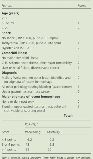
There are several risk-stratification schemes that can help physicians determine the need for urgent interven-tion and predict continued or recurrent bleeding after endoscopic therapy. The Rockall risk scoring system is useful in stratifying patients at higher risk of rebleeding and death and has been prospectively validated in differ-ent populations (Table 530,31).30
In stable patients with gastrointestinal bleeding, potentially ulcerogenic medications should be discon-tinued. A proton pump inhibitor should be administered intravenously; this reduces transfusion requirements, need for surgery, and duration of hospitalization, treatment of Peptic Ulcer Disease
figure 1. Algorithm for the treatment of peptic ulcer disease. (EGD = esophagogastroduodenoscopy; PPI = proton pump inhibitor; NSAID = nonsteroidal anti-inflammatory drug.)
*—Alarm symptoms include evidence of bleeding (e.g., anemia, heme-positive stool, melena), perforation (e.g., severe pain), obstruction (e.g., vomiting), and malignancy (e.g., weight loss, anorexia).
Patient presents with dyspepsia
Alarm symptoms* or age older than 55 years?
EGD (or barium studies if EGD not feasible)
Ulcer present?
Eradicate Helicobacter pylori
Administer antisecretory therapy for 4 to 8 weeks Treat bleeding or other
complications Biopsy gastric ulcer
Consider trial of antisecretory therapy (with PPI preferably, or a histamine H2 blocker)
and follow clinically
Detect and treat H. pylori infection Advise discontinuation of NSAIDS, smoking,
alcohol, and drug abuse
Administer antisecretory therapy (with PPI preferably, or an H2 blocker) for 4 weeks
Persistent symptoms
Continue H2 blocker or
PPI for 4 to 8 weeks
Response?
Consider EGD Recheck and treat
H. pylori, if persistent Check for noncompliance Consider hypersecretory states Observe or consider maintenance therapy with H2 blocker or PPI Good clinical response
Observe; consider long-term maintenance with H2 blocker or PPI
if symptoms recur Consider EGD if red flags or more severe symptoms
Yes No
Yes No
Peptic Ulcer Disease
although it does not reduce mortality.32 EGD should be performed to find characteristics that suggest a high rate of bleeding recurrence (e.g., ulcer larger than 1 cm, vis-ible or actively bleeding vessel).30
Patients whose condition is unstable should undergo fluid or packed-cell resuscitation followed by emergent EGD and coagulation of bleeding sites through endo-scopic ligation; placement of hemoclips; injection of epinephrine, alcohol, or a sclerosant; or a combination of methods.33
Oral antisecretory therapy should be initiated as soon as patients resume oral intake. Treatment of H. pylori infection is more effective than antisecretory therapy without eradication of H. pylori for preventing recurrent bleeding. Therefore, patients with bleeding peptic ulcers should be tested for H. pylori infection and should be prescribed eradication therapy if results are positive.34 If continued administration of aspirin or NSAIDs is required, concurrent administration of misoprostol or a proton pump inhibitor should be considered.35,36
Patients with nonhealing gastric ulcers should have biopsy to rule out cancer.
Angiographic embolization of bleeding vessels or surgery is indicated if a patient’s vital signs or laboratory studies suggest continued or recurrent bleeding.33 Surgi-cal options include gastroduodenotomy and oversew-ing of the blood vessel with or without vagotomy and drainage in duodenal ulcer; and excision of the ulcer with vagotomy and drainage or partial gastrectomy in bleeding gastric ulcers.
PerfOratiOn
Perforation occurs in approximately 2 to 10 percent of peptic ulcers.25 It usually involves the anterior wall of the duodenum (60 percent), although it may also occur in antral (20 percent) and lesser-curve (20 percent) gastric ulcers. Perforation of ulcers in children is rare.
Free peritoneal perforation and resulting chemical and bacterial peritonitis is a surgical emergency caus-ing sudden, rapidly spreadcaus-ing, severe upper abdominal table 4. treatment of Peptic Ulcers
Treatment Comment Options
Eradication of
Helicobacter pylori
Treatment duration is 10 to 14 days (although courses lasting one to seven days have been reported to have comparable effectiveness21,22)
Eradication rates 80 to 90 percent or higher
Omeprazole (Prilosec) 20 mg two times daily or lansoprazole (Prevacid) 30 mg two times daily
plus amoxicillin 1 g two times daily or metronidazole (Flagyl) 500 mg two times daily (if allergic to penicillin)
plus clarithromycin (Biaxin) 500 mg two times daily Ranitidine bismuth citrate (Tritec)* 400 mg two times daily
plus clarithromycin 500 mg two times daily or metronidazole 500 mg two times daily
plus tetracycline 500 mg two times daily or amoxicillin 1 g two times daily Levofloxacin (Levaquin) 500 mg daily
plus amoxicillin 1 g two times daily
plus pantoprazole (Protonix) 40 mg two times daily
Bismuth subsalicylate (Pepto-Bismol) 525 mg (two tablets) four times daily
plus metronidazole 250 mg four times daily
plus tetracycline 500 mg four times daily
plus H2 blocker for 28 days or proton pump inhibitor for 14 days
Histamine H2
blockers
70 to 80 percent healing in duodenal ulcer after four weeks, 87 to 94 percent after eight weeks
Ranitidine (Zantac) 150 mg two times daily or 300 mg at night Famotidine (Pepcid) 20 mg two times daily or 40 mg at night Cimetidine (Tagamet) 400 mg two times daily or 800 mg at night Proton pump
inhibitors
Treatment duration is four weeks for duodenal ulcer and eight weeks for gastric ulcer
80 to 100 percent healing
Omeprazole 20 mg daily Lansoprazole 15 mg daily Rabeprazole (Aciphex) 20 mg daily Pantoprazole 40 mg daily Sucralfate
(Carafate)
Treatment duration is four weeks Effectiveness similar to H2 blockers
1 g four times daily
Surgery Rarely needed Duodenal ulcer: truncal vagotomy, selective vagotomy, highly selective vagotomy, partial gastrectomy
Gastric ulcer: partial gastrectomy with gastroduodenal or gastrojejunal anastomosis
*—Not available in the United States.
pain exacerbated by movement; the pain may radiate to the right lower abdomen or to both shoulders. Fever, hypotension, and oliguria suggest sepsis and circula-tory compromise. Generalized abdominal tenderness, rebound tenderness, board-like abdominal wall rigidity, and hypoactive bowel sounds (clinical signs of perito-nitis) may be masked in older patients and those taking steroids, immunosuppressants, or narcotic analgesics. Upright or lateral decubitus abdominal radiography or erect chest radiography may demonstrate pneumoperi-toneum; however, the absence of this finding does not rule out perforation.17 Sonography, computed tomogra-phy, and gastroduodenography are confirmatory.
Initial resuscitation with large-volume crystalloids; nasogastric suction; and administration of intravenous
broad-spectrum antibiotics against gram-negative rods, anaerobes, and oral flora are usually followed by lapa-rotomy and placement of an omental patch (Graham patch plication) in patients with perforated duodenal ulcers. In otherwise healthy patients with a history of chronic ulcer and minimal peritoneal contamination, a concurrent, definitive, anti-ulcer procedure (e.g., vagot-omy and drainage, highly selective vagotvagot-omy) may also be considered. Perforated gastric ulcers are treated with an omental patch, wedge resection of the ulcer, or a par-tial gastrectomy and reanastomosis. Coexisting H. pylori infection should be eradicated to reduce recurrence and minimize the need for long-term antisecretory therapy and further surgical intervention.25,37 In older patients, mortality rates from perforation and its management may be as high as 30 to 50 percent.1
GastriC OUtlet ObstrUCtiOn
Peptic ulcer disease is the underlying cause in less than 5 to 8 percent of patients presenting with gastric outlet obstruc-tion. Patients with recurrent duodenal or pyloric channel ulcers may develop pyloric stenosis as a result of acute inflammation, spasm, edema, or scarring and fibrosis.
Symptoms suggesting obstruction include recurrent epi-sodes of emesis with large volumes of vomit containing undigested food; persistent bloating or fullness after eat-ing; and early satiety. Weight loss, dehydration, and a hypo-chloremic, hypokalemic metabolic alkalosis may result; a tympanitic epigastric mass representing the dilated stom-ach with visible gastric peristalsis also may be observed.
EGD or gastroduodenography (using diatrizoate meglumine and diatrizoate sodium [Gastrografin] or barium) is recommended to determine the site, cause, and degree of obstruction. Malignancy, a more com-mon cause of obstruction (responsible for more than 50 percent of cases), should be ruled out.38 Obstruction resulting from acute inflammation or edema responds well to nasogastric decompression, administration of H2 blockers or proton pump inhibitors, and eradication of H. pylori. Prokinetic agents should be avoided. Endo-scopic pyloric balloon dilatation or surgery (vagotomy and pyloroplasty, antrectomy, or gastroenterostomy) are options to relieve chronic obstruction.25
the authors
KALYANAKRISHNAN RAMAKRISHNAN, MD, FRCSE, is an associate pro-fessor in the Department of Family and Preventive Medicine, University of Oklahoma Health Sciences Center, Oklahoma City. He received his medical degree and his master’s degree in surgery from the Jawaharlal Institute, Pondicherry, India, and was awarded the Fellowship of the Royal College of Surgeons of Edinburgh, Scotland. Dr. Ramakrishnan completed a family practice residency at the University of Oklahoma Health Sciences Center. table 5. rockall risk scoring system for Patients with Peptic Ulcer Disease Feature Points age (years) < 60 0 60 to 79 1 > 79 2 shock No shock (SBP ≥ 100, pulse < 100 bpm) 0 Tachycardia (SBP ≥ 100, pulse ≥ 100 bpm) 1 Hypotension (SBP < 100) 2 Comorbid illness
No major comorbid illness 0
CHF, ischemic heart disease, other major comorbidity 2 Liver or renal failure, disseminated cancer 3
Diagnosis
Mallory-Weiss tear, no other lesion identified and no stigmata of recent hemorrhage
0 All other pathology causing bleeding (except cancer) 1 Upper gastrointestinal tract cancer 2
Major stigmata of recent hemorrhage
None or dark spot only 0
Blood in upper gastrointestinal tract, adherent clot, visible or spurting vessel
2 total: Score Risk (%)* Rebleeding Mortality < 3 points 6.2 0.2 3 or 4 points 13 6.8 > 4 points 25 20
SBP = systolic blood pressure (mm Hg); bpm = beats per minute; CHF = congestive heart failure.
*—Interpretation using data from two independent validation studies.31
Adapted with permission from Rockall TA, Logan RF, Devlin HB, North-field TC. Risk assessment after acute upper gastrointestinal haemorrhage. Gut 1996;38:318, with additional information from reference 31.
Peptic Ulcer Disease
ROBERT C. SALINAS, MD, is an assistant professor in the Department of Family and Preventive Medicine, University of Oklahoma Health Sciences Center. He received his medical degree from the American University of the Caribbean School of Medicine, Montserrat, British West Indies. Dr. Salinas completed a family medicine residency and a fellowship in geriat-rics at the University of Oklahoma Health Sciences Center.
Address correspondence to Kalyanakrishnan Ramakrishnan, MD, University of Oklahoma Health Sciences Center, Department of Family and Preventive Medicine, 900 NE 10th St., Oklahoma City, OK 73104 (e-mail: kramakrishnan@ouhsc.edu). Reprints are not available from the authors.
Author disclosure: Nothing to disclose.
referenCes
1. University of Michigan Health System. Peptic ulcer disease. Accessed May 4, 2007, at: http://www.cme.med.umich.edu/pdf/guideline/PUD05.pdf. 2. Sonnenberg A, Everhart JE. The prevalence of self-reported peptic ulcer
in the United States. Am J Public Health 1996;86:200-5.
3. Kang JY, Tinto A, Higham J, Majeed A. Peptic ulceration in general practice in England and Wales 1994-98: period prevalence and drug management. Aliment Pharmacol Ther 2002;16:1067-74.
4. Kurata JH, Nogawa AN. Meta-analysis of risk factors for peptic ulcer. Nonsteroidal anti-inflammatory drugs, Helicobacter pylori, and smok-ing. J Clin Gastroenterol 1997;24:2-17.
5. Ziegler AB. The role of proton pump inhibitors in acute stress ulcer prophylaxis in mechanically ventilated patients. Dimens Crit Care Nurs 2005;24:109-14.
6. NIH Consensus Conference. Helicobacter pylori in peptic ulcer disease. NIH Consensus Development Panel on Helicobacter pylori in Peptic Ulcer Disease. JAMA 1994;272:65-9.
7. Nilsson C, Sillen A, Eriksson L, Strand ML, Enroth H, Normark S, et al. Correlation between cag pathogenicity island composition and
Helicobacter pylori-associated gastroduodenal disease. Infect Immun 2003;71:6573-81.
8. Hopkins RJ, Girardi LS, Turney EA. Relationship between Helicobacter pylori eradication and reduced duodenal and gastric ulcer recurrence: a review. Gastroenterology 1996;110:1244-52.
9. Bytzer P, Teglbjaerg PS, for the Danish Ulcer Study Group. Helicobacter pylori-negative duodenal ulcers: prevalence, clinical characteristics, and prognosis—results from a randomized trial with 2-year follow-up. Am J Gastroenterol 2001;96:1409-16.
10. Huang JQ, Sridhar S, Hunt RH. Role of Helicobacter pylori infection and non-steroidal anti-inflammatory drugs in peptic-ulcer disease: a meta-analysis. Lancet 2002;359:14-22.
11. Graham DY. Nonsteroidal anti-inflammatory drugs, Helicobacter pylori, and ulcers: where we stand. Am J Gastroenterol 1996;91:2080-6. 12. Collier DS, Pain JA. Non-steroidal anti-inflammatory drugs and peptic
ulcer perforation. Gut 1985;26:359-63.
13. Lanas A, Serrano P, Bajador E, Esteva F, Benito R, Sainz R. Evidence of aspirin use in both upper and lower gastrointestinal perforation. Gas-troenterology 1997;112:683-9.
14. Spiegelhalter DJ, Crean GP, Holden R, Knill-Jones RP. Taking a calculated risk: predictive scoring systems in dyspepsia. Scand J Gastroenterol Supp 1987;128:152-60.
15. Cappell MS. Gastric and duodenal ulcers during pregnancy. Gastroen-terol Clin North Am 2003;32:263-308.
16. Hilton D, Iman N, Burke GJ, Moore A, O’Mara G, Signorini D, et al. Absence of abdominal pain in older persons with endoscopic ulcers: a prospective study. Am J Gastroenterol 2001;96:380-4.
17. Martinez JP, Mattu A. Abdominal pain in the elderly. Emerg Med Clin North Am 2006;24:371-88.
18. Hassall E. Peptic ulcer disease and current approaches to Helicobacter pylori. J Pediatr 2001;138:462-8.
19. Talley NJ, Vakil NB, Moayyedi P. American Gastroenterological Associa-tion technical review on the evaluaAssocia-tion of dyspepsia. Gastroenterology 2005;129:1756-80.
20. Ables AZ, Simon I, Melton ER. Update on Helicobacter pylori treatment. Am Fam Physician 2007;75:351-8.
21. Lara LF, Cisneros G, Gurney M, Van Ness M, Jarjoura D, Moauro B, et al. One-day quadruple therapy compared with 7-day triple therapy for
Helicobacter pylori infection. Arch Intern Med 2003;163:2079-84. 22. Treiber G, Wittig J, Ammon S, Walker S, van Doorn L, Klotz U.
Clini-cal outcome and influencing factors of a new short-term quadruple therapy for Helicobacter pylori eradication: a randomized controlled trial (MACLOR study). Arch Intern Med 2002;162:153-60.
23. Poynard T, Lemaire M, Agostini H. Meta-analysis of randomized clinical tri-als comparing lansoprazole with ranitidine or famotidine in the treatment of acute duodenal ulcer. Eur J Gastroenterol Hepatol 1995;7:661-5. 24. Vakil N, Fennerty MB. Direct comparative trials of the efficacy of proton
pump inhibitors in the management of gastro-oesophageal reflux dis-ease and peptic ulcer disdis-ease. Aliment Pharmacol Ther 2003;18:559-68. 25. Behrman SW. Management of complicated peptic ulcer disease. Arch
Surg 2005;140:201-8.
26. Lanas AI, Remacha B, Esteva F, Sainz R. Risk factors associated with refractory peptic ulcers. Gastroenterology 1995;109:1124-33. 27. Peura DA. Prevention of non-steroidal anti-inflammatory drug-associated
gastrointestinal symptoms and ulcer complications. Am J Med 2004;177 (suppl 5A):63S-71S.
28. Palanivelu C, Jani K, Rajan PS, Kumar KS, Madhankumar MV, Kadalakat A. Laparoscopic management of acid peptic disease. Surg Laparosc Endosc Percutan Tech 2006;16:312-6.
29. Hernandez-Diaz S, Rodriguez LA. Incidence of serious upper gastro-intestinal bleeding/perforation in the general population: review of epidemiologic studies. J Clin Epidemiol 2002;55:157-63.
30. Rockall TA, Logan RF, Devlin HB, Northfield TC. Risk assessment after acute upper gastrointestinal haemorrhage. Gut 1996;38:316-21. 31. Ebell MH. Prognosis in patients with upper GI bleeding. Am Fam
Physi-cian 2004;70:2348-50.
32. Leontiadis GI, Sharma VK, Howden CW. Systematic review and meta-analysis: proton-pump inhibitor treatment for ulcer bleeding reduces transfusion requirements and hospital stay—results from the Cochrane Collaboration. Aliment Pharmacol Ther 2005;22:169-74.
33. Eisen GM, Dominitz JA, Faigel DO, Goldstein JL, Kalloo AN, Petersen BT, et al., for the American Society for Gastrointestinal Endoscopy. Standards of Practice Committee. An annotated algorithmic approach to upper gastrointestinal bleeding. Gastrointest Endosc 2001;53:853-8. 34. Gisbert JP, Khorrami S, Carballo F, Calvet X, Gene E, Dominguez-Munoz
E. Meta-analysis: Helicobacter pylori eradication therapy vs. antisecre-tory non-eradication therapy for the prevention of recurrent bleeding from peptic ulcer. Aliment Pharmacol Ther 2004;19:617-29.
35. Silverstein FE, Graham DY, Senior JR, Davies HW, Struthers BJ, Bittman RM, et al. Misoprostol reduces serious gastrointestinal complications in patients with rheumatoid arthritis receiving nonsteroidal anti-inflam-matory drugs. A randomized, double-blind, placebo-controlled trial. Ann Intern Med 1995;123:241-9.
36. Rostom A, Dube C, Wells G, Tugwell P, Welch V, Jolicoeur E, et al. Pre-vention of NSAID-induced gastroduodenal ulcers. Cochrane Database Syst Rev 2002;(4):CD002296.
37. Ng EK, Lam YH, Sung JJ, Yung MY, To KF, Chan AC, et al. Eradication of Helicobacter pylori prevents recurrence of ulcer after simple closure of duodenal ulcer perforation: randomized controlled trial. Ann Surg 2000;231:153-8.
38. Shone DN, Nikoomanesh P, Smith-Meek MM, Bender JS. Malignancy is the most common cause of gastric outlet obstruction in the era of H2 blockers. Am J Gastroenterol 1995;90:1769-70.