Final Report Contract N01-LM-0-3508
Human Embryology Digital Library and Collaboratory Support Tools J. Mark Pullen, Principal Investigator
George Mason University 15 June 2003
EXECUTIVE SUMMARY
The “Visible Embryo” project has created advanced software for networked biomedical collaboration in areas of cellular-level databases with voxel-level annotation, distributed embryology education, and clinical case management. The eight member institutions have completed the activities called for in our contract:
1. The Armed Forces Institute of Pathology (AFIP), with the assistance of Dr. Maury Pescitelli and various medical school groups, has completed the final three embryos for the repository funded under this project. Dr. Ray Gasser of Louisiana State University (LSU) served as a consultant to AFIP and periodically as a participant with the biomedical collaboration technology,
2. Eolas Technologies Inc. (ETI) has completed integrated Synchronous and Asynchronous Annotators and finalized their documentation.
3. IRB approval for trans-vaginal ultrasound at John Hopkins Medical Institution (JHMI) has been received. Unfortunately time and funding do not permit completion of the planned study within this project. However, Dr. Paidas is seeking other funding for the project.
4. Lawrence Livermore National Laboratory (LLNL) has provided system engineering and coordination services for transition of the project’s eight sites to the Next-Generation Internet (NGI).
5. Oregon Health Sciences University (OHSU) has led the pilot annotation project for our ground-breaking Carnegie Embryo 836, and participated in additional annotation and embryology education activities.
6. San Diego Supercomputer has placed its new visualization software online, permitting visualized fly-though of three-dimensional embryo models from sites across the NGI. 7. University of Illinois at Chicago (UIC) has completed a sequence of over 20 educational
animations, and also completed a sequence of four recorded “master classes” in embryology, in partnership with the Dr. David Bolender of the Medical College of Wisconsin, using the Network EducationWare (NEW) software developed by GMU.
8. GMU has completed the graphic and movie extensions the Network EducationWare (NEW) whiteboard, upgraded the NEW record/playback system for a video capability, and has improved the audio recording process for use in the UIC master classes. NEW is available as open-source software from GMU.
9. Plans are in progress for a full-scale paper to be submitted to Science, describing advances in biomedical and information technologies achieved by the project, to build on an initial one-page article in Science, September 6, 2002.
10. The project has reported its results in several conferences and publications. These included demonstrations at the Radiological Society of North America 2000 meeting in Chicago,
Illinois; Supercomputing 2001 in Denver, Colorado; and the Internet2 2002 Members Meeting in Arlington, Virginia.
PROJECT OVERVIEW
The Human Embryology Digital Library and Collaboratory Support Tools project, often called the “Visible Embryo” or VE project for short, was begun in 1999 as a demonstration of the biomedical application potential of the Next Generation Internet (NGI). Performers included the eight
organizations listed above, at sites around the continental USA as listed in Table 1, a mix of
medical and information technology organizations. The project undertook three major applications, based on the Carnegie Collection of Embryos at the AFIP’s National Museum of Health and Medicine Human Development Anatomy Center (HDAC), a collection of cellular-level tissue slides that is one of the world’s largest repositories of human embryos.
1. Digitization, curation and annotation of embryo data: We created a production digitization capability, using automated digital microscopy, with data automatically registered for tiling and transmitted to the repository at San Diego Supercomputer, and annotated by teams of biomedical volunteers with expert-level quality control.
2. Distributed embryology education using materials derived from the Carnegie Collection to create animations of embryo development and recorded master classes that can be streamed over the Internet or downloaded to create a portable electronic classroom.
3. Clinical management planning where medical professionals and expectant parent patients can review normal and abnormal development patterns with collaborative consultation from distant experts.
More details on these applications can be found on http://netlab.gmu.edu/visembryo, the project website, which also contains links to websites of all VE team organizations listed below.
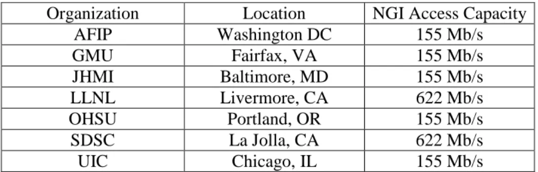
Organization Location NGI Access Capacity
AFIP Washington DC 155 Mb/s GMU Fairfax, VA 155 Mb/s JHMI Baltimore, MD 155 Mb/s LLNL Livermore, CA 622 Mb/s OHSU Portland, OR 155 Mb/s SDSC La Jolla, CA 622 Mb/s UIC Chicago, IL 155 Mb/s
MEMBER ORGANIZATION CONTRIBUTIONS
National Museum of Health and Medicine / Armed Forces Institute of Pathology (American Registry of Pathology)
Human Developmental Anatomy Center staff and a Nikon representative completed the set up of two microscope workstations, each consisting of a Dell Pentium 550 computer, a NIKON E800 compound microscope and a SONY DTK-s5 digital video camera. An off-the-shelf software package was located which could accomplish most requirements. GMU graduate student was assigned to the project (Dave Brooks) to extend its automation capabilities and easily modified this package to address most remaining needs: driving the stage and camera, identifying bounding boxes (AUI) and identifying the orientation coordinates. Each project embryo was imaged at 2X and 20X. The 2x images provide an image for use in creating bounding boxes for tiling,
registration, color and white balance, and correlating the higher magnification image with the original.
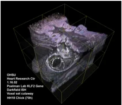
The first embryo selected, Carnegie Number 836, is one that has been extensively studied and was a useful learning process for project participants. Embryo 836 was completed, after dealing with the need to rescan several slides due to problems with the auto focus not handling tissue fluctuations well. The data for 836 were completed and sent to SDSC and LSU in support of ongoing annotation efforts. The auto focus issue was resolved by inserting a pause to allow the technician to verify the focus and correct when necessary. A variability of 2-8 pixels in the 40-pixel overlap of the image tiles, due to the mechanics of stage movement was minimized as much as possible, but some variability will continue to exist. Part of embryo 836, in the process of annotation, is shown in Figure 1.
Embryo 5074 was imaged but it was subsequently determined that the upper portions of the embryo have degraded too much to provide the necessary information for histology or modeling. (This illustrates the biomedical and archival value of the VE project’s digitization efforts.) Embryo 6330 was chosen as a replacement. A high-speed network extension was completed from the NLM to HDAC and the MACAW, imaging and storage machines were migrated to it. The final installation of the automated data transfer was completed, along with improvements in viewing tools. Dave Brooks, though graduated and officially off the project, continued to make improvements to the auto-capture software as his schedule allowed, and aided in the final installation of the transfer software.
With the recognition that annotation staffing remains a critical bottleneck given these higher levels of source automation, AFIP expanded its efforts to work with student help. This included medical students from other participating organizations plus college and high school biology students, supervised by appropriate professionals. Several summer interns worked on various parts of the project. One intern was responsible for scanning photo file images and documents, while another finished 2 models started during the school year. The models are of embryo 5074, one from the bromide data set and the other from the registered tissue sections captured at 10X. Products include a digital animation of the plaster models created for this embryo. These products will provide a baseline for comparing models and images created from the new data sets with models from the
old. A third intern scanned reprints and documents relating to the project embryos, which were added to the collection of documents on the AFIP web site.
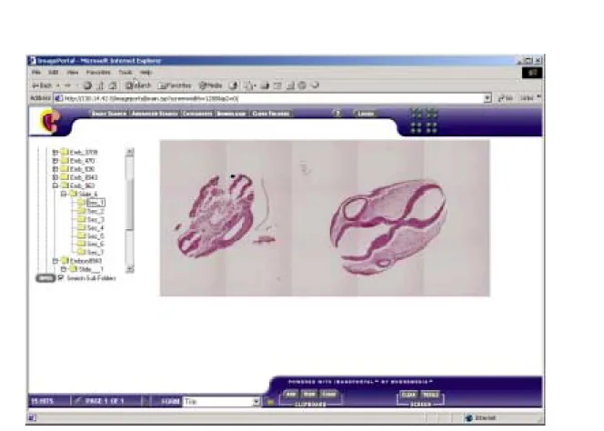
By project end AFIP had placed a total of 15 embryos in the archive at SDSC, of which three are annotated. Annotation using high school students to do outlining in slices that are sandwiched between slices annotated by experienced biomedical professionals has proved quite successful. AFIP has assembled a large collection of documents on their web site, describing work in the project. AFIP is hosting a second copy of the databases from SDSC, to provide a backup source and also in case SDSC is unable to obtain support to maintain the database online. Figure 1 shows a typical Web-based access to the system.
Figure 1: AFIP Website Embryo Access
Eolas Technologies, Inc.
The initial cellular-level annotation software for the VE Project consisted of three major software components:
1. Collaborative image browser/annotator: This allows multiple users to load image(s) over the network, zoom into an image from an entire-section view up to cellular detail, and
allows for outlining and annotation of objects within section images. Zoom integration allows synchronizing the annotation shapes with the zoom/pan parameters, so that
annotation polygons stay in register with the section images as the user is navigating around the image. The groupware is synchronized across multiple clients. It allows multiple users to view, zoom and pan around, outline and annotate structures in a synchronized (across users) fashion.
2. GroupVisualization: This software allows multiple users over the net to explore 3D annotated datasets, where the visualization sessions are synchronized among the users. Multiple users can connect to the VIS server and each user sees the same visualization images as the other user(s). When one user manipulates the interface, the other user(s) experience those manipulations in their own GUIs and see the resultant visualizations simultaneously.
3. Multimedia Collaboration Tools: GMU provided multi-session video, text and audio teleconferencing capabilities based on the “MBone tools,” which are open-source software developed by the multimedia multicast community; the Windows NT version came from University College, London UK. Because high performance networks don’t all have
multicast, we use a multicast overlay built from unicast tunnels created by GMU. Integrated in the Visible Embryo collaboration environment, the tools provide a strong multimedia capability: full motion video (if you turn the knobs up high), full duplex audio, shared whiteboard with color graphics import, and a text tool for chatting. They are implemented in TCL/TK, which meant they were easy to integrate with the software Eolas developed. Eolas also provided extremely useful support for project coordination in the form of a web-based conference board. Dr. Michael Doyle of Eolas also provided supervision for GMU doctoral student Yongjian Guo, who developed techniques of warping three-dimensional datasets for investigation of gene expression in embryo datasets.
The final version of the software developed by Eolas consists of five major components:
1. a database server that provides navigation through and management of the database records for the project
2. the annotation and zoom server, which manages synchronized zooming/panning/annotation of section images
3. the visualization server, which provides navigation and visualization of embryo 3D volume reconstructions
4. the session manager, which coordinates the group-wise synchronization of the annotation and visualization client modules, and
5. the client GUI, which integrates into a single easy-to-use interface the client modules that provide access to the various server resources.
Eolas has refined the annotation tools to enable production-level annotation of embryo image data. Working with Dr. Pescitelli, both the OHSU and Johns Hopkins teams have used the tool to
annotate embryo image data. The updated annotation tool was integrated with the SDSC
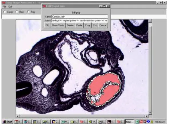
visualization of geographically distributed groups working from a common database. Figure 2 shows an embryo section in the process of annotation.
Figure 2: Annotation of Embryo
Johns Hopkins Medical Institution
The initial JHMI challenge under the VE project was obtaining the network conenctivity required to participate. Their PI, Dr. Paidas, obtained a commitment from our Networking and
Telecommunications Services as well as dedicated secured space for project equipment. With limited initial connectivity, JHMI was still an active VE collaborator through the WebBoard, conference calls and strategy sessions in San Diego and GMU, thus staying abreast of the latest technological developments and project planning. They guaranteed a commitment to the annotation, embryology education and clinical planning applications for the project. Also they provided a digitized Stage 13 human embryo that was be used as a model for the first project demonstration. Medical illustrator Bob Morreale was instrumental in preparing these and many other graphics used in the project.
JHMI led the Clinical Management Planning application from Summer, 2001. Planning for this application was accelerated in order to begin work somewhat earlier with the intention to have more significant results by the end of the project. At the second annual project meeting, PI Chuck Paidas presented a plan for this application, which was adopted by the overall team after
discussion. The concept is to use transvaginal patient ultrasound images in the collaboration environment as a basis for clinical comparisons with our annotated Carnegie embryo
reconstructions. The plan called for scanning four in-vitro fertilized patients every other day for the first trimester of their pregnancy. The transvaginal scan was to consist of a standard embryo
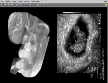
evaluation using currently accepted probes and focal distances. A minimum of 8 morphological landmarks was to be evaluated and compared to existing Carnegie embryos. Unfortunately, Internal Review at the Johns Hopkins Hospital turned out to be extremely slow, although no reasons was expressed for rejection of the first trimester sonography because it is accepted as a safe method in the literature. The project team agreed that this approach was sufficiently desirable to reallocate a small amount of the project's equipment budget if necessary. While waiting for IRB approval, JHMI supported the project by annotating the urogenital region of embryo 836. Dr. Paidas did not receive has received an initial response from the Johns Hopkins IRB until Spring, 2003. At this point he worked to deal with their questions so the study could proceed receiving final approval just at the end of the project. He is seeking support to carry out this promising study. Figure 3 contains a comparison of ultrasound and Carnegie images developed as part of the JHMI effort.
Figure 3: UFL volume rendering of MRM image from a Stage 14 embryo And JHMI Rhombencephalon Saggital View Stage 18
Lawrence Livermore National Laboratory (LLNL)
The architecture of the “Visible Embryo” networked collaboration is depicted in Figure 4. LLNL was responsible for coordination and facilitation of high performance networking in the project, to leverage the on-going Next Generation Internet (NGI) and NGI Supernet research programs at minimum expense and position the collaboration for anticipated developments in commercial telecommunications infrastructure. (LLNL’s contract is with NLM directly -- that is, they are not a subcontractor to GMU -- due to policy issues between NLM and their primary sponsor, the
UIC were connected to high-performance networks. We did not find a cost-effective way to connect the Eolas site near Chicago at comparable data rates because the Abilene will not connect for-profit organizations. However, by placing all software requiring higher performance at AFIP, GMU and SDSC there has been no impact on performance at other locations. We had pursued establishing a connection to Eolas via nearby DePaul University and would re-examine that option for a future research collaboration.
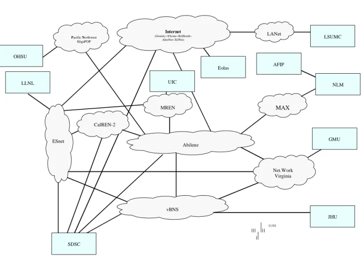
With help from NLM personnel, particularly Network Engineer Mike Gill, the project developed a special communications path to AFIP via NLM. The campus high-speed research network was changed from an ATM connection via Bell Atlantic into ATDnet, to a dedicated fiber from AFIP to NLM as an extension of NLM’s high performance network connection. The new path offers higher performance and overall lower cost. The initial connection uses Gigabit Ethernet between AFIP and NLM and uses NLM’s existing 155 Mbps connection to the national research network infrastructure. OHSU LLNL UIC JHU Eolas GMU Abilene vBNS Net.Work Virginia AFIP SDSC Internet (Genuity+@home+BellSouth+ AlterNet+XONet) Pacific Northwest GigaPOP CalREN-2 MREN MAX LANet LSUMC NLM ESnet 11/01
Figure 4: Project NGI Connectivity
Our overall project goal had been to reach OC3 (155 Mbps) network capacity at all sites within 2001, and continue to press on toward OC12 (622 Mbps) for as many sites as possible by the end of the project. It now appears that connections beyond OC3 will not take place within the project
lifetime at sites other than LLNL and SDSC, which were NGI hubs. The reason for this is that participating academic campuses have found costs of the next higher data rate (OC12) to outweigh the potential benefits at this time. When the OC3 network pipes fill up, we expect to see investment in higher capacity.
LLNL also provided the planning and coordination for the project’s Supercomputing and
Communications 2001 conference (SC2001) presentation over the Access Grid. Sites used on the Grid were Arlington, VA; Argonne, IL; SC2001 Denver; and LLNL. The 40-minute presentation featured full-motion video from all sites and network delivery of the image from the project’s 2D and 3D collaboration software. Observers from all over the world joined the multi-cast presentation to see a geographically distributed presentation of a geographically distributed collaboration
including the interactive annotation of embryology images.
Oregon Health Sciences University
OHSU spearheaded to VE project’s effort be to label selected regions of embryo 836. As such, they deal patiently with all the pains of being an “early adopter” of the collaborative annotation
technology and helped to shape its development into the current, much more mature, form. Using digitized histologic sections from the Carnegie Collection, they applied our annotation process to cardiac and lung tissues, supporting structures, and differentiating tissue types in these regions. Dr. Kent Thornburg of OHSU has a compelling interest in gene expression, and has developed
exceptional visualizations in this area, as represented by Figure 5.
In the annotation application, OHSU also coordinated the annotation process by UIC and JHMI and played a lead role in establishing consistent nomenclature for the annotations. The nomenclature used deals only with components that can be distinguished morphologically and follows a scheme developed collaboratively with researchers at the University of Washington (Rosse, Brinkley). Eventually it became apparent that one individual with an expert overview of embryology should organize the annotation. Dr. Maury Pescitelli, who was at the time serving as a consultant to Eolas, took on this role in close coordination with OHSU. Collaborative leadership of Dr. Pescitelli, teamed with Dr. Jeff Pentecost of OHSU, proved so productive that GMU extended Dr. Pescitelli’s availabilty for the duration of the project, as a direct consultant to GMU. Dr. Pentecost also is leading an ongoing effort by team members to document the project’s accomplishments in the professional literature, hopefully by a paper in Science or Nature.
Figure 5: OHSU Gene Expression Visualization
San Diego Supercomputer Center
SDSC tasks within the VE team included archival storage support, installation and support for the distributed data handling system, support for the Digital Embryo collection catalog, and support for advanced 3D visualization.
In archiving, SDSC has provided online repository storage with tape cartridge backup, and arranged for offsite storage of a second copy at CalTech against the possibility of disaster.
For the data handling system, SDSC made available their Storage Resource Broker, a
meta-database developed under other projects that organizes access to repository assets distributed across the Internet: SDSC installed the SRB at George Mason University AFIP. The tests show that over a suitably tuned network, data transfer rates of 8 MB/second can be sustained through use of two I/O streams. The tests were conducted using the SRB container technology, in which multiple data sets were written into a 300 MB container on disk cache. When the container was full, it was then written into the archival storage system. Transaction processing tests at SDSC were conducted on the sustainable data ingestion rate. Over one million files can be added to the SRB metadata catalog on a daily basis. SRB supports the Grid Security Infrastructure, using a challenge-response based authentication system in addition to the PKI based Grid Security Infrastructure system. It also provides for XML-formatted metadata retrieval and ingestion, and has had a continuing record of performance enhancements. This version supports. The SRB data handling system has been integrated into the image management scripts at SDSC, automating the movement of images from AFIP to SDSC. Access to the VE digital library is primarily through the AFIP web site. Images are retrieved from SDSC by AFIP for non-project access. The SDSC SRB Windows NT/2000 browser is used for retrieval of images by project partners, directly from SDSC. The latest release of the
SRB technology, developed under NSF, NASA, and DOE funding, provides the metadata access controls requested by the VE Project, as well as support for bulk metadata load, and tape access. In the VE catalog system, SDSC worked with AFIP on the organization of metadata attributes, and the specification of the catalog structure using XML tags, resulting in creation of a metadata catalog at SDSC and a backup catalog at AFIP.
In advanced visualization, as part of the NPACI project, a 3D hierarchical image compositing system has been created based upon the MPIRE volume rendering system developed at SDSC. SDSC has been very effective in adapting available software for the purpose of embryo
visualization. Using the Vista 3D rendering system, they are able to access data stored in a Storage Resource Broker collection on a remote storage repository. This makes it possible to visualize registered images from the Digital Embryo collection, from either disk cache, or directly from the archive. Tests supported by NPACI used the MPIRE system to conflate 3D volumes from differing modalities with the visible human data set. The same techniques can be used to support conflation of digitized section data and ultrasound images. A Java user interface for the MPIRE system was provided to the VE Project and used at the CENDI demonstration in January, 2003. Figure 6 shows a fly-through graphic from SDSC’s work.
SDSC supported storage of images created by AFIP into an archival storage system at SDSC. The total number of embryos stored is now 15 embryos. The embryo Carnegie Collection numbers are 1380, 1878, 3709, 391, 470, 5074, 5960, 6330, 6344, 779, 792, 836, 8943, 963, and 966. The bulk of the data stored in TIF images, comprising 43,099 files, and the total data file size is about 612 GB. The images are replicated from the SDSC HPSS archive into the Caltech HPSS archival storage system. The total number of files stored at SDSC is twice as large, since a compressed browse image is stored in addition to the full resolution TIF image.
The rate of storage/access from AFIP to for large data transfers was an issue during most of the project. For the first two year, the highest rates achieved were around 150 KB/s. By the end of the project, rates up to 11 MB/s were achieved.
Figure 6: SDSC Embryo Fly-Through Rendering
University of Illinois at Chicago
Under leadership of Dr. Mark Holterman, UIC led the distributed embryology education application in the VE project, in addition to participating as a member of the core management team and assisting with annotation of the foregut area of embryos. An early achievement in
embryology education was recruitment Dr. David Bolender of the Department of Anatomy and Cell Biology at the Medical College of Wisconsin to help with the annotation and educational materials. Dr. Bolender’s experience in teaching embryology for many years was invaluable, as was his research interest in cardiac development. In addition, Dr. Oladapo Ashiru who teaches embryology at the UIC and the Rush Schools of Medicine and Dr. Ra-id Abdulla, who is a pediatric
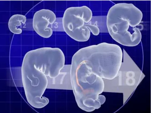
cardiologist, have supported UIC’s role in the project. UIC also brought to the project computer animator Greg Blew who assisted in the preparation of the educational animations and interactive educational programs that have achieved pivotal effect in embryology education. Images from one of the more compelling animations are shown in Figure 7.
Dr. Holterman also has served as a highly effective advocate for the VE project, making presentations to a joint meeting of the American Clinical Anatomy Association and the British Clinical Anatomy Association in July of 2000 and the American Association of Clinical Anatomists in June 2002 that sparked considerable interest in the medical community.
George Mason University
GMU provided overall organization, management, logistical support, and networked collaboration technology for the project. This has included:
The project has held a series of planning and coordinating meetings, roughly every six months, at various member organization locations. The format has been one half day of status reports followed by another half day of plan review aimed at enhancing project results through more effective
collaboration.
The project has held teleconferences roughly every two weeks to track plan execution and support detailed cooperation among the member organizations in achieving our goals.
The project has held a major demonstration yearly at the National Library of Medicine (NLM). The initial demonstration consisted of “local” technology, supportable in a single workstation plus network-based services. The second demonstration consisted of “regional” technology, supportable in two workstations, with synchronous collaborative annotation as a specific example. The third demonstration consisted of “national” technology, representing the capability of three or more workstations distributed around the USA to collaborate effectively over high resolution data for annotation, research, teaching, and clinical activity. The final demonstration was included in a presentation to the CENDI group at NLM, showcasing the capabilities we have developed and demonstrating a combination of synchronous and asynchronous annotation, synchronous and asynchronous distributed embryology education, and high-potential clinical activities.
The project also developed a CDROM with vignettes, recorded as presented “over the net,” by each member organization. We also presented a live version of the same materials at two national
conferences: Supercomputing and Communications 2001 in Denver, CO and Internet2 Spring 2002 in Arlington, VA.
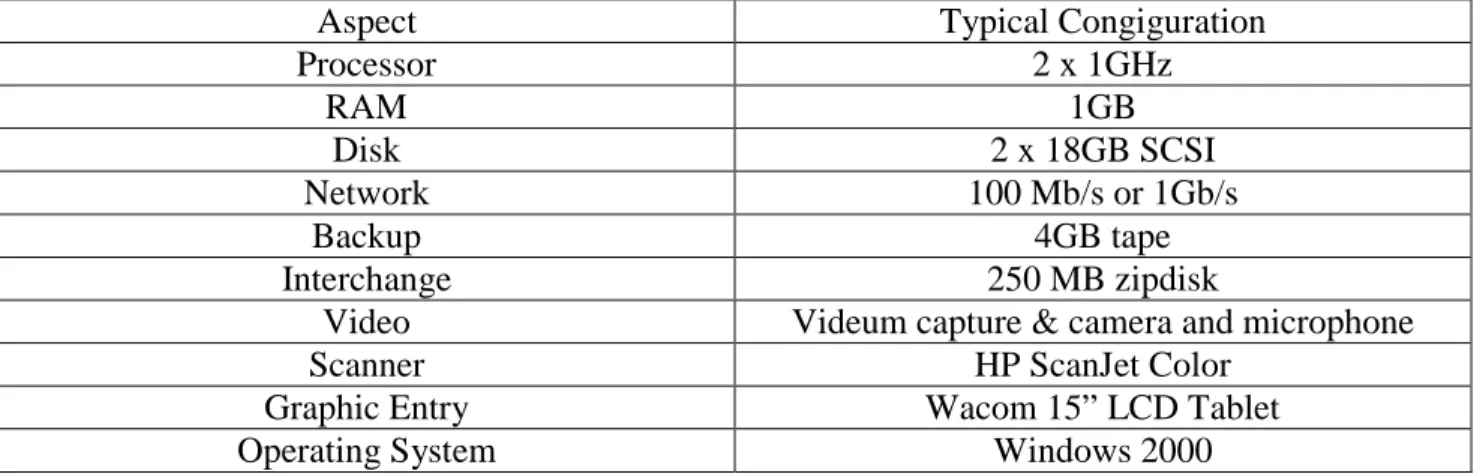
Aspect Typical Congiguration
Processor 2 x 1GHz RAM 1GB Disk 2 x 18GB SCSI Network 100 Mb/s or 1Gb/s Backup 4GB tape Interchange 250 MB zipdisk
Video Videum capture & camera and microphone
Scanner HP ScanJet Color
Graphic Entry Wacom 15” LCD Tablet
Operating System Windows 2000
Table 2: MACAW Workstation Configuration
GMU has been responsible for purchasing, maintaining, and administering common software on Modeling and Collaborative Annotation Workstations (MACAWs) across the project’s eight locations. This includes both original MACAWs and the mini-MACAWs purchased at the
beginning of project year three, which have very nearly the same capability at one-fourth the cost. The workstation configuration is listed in Table 2. It was specifically chosen to be available off-the-shelf at reasonable price by the end of the project, so our software would be accessible. During the course of the project, we have entirely rebuilt each of the original MACAWs an average of one time, and upgraded the operating systems twice.
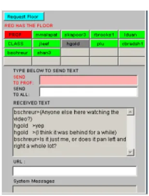
Network EducationWare (NEW) is a continuing project of the Networking and Simulation Laboratory in GMU’s C3I Center. NEW was assembled from a number of open-source software components available from the Internet multicasting community, most notably the Multicast Backbone (Mbone) tools VIC (video) and WBD (whiteboard) and the audio tool Speak Freely. GMU student and volunteer projects have expanded NEW with a record/playback capability and the VE project provided the Transport Layer Multicaster, a multi-site switching and floor control capability necessary to coordinate conferences involving over three sites with multiple tools. The floor control panel is shown in Figure 8. Most recently the VE project enhanced the WBD with new graphics formats: annotatable JPEG still images and auto-launched digital movies (Quicktime, AVI and MPEG) for use with the Distributed Embryology Education subproject conducted by UIC. Details and software (both source and executables) are available at http://netlab.gmu.edu/NEW. On the general educational from, GMU is now using NEW for eleven courses per semester in the Information Technology and Health Sciences areas, and the software is now is use for trial classes at West Virginia University and the Naval Postgraduate School, with several other institutions in planning stages for adoption.
CONCLUSION
In summary, we are proud of the tools we have developed for network-based biomedical
collaboration, and of the close collaboration we achieved in developing and demonstrating these tools. This has been a technically and professionally rewarding project for our entire team, and we believe it has produced both tools and instances of their use that represent a significant advance for networked biomedical collaboration. We are looking for additional sources of funding that would provide for using the tools we have developed to bring the Visible Embryo repository to a state of completion that will make it a functionally useful tool for the scientific community.
BIBLIOGRAPHY
Abdulla R, Blew G.A., Holterman M.J., Cardiovascular Embryology, Journal Pediatr Cardiol
volume 24 (3) 2003 (in-press).
Boisvert R. and Tang P., The Architecture of Scientific Software, pp. 273- 284, Data Management Systems for Scientific Applications, Kluwer Academic Publishers, 2001. Bolender D. and Holterman M.J., Animated thoughts on teaching human development, presentation to FASEB, Washington DC, May 2001, FASEB Journal 15(5)Abstract #793.1, 2001.
Bolender D.L., Holterman M.J., Blew G., and Holcomb J., Animation of human foregut development, presentation to American Association of Clinical Anatomists Annual Meeting, Gainesville, FL, June 2002, J Clin. Anat. 15:420, 2002.
Breitsprecher L., Fanghanel J., Steding G., Gasser R., Gibt es neue Erkenntnisse zur Embryologie und funktionelln Anatomie der humanen mimischen Muskulatur und der Oberlippe?, Mund Keifer GesichtChir 2002-6:102-110, Jan 23, 2002.
Breitsprecher L., Fanghanel J., Noe A., Lockett E.C. and Raab U., The functional anatomy of the muscles of facial expression in humans with and without Cleft Lip and Palate, Ann Anat (2002) 184: 27-34.
Chen C., Global Digital Library Development, pp. 197-204, Knowledge-based Data Management for Digital Libraries, Tsinghua University Press, 2001.
Cohen, J., Embryo Development at the Click of a Mouse, Science 2002 September 6; 297: 1629 (in News of the Week).
Degroff C., Sahn D.J., Thornburg K.L., Pentecost J.O., Gharib M. and Baptista A., Flow in the early embryonic human heart: a numerical study, Pediatric Cardiology (accepted for
Doyle, MD, et al., MultiVIS: A Web-based Interactive Remote Visualization Environment and Navigable Volume Imagemap System, in 28th AIPR Workshop: 3D Visualization for Data Exploration and Decision Making, 2000.
Doyle, M.D., The Visible Embryo Next Generation Internet Project, IEEE Engineering in Medicine and Biology Society WC2000 - World Congress on Medical Physics and Biomedical Engineering - Chicago, July 23-28, 2000.
Doyle, MD, Noe A.,et al., The Visible Embryo Project: A Platform for Spatial Genomics, presentation to 28th AIPR Workshop: 3D Visualization for Data Exploration and Decision Making, 2000.
Doyle, M.D., Telepathology and Medical Imaging for the Masses, Supercomputing 1999, Portland, OR.
Duenes, S, The Stem Cell Debate: The Embryonic Journey and Its Milestones, New York Times
Science Section, Tuesday, December 18, 2001: F4-5.
Gasser R.F., Cork R.J., and Brooks D.A., Digital images of serial sections of human embryos on DVDs, presentation to FASEB, Washington DC, May 2001, FASEB Journal, Vol., 15, p.A1105.
Gasser R.F. and Cork R.J., Digitally Reproduced Embryonic Morphology (DREM) available on CD’s and DVD’s, presentation to FASEB, New Orleans LA, May 2002, FASEB Journal.
Gasser R.F. and Cork R.J., Digital image processing as a means of preserving historic collections of serially sectioned human embryos, poster presentation to FASEB, Washington DC, May 2001, FASEB Journal 15, p. A382.
Gasser, R.F. and Cork, R.J., Digital images of sectioned human embryos on DVD’s, invited presentation International Symposium of Morphologist Sciences, Sun City, South Africa, 2002. Holterman M.J., Ashiru O., Abdulla R., Blew G.A., Sundararajan S., Rao S. and
Radhakrishnan, J., Clinically relevant embryology: new approaches to education, presentation to American Academy of Pediatrics Surgical section meeting, October 1999.
Holterman M.J et.al., The Human Embryology Digital Library: a Valuable Resource for Embryology Research and Education, presentation by M. Holterman to Biannual joint meeting of the American Association of Clinical Anatomists and the British Association of Clinical Anatomists, Cambridge, England, July 2000, J. Clin. Anat. 14:74-75.
Holterman M.J. et. al., The Visible Embryo Project: New Approaches to Embryology
Education, presentation to the biannual joint meeting of the American Association of Clinical Anatomists and the British Association of Clinical Anatomists, Cambridge UK, July 2000.
Holterman M.J., et al. Embryology and the Next Generation Internet, presentation to Annual meeting of the American Academy of Pediatrics, Chicago IL, October 2000.
Holterman M.J., Blew G.A., Bolender D. and Abdulla, R., Clinical Education Development Using Multimedia, presentation to Embryology Imaging and Education Conference, National Museum of Health and Medicine, Washington DC, May 2001.
Holterman M.J., Bolender D.L., Pesticelli M., Blew G., Lockett E.C., Noe A., Paidas C., Pentecost J. and Pullen J.M., Embryology Distance Learning: calling all hands, presentation to Association of Clinical Anatomists Annual Meeting, Gainesville, FL, June 2002, J. Clin. Anat. 15:425, 2002. Holterman M.J. et al, Distance learning for the study of embryology, presentation to Joint meeting of the British Association of Clinical Anatomy and the Society of Spanish Anatomists, Barcelona, Spain, July 2002.
Holterman M.J., Bolender D., Pescitelli M.J., Ashiru O.A., and Blew, G.A., Human
Embryology Education: Computer Models and Animations Provide Valuable Teaching Tools, presentation to XVII International Symposium on Morphological Sciences Timisoara,
Romania, September 2002.
Lennon W., Pullen J.M., Noe A., Pentecost J., and Doyle M., The Visible Embryo Project, presentation and demonstration to Supercomputing 2001, Denver, CO, Nov 14, 2001.
Lockett E.C., An Exploration of Interfaces for the Human Embryology Digital Library, poster presentation to Applied Imaging and Pattern Recognition Conference ’99, Washington, DC, July 1999.
Lockett E.C., Noe A., Sweet D., Human Embryology Digital Library and Collaboratory Support Tools, poster presentation to Uniform Health Sciences University, Bethesda, MD, March 23, 2000.
Lockett E.C., Sweet D., Noe A., Human Embryology Digital Library and Collaborative Support Tools, poster presentation by William F. Discher, American Association of Clinical
Anatomists/ British Association of Clinical Anatomists Joint Meeting, Cambridge, England, July 19, 2000.
Lockett E.C., Noe A., Pullen J.M., Rajasekar A., Doyle M., Brooks D., Holterman M., Paidas C., Pentecost J., The Visible Embryo Project, presentation by E.C. Lockett to The Museums and the Web Conference, Seattle WA, March 15, 2001.
Mansouri J., Pescitelli M.J. and Holterman M.J. Integrating diffusion, T1 and T2 weighted images into composite color representations, presentation to First Annual Digital Biology Symposium at the NIH, 2003.
Moore R.W., Knowledge-based Grids, presentation to 18th IEEE Symposium on Mass Storage Systems: Large Scale Storage in the Web, San Diego CA, April 17-20, 2001.
Moore R.W., Building a Persistent Archive” Technology Transfer to Federal Agencies, presentation to Digital Government Program, National Science Foundation, 2002,
http://ww.diggov.org/archive/library/pdf/moore.pdf.
Moore, R. and Baru, C., Virtualization Services for Data Grids, Book chapter in Grid Computing: Making the Global Infrastructure a Reality, John Wiley & Sons Ltd, 2003.
Nash, J.M., Inside the Womb, Time 2002 November 11; 160:20 68-78 (in News of the Week). NPACI and SDSC, SDSC Plays a Role in the National Library of Medicine Project, ONLINE, Vol. IV Issue 6, March 22, 2000.
NPACI and SDSC, Powerful Data Management Tool Comes of Age, Quarterly Science Magazine, Vol. 18 No. 3, ENVision on-line, July-Sept 2002.
Paidas C.N., Morreale R.F., Holoski K.M., Lund R.E., Hutchins G.M., Septation and differentiation of the embryonic human cloaca, J Ped Surg 34(5):1-10, May,1999.
Paidas C.P., Morreale, R. and Holterman M.J., The Visible Embryo: Correlation of Carnegie Morphological Landmarks with first trimester ultrasonography- a preliminary report,
presentation to Annual meeting of the American Pediatric Surgery Association, Naples FL, May 2001.
Painter, K., The miracle of life unfolding, Special for USA Today:11/07/02. Pentecost J.0., Thornburg K.L., 3D Computer Modeling of Human Cardiogenesis,
Computerized Medical Imaging and Graphics 23:45-49 1999.
Pentecost J., Sahn D.J., Gharib M., Baptista A. and Thornburg K.L., Graphical and stereolithographic models of the developing human heart lumen. Computerized Medical Imaging and Graphics 25: 459-463, 2001.
Pullen, J. M., The Internet Lecture: Converging Teaching and Technology, ACM Special Interest Group on Computer Science Education (SIGCSE) Bulletin Vol 32 No 3 pp 101-104, September 2000.
Pullen, J.M., Collaborative Online Embryology, presentation to MAX Day, University of Maryland, Beltsville MD, Sept 14, 2001.
Pullen J.M., Synchronous Internet Distance Education: Wave of the Future or Wishful
Thinking?, Proceedings of the United Engineering Foundation Workshop on E-Technologies in Engineering Education, Davos, Switzerland, August 2002.
Pullen, J.M., System Design of Network EducationWare: Open-Source Software for
Synchronous Internet Teaching and Learning, Proceedings of the ASEE/SEFI/TUB Colloquium on Technology in Engineering Education, Berlin, Germany, September 2002.
Pullen, J.M., A Software System for Cost-Effective Internet Delivery of Synchronous Distance Education, Proceedings of IASTED International Conference on Computers and Advanced Technology in Education 2003, IASTED, Calgary, AB, June 2003.
Rajasekar A., Wan M., Moore R., Jagatheesan A. and Kremenek G., Real Experiences with Data Grids - Case studies in using the SRB, International Symposium on High-Performance Computer Architecture, Kyushu, Japan, December, 2002.
Rajasekar A., Moore R., Ludäscher B. and Zaslavsky I., The Grid Adventures: SDSC's Storage Resource Broker and Web Services in Digital Library Applications:, 4th Russian Conference on Digital Libraries, Dubna, Russia, October, 2002.
Rajasekar A., Wan M., Moore R., mySRB and SRB, Components of a Data Grid, 11th High Performance Distributed Computing conference, Edinburgh, Scotland, July 2002.
ROADNet, ROADNet and the STORAGE RESOURCE BROKER: Collaboration Architecture for Real-time Data, University of California San Diego, La Jolla, CA, 2002,
http://roadnet.ucsd.edu/srb.html.
Rogers D.S., Paidas C.N., Morreale R.F., Hutchins G.M., Septation of the anorectal and genitourinary tracts in the human embryo: crucial role of the catenoidal shape of the urorectal sulcus, Teratology 66(2):144-152, 2002.
Sahn D.J., Thornburg K.L., Thornburg B.L., Pentecost J.O., Gharib M., Shaviv H., Martin S.L., Studies of flow in accurate models of early embryonic human heart, abstract in Am Coll Cardiol
2000, 35.
Versweyveld, L., National Library of Medicine project to create the largest-ever digital medical image library, Virtual Medical Worlds Monthly, San Diego, CA, March 22, 2000.