by
Kate Sendin
Thesis presented in partial fulfilment of the requirements for the
degree of Master of Science
in the Faculty of AgriScience at Stellenbosch University
Supervisor: Prof. Marena Manley
Co-supervisor: Dr Paul J. Williams
March 2017
DECLARATION
By submitting this thesis electronically, I declare that the entirety of the work contained therein is my own, original work, that I am the sole author thereof (save to the extent explicitly otherwise stated), that reproduction and publication thereof by Stellenbosch University will not infringe any third party rights and that I have not previously in its entirety or in part submitted it for obtaining any qualification.
Kate Sendin March 2017
Copyright © 2017 Stellenbosch University All rights reserved
ACKNOWLEDGMENTS
I would like to express my sincere gratitude to my supervisors Prof. Manley and Dr. Williams for the continuous support of my research. Their patience, motivation, and vast knowledge were greatly appreciated whilst conducting the research and throughout the writing of the thesis. Under your guidance I have grown immeasurably as a researcher.
I would like to extend my most sincere thanks to the following people and institutions for their contribution to the successful completion of this study:
Prof. Geladi (Swedish University of Agricultural Sciences) and Prof. Linderholm (Umeå Univeristy) for the use of their Videometer and Sisuchema instruments, and for their expert advice while introducing me to the practical side of hyperspectral imaging;
Wiana Louw and the graders at the South African Grain Laboratory for going out of their way to gather samples on my behalf;
All staff and post-graduate students in the Food Science Department for a warm and friendly working environment. Special thanks to the head of our department, Prof. Sigge;
Megan van der Merwe for kindly translating my thesis abstract to Afrikaans – jy’s ‘n engel; and The National Research Fund (NRF) (Thutuka Bursary, 2015, and Scarce Skills Bursary, 2016) are hereby acknowledged for financial support (any opinion, findings and conclusions or recommendations expressed in this material are those of the author and therefore the NRF does not accept any liability in regard thereto).
Last but not least, I would like to thank my parents Billy and Heidi, my brother William, and Charles Peek for their continued support and encouragement throughout writing this thesis.
ABSTRACT
Maize (Zea mays L.) is the most important cereal crop grown in South Africa. It is produced widely across the country under diverse environments, and thus a variety of defects tend to occur. Grading is an important quality and safety control step where these defective materials are identified and quantified. This study considered the most important defective material classes, namely 6 types of defective white maize kernels, 5 types of foreign matter, other colour kernels (yellow maize) and pinked white maize kernels. Current maize grading is manual and tedious, and modern analytical methods could improve this process. This study aimed to investigate the viability of using spectral imaging with multivariate data analysis for maize grading by separating sound maize from the 13 defective materials classes.
NIR hyperspectral imaging with pixel-wise and object-wise data analysis were used for two-way discrimination of the sound and defective material classes. The average spectra indicated prominent bands at 1219 and 1476 (related to starch), 1941 (related to moisture), and 2117 nm (related to protein). The loadings of principal component (PC) 1 exhibited similar bands. The object-wise approach performed superiorly to the pixel-object-wise approach across all 13 analyses. Little separation was observed in the principal component analysis (PCA) score plots in the pixel-wise results due to a large similarity between classes. The object-wise approach utilised the average spectrum for each maize kernel, and the overlap was reduced. Partial least squares discriminant analysis (PLS-DA) models were calculated and used to classify an independent validation set of 30 sound kernels and 30 defective materials. The pixel-wise analyses achieved classification accuracies ranging 75-99%. This approach was not able to accurately distinguish closely related classes. The object-wise analyses performed well, with 8 of the 13 achieving 100% classification accuracy, and the remaining 5 classes incurring only one error per analysis of 60 kernels.
Multispectral imaging followed to compare the two imaging techniques. Pixel-wise PCA was applied to pre-process the spectral imaging data, followed by object-wise two-way PLS-DA modelling using 17 sound kernels and 18 defective material objects. The PCA loadings revealed that colour played a role in separating the classes, with a wide band appearing across 505, 525, 570 and 590 nm. Classification accuracies of 83-100% were achieved, and were generally slightly lower than the results obtained for all classes using the NIR hyperspectral imaging instrument.
Spectral imaging was shown to be capable of separating white maize from 13 commonly occurring defective materials. NIR hyperspectral imaging performed superiorly to multispectral imaging, and the use of an object-wise data analysis approach further improved the accuracy of the separations. These techniques have the potential to offer the maize industry a rapid, accurate and objective alternative grading method.
UITTREKSEL
Mielies (Zea mays L.) is die belangrikste graangewas wat tans in Suid Afrika geproduseer word. Dit word landwyd geproduseer, en deurdat dit in soveel diverse omgewings groei, word verskeie defekte gereeld opgespoor. Gradering is ‘n baie belangrike kwaliteit- en veiligheidsmaatreël waardeur hierdie foutiewe materiaal uitgesonder en dan gekwantifiseer word. Hierdie studie oorweeg die mees prominente foutiewe materiaal klasse, naamlik: ses tipes foutiewe wit mieliepitte, vyf tipes vreemde materiaal, anderskleurige mieliepitte (geel mielies) en verpienkte wit mieliepitte. Huidige mieiliegradering is ‘n duur en tydsame proses, en moderne analitiese metodes kan hierdie proses verbeter. Hierdie studie stel ondersoek in om te bepaal asof die gebruik van spektrale beelding met meerveranderlike data ontleding vir mieliegradering lewensvatbaar is, deur gesonde mielies van die 13 foutiewe materiaal klasse te skei.
Naby infrarooi (NIR) hiperspektrale beelding met pixel- en voorwerp-wyse data analiese is gebruik vir ‘n tweerigting diskriminasie van die gesonde en foutiewe materiaal klasse. Die gemiddelde spektra het prominente bande aangedui by 1219 en 1476 (stysel-verwant), 1941 (proteien-verwant) en 2117 nm (vog-verwant). Die lading-stip van hoofkomponent (HK) 1 het soortgelyke bande gewys. Die voorwerp-wyse benadering het regoor al 13 analises beter as die pixel-wyse benadering presteer. As gevolg van ‘n groot ooreenkomste tussen die verskillende klasse, was min skeiding geobserveer in die hoofkomponent analise (HKA) telling-beelde in die pixel-wyse resultate. Die voorwerp-wyse benadering het van die gemiddelde spektrum van elke mieliepit gebruik gemaak, en die oorvleuling was so verminder. Parsiële kleinste waarde diskriminantanalise (PKW-DA) modelle was bereken om 30 gesonde- en 30 foutiewe materiale te klassifiseer. Die pixel-wyse analises het klassifikasie akkuraatheid tussen 75-99% bereik. Hierdie benadering kon nie akkuraat tussen die verwante klasse onderskei nie. Die voorwerp-wyse analise het goed presteer, waar 8 van die 13, 100% klasifikasie akkuraatheid bereik het, en die oorblywende 5 klasse net een fout per analise van 60 pitte aangegaan het.
Multispektrale beelding het gevolg om die twee beeldingstegnieke te vergelyk. Pixel-wyse HKA was bereken om skoonmaak van die beeld te akkomodeer, en was vervolg deur voorwerp-wyse tweerigting PKW-DA modellering wat van 17 gesonde pitte en 18 voorwerpe van foutiewe materiaal gebruik gemaak het. Die HKA lading-stippe het onthul dat kleur ‘n massiewe rol in die skeiding van die klasse gespeel het, met ‘n wye band wat oor 505, 525, 570 en 590 nm verskyn het. Klassifikasie akkuraatheid van 83-100% was bereik, en was oor die algemeen iewat laer as die resultate wat in alle klasse bereik is tydens die gebruik van die NIR hiperspektrale beelding instrument.
Dit was daardeur gewys dat spektrale beelding bekwaam was om wit mielies van 13 bekende foutiewe materiale te skei. NIR hiperspektrale beelding het beter gedoen as multispektrale beelding, en die gebruik van ‘n voorwerp-wyse data analise benadering het verder die akkuraatheid van die skeidings verbeter. Hierdie tegnieke het die potensiaal om vir die mielie industrie ‘n vinnige, akkurate en objektiewe alternatiewe graderings metode aan te bied.
TABLE OF CONTENTS
DECLARATION ... i
ACKNOWLEDGMENTS ... ii
ABSTRACT ... iii
UITREKSEL ... iv
LIST OF FIGURES ... vii
LIST OF TABLES ... xii
LIST OF ABBREVIATIONS USED ... xiii
CHAPTER 1: INTRODUCTION ... 1
References ... 4
CHAPTER 2: LITERATURE REVIEW ... 8
Introduction ... 9
Maize Grading in South Africa ... 11
Brief History of Hyperspectral Imaging ... 14
Principles and Theoretical Background of Hyperspectral Imaging ... 14
Fundamentals of Hyperspectral Imaging ... 14
Components of Hyperspectral Imaging Systems ... 16
Acquisition of Hyperspectral Images ... 17
Analysis of Hyperspectral Images ... 18
Applications of Hyperspectral Imaging in Cereal Evaluation ... 21
Quality ... 21
Safety ... 30
Conclusion ... 34
References ... 34
CHAPTER 3: CHARACTERISATION OF WHITE MAIZE KERNELS USING NIR
HYPER-SPECTRAL IMAGING ...
42
Introduction ... 43
Materials and Methods ... 44
Samples ... 44
NIR Hyperspectral System ... 45
Image Acquisition ... 45
Hyperspectral Image Analysis ... 46
Spectral Analysis ... 49
Multivariate Data Analysis ... 51
Conclusion ... 68
References ... 68
CHAPTER 4: CHARACTERISATION OF WHITE MAIZE KERNELS USING
MULTI-SPECTRAL IMAGING ... 72
Introduction ... 73
Materials and Methods ... 74
Samples ... 74
Multispectral System ... 74
Image Acquisition ... 75
Multispectral Image Analysis ... 75
Results and Discussion ... 78
Multivariate Data Analysis ... 78
Conclusion ... 90
References ... 91
CHAPTER 5: GENERAL DISCUSSION AND CONCLUSIONS ... 94
References ... 96
LIST OF FIGURES
Figure 2.1 A NIR hyperspectral imaging hypercube comprises of one wavelength (z) and two spatial (x and y) dimensions. One can view the data as a spectrum of a pixel in the sample, or as an image plane of the entire sample at a chosen wavelength (e.g. at 1000 nm). ... 15
Figure 2.2 Schematic of a pushbroom NIR hyperspectral imaging system, illustrating the line-by-line data acquisition of the samples on the linear translation stage. ... 16
Figure 2.3 (a) Scores images of PC2 and PC5 for whole yellow maize kernels enabling visualisation of similarity in chemical composition (similar colours indicate similar chemical composition, in this case similar endosperm texture). (b) Scores plot of PC2 vs. PC5 with three clusters. (c) Classification plot based on clusters identified in the PC scores plot. (d) Classification image after projection of the classes identified in the scores plot onto the scores image [Copyright 2014 Royal Society of Chemistry. Reproduced with permission from Manley (2014)]. ... 19
Figure 2.4 (a) PC5 score image for the barley cultivar Erica image dataset where dark blue indicates viable kernels and green non-viable kernels. (b) The corresponding PC5 vs. PC1 score plot showing density and clustering of pixels. Positive PC5 scores values (green box) were identified and assigned to the viable class and negative PC5 scores values (blue box) to the non-viable class. Brushing between score plot and score image was used. (c) Classification image showing viable and non-viable regions. [Copyright 2011 Springer. Reproduced with permission from McGoverin et al. (2011)]. ... 29
Figure 2.5 (a) PCA score plot of PC4 vs. PC5 (0.49% and 0.34%) showing three clusters and (b) corresponding score image of PC4 showing decrease in score values from left to right, where warm colours (yellow to red) correspond with positive score values and cold colours (blue) correspond to negative score values; (c) classification plot of PC4 vs. PC5 with classes and degree of infection in the direction of PC4 and (d) classes projected onto score image showing the control (green), T0&T1 (black) and the remaining time intervals up to 90 h post inoculation (red). [Copyright 2012 Elsevier. Reproduced with permission from Williams et al. (2012)]. ... 31
Figure 3.1 Digital image of (a) sound white maize and the 13 undesirable materials: (b) Fusarium damage; (c) Diplodia damage; (d) pinked maize; (e) water damage; (f) rodent damage; (g) heat damage; (h) plant material; (i) screenings; (j) wheat; (k) sorghum; (l) soy; (m) sunflower; and (n) yellow maize. ... 45
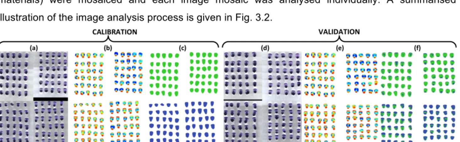
Figure 3.2 Summary of image analysis process, illustrated using sound class (top) vs. yellow class (bottom) with the pixel-wise approach (object-wise analysis followed the same method). The left half of each section (a–f) is germ-up, and the right half is germ-down. Each half contains the same kernels in the same positions, to allow for inspection of both sides. (a) Raw mosaic image (at 1426 nm) of calibration samples arranged in known order of classes; (b) PCA score image of calibration samples after pre-processing; (c) Samples were assigned classes (sound as green and defect as blue) and PLS-DA model was calibrated; (d) Raw mosaic image (at 1426 nm) of independent validation samples arranged in known order of classes; (e) PCA score image of validation samples after pre-processing; and (f) PLS-DA model applied to generate classification image. ... 46
Figure 3.3 Method used for totalling the object-wise classification results, illustrated using yellow maize class vs. sound class. (a) Unaltered object-wise classification image; (b) Assignment of overall classification; and (c) Overall object-wise classification image. ... 48
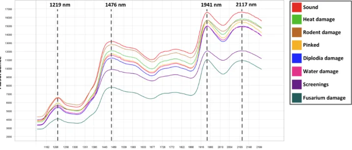
Figure 3.4 Unprocessed average pseudo-absorbance spectra for sound white maize; white maize defects (heat damage, rodent damage, Diplodia damage, water damage, screenings and Fusarium damage); and pinked white maize. ... 50
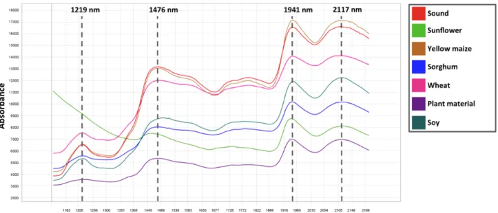
Figure 3.5 Unprocessed average pseudo-absorbance spectra for sound white maize; yellow maize; and foreign matter (sunflower seeds, sorghum, wheat, plant material and soy). ... 51
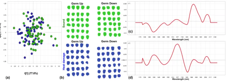
Figure 3.6 Pixel-wise PCA of heat damage class vs. sound class. Minimal separation of classes was observed. Scores given as (a) PCA score plot of PC1 (79% SS) vs. PC2 (8% SS); and (b) PCA score image (PC1). Loadings line plots given for (c) PC1; and (d) PC2. ... 51
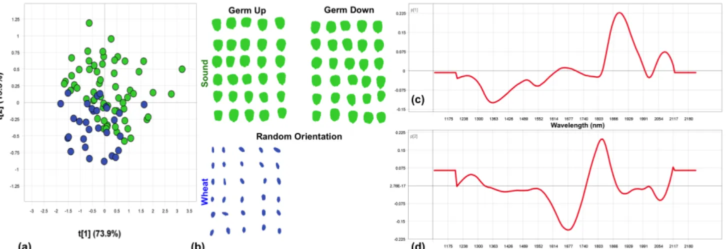
Figure 3.7 Pixel-wise PCA of wheat class vs. sound class. Slight separation of classes was observed. Scores given as (a) PCA score plot of PC1 (81% SS) vs. PC2 (7% SS); and (b) PCA score image (PC1). Loadings line plots given for (c) PC1; and (d) PC2. ... 52
Figure 3.8 Pixel-wise PCA of yellow maize class vs. sound class. Minimal separation of classes was observed. Scores given as (a) PCA score plot of PC1 (82% SS) vs. PC2 (7% SS); and (b) PCA score image (PC1). Loadings line plots given for (c) PC1; and (d) PC2. ... 52
Figure 3.9 Pixel-wise PCA of screenings class vs. sound class. Minimal separation of classes was observed. Scores given as (a) PCA score plot of PC1 (79% SS) vs. PC2 (6% SS); and (b) PCA score image (PC1). Loadings line plots given for (c) PC1; and (d) PC2. ... 52
Figure 3.10 Pixel-wise PCA of Diplodia damage class vs. sound class. Minimal separation of classes was observed. Scores given as (a) PCA score plot of PC1 (82% SS) vs. PC2 (6% SS); and (b) PCA score image (PC1). Loadings line plots given for (c) PC1; and (d) PC2. ... 53
Figure 3.11 Pixel-wise PCA of pinked class vs. sound class. Minimal separation of classes was observed. Scores given as (a) PCA score plot of PC1 (81% SS) vs. PC2 (8% SS); and (b) PCA score image (PC1). Loadings line plots given for (c) PC1; and (d) PC2. ... 53
Figure 3.12 Object-wise PCA of heat damage class vs. sound class. Minimal separation of classes was observed. (a) PCA score plot of PC1 (78% SS) vs. PC2 (12% SS). (b) Classes of sound (green) and heat damaged (blue) objects. Loadings line plots given for (c) PC1; and (d) PC2. ... 54
Figure 3.13 Object-wise PCA analysis of wheat class vs. sound class. Fair separation of classes was observed. (a) PCA score plot of PC1 (74% SS) vs. PC2(15% SS). (b) Classes of sound (green) and wheat (blue) objects. Loadings line plots given for (c) PC1; and (d) PC2. ... 55
Figure 3.14 Object-wise PCA analysis of yellow maize class vs. sound class. Fair separation of classes was observed. (a) PCA score plot of PC1 (80% SS) vs. PC2 (13% SS). (b) Classes of sound (green) and yellow maize (blue) objects. Loadings line plots given for (c) PC1; and (d) PC2. ... 55
Figure 3.15 Object-wise PCA analysis of screenings class vs. sound class. Good separation of classes was observed. (a) PCA score plot of PC1 (70% SS) vs. PC2 (15% SS). (b) Classes of sound (green) and screenings (blue) objects. Loadings line plots given for (c) PC1; and (d) PC2. ... 54
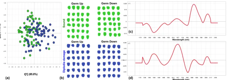
Figure 3.16 Object-wise PCA analysis of Diplodia damage class vs. sound class. Slight separation of classes observed. (a) PCA score plot of PC1 (86% SS) vs. PC2 (8% SS). (b) Classes of sound (green) and Diplodia damage (blue) objects. Loadings line plots given for (c) PC1; and (d) PC2. ... 56
Figure 3.17 Object-wise PCA analysis of pinked class vs. sound class. Fair separation of classes was observed. (a) PCA score plot of PC1 (85% SS) vs. PC2 (9% SS). (b) Classes of sound (green) and pinked (blue) objects. Loadings line plots given for (c) PC1; and (d) PC2. ... 56
Figure 3.18 Pixel-wise PLS-DA classification resulted in well classified heat damage class vs. sound class (90.72% classification accuracy). Fair separation of classes was observed. (a) PLS-DA score plot of PLS factor 1 (78% SS) vs. 2 (6% SS); (b) Classification image. ... 57
Figure 3.19 Pixel-wise PLS-DA classification resulted in extremely poorly classified wheat class vs. sound class (90.87% classification accuracy; 2.38% sensitivity). Minimal separation of classes was observed. (a) PLS-DA score plot of PLS factor 1 (72% SS) vs. 2 (23% SS); (b) Classification image. ... 59
Figure 3.20 Pixel-wise PLS-DA classification resulted in poorly classified yellow maize class vs. sound class (75.32% classification accuracy). Minimal separation of classes was observed. (a) PLS-DA score plot of PLS factor 1 (81% SS) vs. 2 (7% SS); (b) Classification image. ... 59
Figure 3.21 Pixel-wise PLS-DA classification resulted in well classified screenings class vs. sound class (91.58% classification accuracy). Fair separation of classes was observed. (a) PLS-DA score plot of PLS factor 1 (64% SS) vs. 2 (14% SS); (b) Classification image. ... 60
Figure 3.22 Pixel-wise PLS-DA classification resulted in well classified Diplodia damage class vs. sound class (85.22% classification accuracy). Slight separation of classes was observed. (a) PLS-DA score plot of PLS factor 1 (81% SS) vs. 2 (5% SS); (b) Classification image. ... 60
Figure 3.23 Pixel-wisePLS-DA classification resulted in extremely poorly classified pinked class vs. sound class (62.94% classification accuracy). Slight separation of classes was observed. (a) PLS-DA score plot of PLS factor 1 (74% SS) vs. 2 (12% SS); (b) Classification image. ... 61
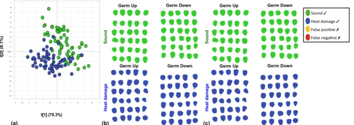
Figure 3.24 Object-wisePLS-DA classification resulted in perfectly classified heat damage class vs. sound class (100% classification accuracy). (a) PLS-DA score plot of PLS factor 1 (75% SS) vs. 2 (8% SS); (b) Unprocessed classification image; and (c) Overall classification image. ... 63
Figure 3.25 Object-wisePLS-DA classification resulted in perfectly classified wheat class vs. sound class (100% classification accuracy). (a) PLS-DA score plot of PLS factor 1 (71% SS) vs. 2 (14% SS); (b) Unprocessed classification image; and (c) Overall classification image. ... 64
Figure 3.26 Object-wise PLS-DA classification resulted in well classified yellow maize class vs. sound class (98.33% classification accuracy). (a) PLS-DA score plot PLS factor 1 (79% SS) vs. 2 (12% SS); (b) Unprocessed classification image; and (c) Overall classification image. ... 64
Figure 3.27 Object-wise PLS-DA classification resulted in perfectly classified screenings class vs. sound class (100% classification accuracy). (a) PLS-DA score plot PLS factor 1 (47% SS) vs. 2 (25% SS); (b) Unprocessed classification image; and (c) Overall classification image. ... 64
Figure 3.28 Object-wise PLS-DA classification resulted in perfectly classified Diplodia damage class vs. sound class (100% classification accuracy). (a) PLS-DA score plot PLS factor 1 (83% SS) vs. 2 (9% SS); (b) Unprocessed classification image; and (c) Overall classification image. ... 65
Figure 3.29 Object-wise PLS-DA classification resulted in well classified pinked class vs. sound class (98.15% classification accuracy). (a) PLS-DA score plot of PLS factor 1 (73% SS) vs. 2 (12% SS); (b) Unprocessed classification image; and (c) Overall classification image. ... 65
Figure 4.1 Summary of image analysis, illustrated using sound class (first 17 kernels) vs. yellow class (second 18 kernels). The top half of each section (a–f) is germ-up, and the bottom half is germ-down. Each half contains the same kernels in the same positions, to allow for inspection of
both sides. (a) Digital image of calibration samples arranged in known order of classes; (b) PCA score image of calibration samples after pre-processing; (c) Samples were assigned classes (sound as green and defect as blue) and PLS-DA model was calibrated; (d) Digital image of independent validation samples arranged in known order of classes; (e) PCA score image of validation samples after pre-processing; and (f) PLS-DA model applied to generate classification image. ... 76
Figure 4.2 Method used for totalling results, illustrated using Diplodia class vs. sound class. (a) Unprocessed classification image; (b) Totalling procedure for determining overall classification; and (c) Overall classification image. ... 77
Figure 4.3 Well separated heat damage class vs. sound class in (a) the PCA scores plot (PC1 vs. PC2) and (b) the PCA scores image (PC1). Loadings line plots for (c) PC1; and (d) PC2 represent the general loadings line trend observed over most of the 13 PCA analyses. ... 78
Figure 4.4 Well separated wheat class vs. sound class in (a) the PCA scores plot (PC1 vs. PC2) and (b) the PCA scores image (PC1). Loadings line plots for (c) PC1; and (d) PC2 represent the general loadings line trend observed over most of the 13 PCA analyses. ... 78
Figure 4.5 Well separated yellow maize class vs. sound class in (a) the PCA scores plot (PC1 vs. PC2) and (b) the PCA scores image (PC1). Loadings line plots for (c) PC1; and (d) PC2 do not represent the general loadings line trend observed over most of the 13 PCA analyses. ... 79
Figure 4.6 Moderately separated screenings class vs. sound class in (a) the PCA scores plot (PC1 vs. PC2) and (b) the PCA scores image (PC1). Loadings line plots for (c) PC1; and (d) PC2 represent the inverse of the general loadings line trend observed over most of the 13 PCA analyses. ... 79
Figure 4.7 Not separated Diplodia damage class vs. sound class in (a) the PCA scores plot (PC1 vs. PC2) and (b) the PCA scores image (PC1). Loadings line plots for (c) PC1; and (d) PC2 represent the general loadings line trend observed over most of the 13 PCA analyses. ... 80
Figure 4.8 Not separated pinked class vs. sound class in (a) the PCA scores plot (PC1 vs. PC2) and (b) the PCA scores image (PC1). Loadings line plots for (c) PC1; and (d) PC2 do not represent the general loadings line trend observed over most of the 13 PCA analyses. ... 80
Figure 4.9 Perfectly classified heat damage class vs. sound class (100% classification accuracy). (a) PLS-DA scores plot (PLS factor 1 vs. 2); (b) Unprocessed classification image; and (c) Overall classification image. ... 85
Figure 4.10 Perfectly classified wheat class vs. sound class (100% classification accuracy). (a) PLS-DA scores plot (PLS factor 1 vs. 2); (b) Unprocessed classification image; and (c) Overall classification image. ... 85
Figure 4.11 Perfectly classified yellow maize class vs. sound class (100% classification accuracy). (a) PLS-DA scores plot (PLS factor 1 vs. 2); (b) Unprocessed classification image; and (c) Overall classification image. ... 86
Figure 4.12 Perfectly classified screenings class vs. sound class (100% classification accuracy). (a) PLS-DA scores plot (PLS factor 1 vs. 2); (b) Unprocessed classification image; and (c) Overall classification image. ... 86
Figure 4.13 Poorly classified Diplodia damage class vs. sound class (82.86% classification accuracy). (a) PLS-DA scores plot (PLS factor 1 vs. 2); (b) Unprocessed classification image; and (c) Overall classification image. ... 87
Figure 4.14 Moderately well classified pinked class vs. sound class (91.43% classification accuracy). (a) PLS-DA scores plot (PLS factor 1 vs. 2); (b) Unprocessed classification image; and (c) Overall classification image. ... 87
LIST OF TABLES
Table 2.1 Standards for grades of white maize (per category of undesirable material and maximum allowed weight) according to South African grading regulations. ... 12
Table 2.2 Applications of hyperspectral imaging and chemometrics for quality and safety evaluation of cereals and cereal products. ... 22
Table 3.1 Method used to determine the overall classification of a kernel based on the classification image generated from the germ-up and -down image data. ... 48
Table 3.2 Results of pixel-wise PLS-DA model calibration and validation for the separation of sound white maize class from 13 undesirable material classes. ... 62
Table 3.3 Results of object-wise PLS-DA model calibration and validation for the separation of sound white maize class from 13 undesirable material classes. ... 66
Table 4.1 The wavebands utilised by the VideometerLab2 instrument and the associated region in the electromagnetic spectrum. ... 75
Table 4.2 Results of PLS-DA model calibration and validation for the separation of sound white maize class from 13 undesirable material classes. ... 88
LIST OF ABBREVIATIONS USED
3D three dimensional
ANN artificial neural networks AOTF acousto-optic tunable filter
AVRIS airborne visible/infrared imaging spectrophotometer BPNN back propagation neural network
CCD charged coupled device
CMOS complementary metal oxide semiconductor FDA factorial discriminant analysis
HgCdTe mercury-cadmium-telluride InGaAs indium-gallium-arsenide LCTF liquid crystal tunable filter LDA linear discriminant analysis LDC linear discriminant classifier LED light emitting diode
MIR mid infrared
MLR multiple linear regression MNF minimum noise fraction
MSC multiplicative scatter correction MSEP mean square errors of prediction
NASA/JPL National Aeronautics and Space Administration Jet Propulsion Laboratory
NIR near infrared
PC principal component
PCA principal component analysis PCR principal component regression PDA penalised discriminant analysis PLS partial least squares
PLS-DA partial least squares discriminant analysis
PLS-FDA partial least squares factorial discriminant analysis PLSR partial least squares regression
Q2 cross-validated coefficient of determination QDA quadratic discriminant analysis
R2 coefficient of determination
RMSEP root mean square error of prediction SAGL South African Grain Laboratory SAM spectral angle mapping
SECV standard error of cross-validation SNV standard normal variate
SS sum of squares
SVM support vectors machine SWIR short wave infrared
UV ultra-violet
WM1 white maize grade 1 WM2 white maize grade 2 WM3 white maize grade 3
CHAPTER 1
INTRODUCTION
Maize (Zea mays L.) is a widely consumed and grown cereal crop. It can be found in many forms such as minimally processed staple foods (porridges and breads), processed foods (tortillas, chips and breakfast cereals), and even in alcohol production (Fox and Manley, 2009). Generally, developing regions, such as in Africa, South America and Asia, consume maize directly as staple diet (Fox and Manley, 2009). In developed countries, maize is more often consumed as second-cycle produce, in the form of meat, eggs and dairy (Scanes, 2010). Maize, particularly white maize, is a vital food source for African people, including the majority of the South African population (Gouse et al., 2006). In South Africa, white maize is generally consumed as a minimally processed unfermented porridge, which is commonly the base of all meals. Although maize lacks one essential amino acid, lysine, it is a good source of protein and carbohydrates, and can constitute a large proportion of a healthy diet (Eckhoff and Paulsen, 2012). The popularity of maize as a food crop is a combination of its ease of cultivation, large yields, storage ability, and high content of easily metabolised starch (Eckhoff and Paulsen, 2012). Furthermore, the low cost, reliable supply, high digestibility, and close proximity of maize growing and livestock rearing regions contribute to maize products generally being the most cost-effective animal feed available.
During maize classification several physical properties of a sample are evaluated to give an indication of a sample’s chemical composition, functionality and appropriate end-uses (Serna-Saldivar, 2010). First, the class is determined by maize kernel colour, white or yellow. Each class is then graded in accordance with a class-specific grading regulation. During grading, the main aim is to determine the grain condition and health. Regardless of the specific product being produced, certain grading quality and safety characteristics are important for all human food uses (Eckhoff and Paulsen, 2012). The maize must not exhibit visual insect, rodent or fungal deterioration, as the palatability, sanitation and shelf life of the resulting products would be decreased. Furthermore, mycotoxins originating from fungal contamination could cause sickness or death if consumed (Yoshizawa et al., 1994; Sydenham et al., 1990). The kernels should be intact, and the pericarp should not exhibit obvious damage. Broken and damaged maize will have different water absorbing properties, leading to less uniform cooking characteristics and final product consistency. The grading of maize facilitates fair marketing between buyers and sellers by providing an indication of its intrinsic value. Subsequent to grading, further classification provides more insight into industrial use or functionality. Factors such as test weight (hectolitre mass), hardness, or chemical composition are not considered during grading, but may be agreed upon in an additional trade agreement.
While many manual methods are utilised in the cereal industry, especially in grading, much research has focused on alternative analytical methods that aim to avoid human error and subjectivity. Considerable work has focused on the development of invasive and non-destructive techniques. These include reflectance and transmittance spectroscopy (including ultraviolet, visible, near infrared (NIR) and mid infrared (MIR)), acoustic response (Pearson et al., 2007; Stasiak et al., 2007), and X-ray computed tomography (Neethirajan et al., 2008; Zhu et al., 2012). Spectroscopic techniques, most commonly utilising the NIR region, have shown to be promising for the non-destructive and non-invasive inspection of cereals (Fox and Manley, 2014; Sendin et al., 2016). NIR spectroscopy is based on the bond vibrations within organic molecules consisting mainly of C–H, C=O, O–H and N–H, which occur due to excitement by light with wavelengths of 780 to 2500 nm (Manley, 2014). NIR spectroscopy is widely implemented in the cereal industry, and has been found useful in qualitative and quantitative measuring of major components, including dry matter, moisture, protein and starch, as well as minor components, such as gluten (Huang et al., 2008). Applications include inspection pre- and during harvest (Engel et al., 1997; Maertens et al., 2004; Montes et al., 2006), during grain processing (Kawamura et al., 2003), and in cereal end-product quality (De Temmerman et al., 2007; Kays et al., 1996; Weeranantanaphan and Downey, 2010). Although studies have demonstrated that conventional spectroscopy is capable of evaluating a range of maize properties, it has some limitations. Foods and biomaterials are, in general, heterogeneous and it is often a challenge to obtain spectral data that are representative for the chemical composition and distribution. No information is gathered in the spatial dimension, and thus measurements may only cover a small area of the kernel. As maize kernels are heterogeneous, measurements of the entire kernel are necessary. In contrast, spectral imaging offers localisation, allowing entire samples to be analysed rapidly and accurately.
Spectral imaging, known also as chemical imaging, integrates conventional imaging and spectroscopy to attain both spatial and spectral information from the sample (Gowen et al., 2007). A spectral image, commonly referred to as a three-dimensional (3D) hypercube, comprises numerous adjacent wavelengths for each spatial point of the object. Thus, each pixel in the image contains the spectrum at that exact point (Burger and Geladi, 2006). Multispectral imaging and hyperspectral imaging are two spectral imaging techniques based on the same principles. The difference between the two is simply the complexity of the spectral data collected. Hyperspectral imaging systems acquire spectra in hundreds of narrow bands that are separated by regular intervals (e.g. one band every 5 nm). The resulting spectra are smooth and closely resemble the spectra of conventional techniques. Multispectral imaging systems often utilise a handful of wavebands, ca. 20, which are irregularly spaced across the chosen range, and thus a jagged spectrum is achieved. Multispectral imaging systems are simpler, and thus offer faster acquisition times and lower cost of the camera. Hyperspectral imaging is more suited to detailed research and laboratory applications, while
multispectral imaging offers a more practical solution to industry requirements. Both techniques will be explored in this study.
To establish if spectral imaging has the potential for replacing manual maize grading, one must understand the current demands of the maize industry in South Africa. Refer to Addendum 1 for the relevant maize grading legislation. In manual grading, a sample (ca. 150 g) of a maize consignment is visually inspected to identify any content other than sound white maize, such as defective or foreign material (Department of Agriculture, 2009). The method involves spreading the kernels onto a flat surface and slowly inspecting each kernel. Any undesirable materials are weighed and referenced to maximum levels stipulated for each material’s category. There are three white maize classes, WM1 (best), WM2 and WM3. The grade is allocated solely on the contents of undesirable materials within a sample. Unfortunately, evaluating physical features by human inspection requires significant training and is very tedious, even on small samples. To emulate these current practices and demonstrate that sound (healthy) maize could be separated from all of the undesirable materials using an objective method such as spectral imaging, two-way comparisons must be made. The two classes, sound white maize and a single defect, must be separated through a series of analyses. Furthermore, the grader inspects both sides (germ-up and germ-down) of every kernel to detect defects during manual grading, as the extent and presence of a defect on individual kernels is extremely variable and irregular. Thus, identical up and germ-down image pairs must be collected for assessing each side of all kernels.
The analytical capability of the near infrared (NIR) spectroscopy component of spectral imaging relies on absorption by wavebands that often overlap, making interpretation of spectra difficult. In addition, the sheer size of the hypercube further complicates data analysis and interpretation. By using multivariate data analysis, the composition of the object may be charaterised by relating the varying spectra to their spatial distributions. Accurate calibration equations can be calculated for various cereal quality and safety attributes. Before being subjected to these chemometric analyses, data must first be mathematically pre-processed. Analytical information is extracted from spectra, and non-chemical biases, such as scattering effects, are removed (Del Fiore et al., 2010). Principal component analysis (PCA) is arguably one of the most useful and widely used exploratory data analysis techniques (Amigo et al., 2013; Cowe and McNicol, 1985). It is used to generate score and loading plots that may be visually assessed. Once data has been explored using PCA, a popular supervised classification technique, partial least squares discrimination analysis (PLS-DA), may be applied. This combination of PCA and PLS-DA has been used effectively to identify, differentiate or evaluate a variety of cereal properties (Cogdill et al., 2004; Del Fiore et al., 2010; Manley et al., 2009; McGoverin et al., 2011; Vermeulen et al., 2012; Wallays et al., 2009; Williams et al., 2009).
Multivariate data analysis traditionally folllows a pixel-wise approach (Williams et al., 2012; McGoverin and Manley, 2012). However, an object-wise approach has emerged in recent years
(Kucheryavskiy, 2013; Williams and Kucheryavskiy, 2016). Pixel-wise analysis simply utilises each pixel’s spectrum as a sample for classification. However, during object-wise image analysis an average spectrum from all pixels that fall within a single object is calculated (i.e. each maize kernel). The average spectrum is used for classification instead of original spectra of individual pixels. When different classes of samples are closely related, such as in maize kernels, the majority of pixels exhibit similar spectral responses. A large number of misclassified pixels often occur when using a pixel-wise approach. This can be avoided by classifying objects to significantly decrease the amount of data and calculate more stable classification models. As few studies have compared the two approaches, the best approach for separating closely related healthy and defective maize, as well as other cereal commodities and foreign matter, must be identified.
This study investigated the potential for maize grading by spectral imaging. Two imaging systems, NIR hyperspectral imaging (1118–2425 nm) and multispectral imaging (375–970 nm), as well as two multivariate data analysis approaches, pixel-wise and object-wise, were compared. Specific objectives were to establish capability of:
• NIR hyperspectral imaging to separate sound white maize kernels from common grading defects; and
• multispectral imaging to achieve comparable results for separating sound white maize kernels from common grading defects.
REFERENCES
Amigo, J. M., Martí, I. & Gowen, A. (2013). Hyperspectral imaging and chemometrics: a perfect combination for the analysis of food structure, composition and quality. In: Data Handling in Science and Technology. Pp. 343-370. Amsterdam, The Netherlands: Elsevier Science. Burger, J. & Geladi, P. (2006). Hyperspectral NIR imaging for calibration and prediction: a
comparison between image and spectrometer data for studying organic and biological samples. Analyst, 131, 1152-1160.
Cogdill, R. P., Hurburgh, C. R., Rippke, G. R., Bajic, S. J., Jones, R. W., McClelland, J. F., Jensen, T. C. & Liu, J. (2004). Single-kernel maize analysis by near-infrared hyperspectral imaging. Transactions of the ASAE, 47, 311.
Cowe, I. A. & McNicol, J. W. (1985). The use of principal components in the analysis of near-infrared spectra. Applied Spectroscopy, 39, 257-266.
De Temmerman, J., Saeys, W., Nicolaï, B. & Ramon, H. (2007). Near infrared reflectance spectroscopy as a tool for the in-line determination of the moisture concentration in extruded semolina pasta. Biosystems Engineering, 97, 313-321.
Del Fiore, A., Reverberi, M., Ricelli, A., Pinzari, F., Serranti, S., Fabbri, A. A., Bonifazi, G. & Fanelli, C. (2010). Early detection of toxigenic fungi on maize by hyperspectral imaging analysis. International Journal of Food Microbiology, 144, 64-71.
Department of Agriculture (2009). Regulations relating to the grading, packing and marking of maize intended for sale in the Republic of South Africa. In: Agricultural Product Standards Act (Act No. 119 of 1990).
Eckhoff, S. R. & Paulsen, M. R. (2012). Maize. In: Cereal Grain Quality (edited by R. Henry & P. Kettlewell). Pp. 77-112. London, UK: Springer Science & Business Media.
Engel, R., Long, D. & Carlson, G. (1997). On-the-go grain protein sensing is near. Better Crops with Plant Food, 81, 20-23.
Fox, G. & Manley, M. (2009). Hardness methods for testing maize kernels. Journal of Agricultural and Food Chemistry, 57, 5647-5657.
Fox, G. & Manley, M. (2014). Applications of single kernel conventional and hyperspectral imaging near infrared spectroscopy in cereals. Journal of the Science of Food and Agriculture, 94, 174-179.
Gouse, M., Pray, C., Schimmelpfennig, D. & Kirsten, J. (2006). Three seasons of subsistence insect-resistant maize in South Africa: have smallholders benefited? AgBioForum, 9, 15-22. Gowen, A. A., O'Donnell, C. P., Cullen, P. J., Downey, G. & Frias, J. M. (2007). Hyperspectral
imaging–an emerging process analytical tool for food quality and safety control. Trends in Food Science & Technology, 18, 590-598.
Huang, H., Yu, H., Xu, H. & Ying, Y. (2008). Near infrared spectroscopy for on/in-line monitoring of quality in foods and beverages: A review. Journal of Food Engineering, 87, 303-313.
Kawamura, S., Natsuga, M., Takekura, K. & Itoh, K. (2003). Development of an automatic rice-quality inspection system. Computers and Electronics in Agriculture, 40, 115-126.
Kays, S. E., Windham, W. R. & Barton, F. E. (1996). Prediction of total dietary fiber in cereal products using near-infrared reflectance spectroscopy. Journal of Agricultural and Food Chemistry, 44, 2266-2271.
Kucheryavskiy, S. (2013). A new approach for discrimination of objects on hyperspectral images. Chemometrics and Intelligent Laboratory Systems, 120, 126-135.
Maertens, K., Reyns, P. & De Baerdemaeker, J. (2004). On-line measurement of grain quality with NIR technology. Transactions of the ASAE, 47, 1135.
Manley, M. (2014). Near-infrared spectroscopy and hyperspectral imaging: non-destructive analysis of biological materials. Chemical Society Reviews, 43, 8200-8214.
Manley, M., Williams, P., Nilsson, D. & Geladi, P. (2009). Near infrared hyperspectral imaging for the evaluation of endosperm texture in whole yellow maize (Zea maize L.) kernels. Journal of Agricultural and Food Chemistry, 57, 8761-8769.
McGoverin, C. & Manley, M. (2012). Classification of maize kernel hardness using near infrared hyperspectral imaging. Journal of Near Infrared Spectroscopy, 20, 529.
McGoverin, C. M., Engelbrecht, P., Geladi, P. & Manley, M. (2011). Characterisation of non-viable whole barley, wheat and sorghum grains using near-infrared hyperspectral data and chemometrics. Analytical and Bioanalytical Chemistry, 401, 2283-2289.
Montes, J., Utz, H., Schipprack, W., Kusterer, B., Muminovic, J., Paul, C. & Melchinger, A. (2006). Near‐infrared spectroscopy on combine harvesters to measure maize grain dry matter content and quality parameters. Plant Breeding, 125, 591-595.
Neethirajan, S., Jayas, D. S., White, N. D. G. & Zhang, H. (2008). Investigation of 3D geometry of bulk wheat and pea pores using X-ray computed tomography images. Computers and Electronics in Agriculture, 63, 104-111.
Pearson, T. C., Cetin, A. E., Tewfik, A. H. & Haff, R. P. (2007). Feasibility of impact-acoustic emissions for detection of damaged wheat kernels. Digital Signal Processing, 17, 617-633. Scanes, C. (2010). Animal Nutrition: Feeds and Feeding. In: Fundamentals of Animal Science. Pp.
261-286. New York, USA: Cengage Learning.
Sendin, K., Williams, P. J. & Manley, M. (2016). Near infrared hyperspectral imaging in quality and safety evaluation of cereals. Critical Reviews in Food Science and Nutrition.
Serna-Saldivar, S. O. (2010). Physical Properties, Grading, and Speciality Grains. In: Cereal grains: properties, processing, and nutritional attributes. Pp. 43-80. Boca Raton, USA: CRC Press. Stasiak, M., Molenda, M. & Horabik, J. (2007). Determination of modulus of elasticity of cereals and
rapeseeds using acoustic method. Journal of Food Engineering, 82, 51-57.
Sydenham, E. W., Thiel, P. G., Marasas, W. F., Shephard, G. S., Van Schalkwyk, D. J. & Koch, K. R. (1990). Natural occurrence of some Fusarium mycotoxins in corn from low and high esophageal cancer prevalence areas of the Transkei, Southern Africa. Journal of Agricultural and Food Chemistry, 38, 1900-1903.
Vermeulen, P., Pierna, J. A. F., Van Egmond, H. P., Dardenne, P. & Baeten, V. (2012). Online detection and quantification of ergot bodies in cereals using near infrared hyperspectral imaging. Food Additives & Contaminants: Part A, 29, 232-240.
Wallays, C., Missotten, B., De Baerdemaeker, J. & Saeys, W. (2009). Hyperspectral waveband selection for on-line measurement of grain cleanness. Biosystems Engineering, 104, 1-7.
Weeranantanaphan, J. & Downey, G. (2010). Identity confirmation of a branded, fermented cereal product by UV spectroscopy: A feasibility study involving a Trappist beer. Journal of the Institute of Brewing, 116, 56-61.
Williams, P., Geladi, P., Fox, G. & Manley, M. (2009). Maize kernel hardness classification by near infrared (NIR) hyperspectral imaging and multivariate data analysis. Analytica Chimica Acta, 653, 121-130.
Williams, P. J., Geladi, P., Britz, T. J. & Manley, M. (2012). Investigation of fungal development in maize kernels using NIR hyperspectral imaging and multivariate data analysis. Journal of Cereal Science, 55, 272-278.
Williams, P. J. & Kucheryavskiy, S. (2016). Classification of maize kernels using NIR hyperspectral imaging. Food Chemistry, 209, 131-138.
Yoshizawa, T., Yamashita, A. & Luo, Y. (1994). Fumonisin occurrence in corn from high-and low-risk areas for human esophageal cancer in China. Applied and Environmental Microbiology, 60, 1626-1629.
Zhu, L. J., Dogan, H., Gajula, H., Gu, M.-H., Liu, Q. Q. & Shi, Y. C. (2012). Study of kernel structure of high-amylose and wild-type rice by X-ray microtomography and SEM. Journal of Cereal Science, 55, 1-5.
CHAPTER 2
LITERATURE REVIEW
Current maize grading is a tedious manual process that could be improved by utilising modern analytical techniques. Other manual procedures, for example used in cereal research, have further encouraged the development of non-destructive, rapid and accurate analytical techniques to evaluate the grain quality and safety. Near infrared (NIR) hyperspectral imaging integrates spectroscopy and imaging techniques in one analytical system, allowing the identification of chemical components and their spatial distribution within the sample. Individual kernels can be analysed non-destructively, enabling one to follow changes in the same sample over time. NIR hyperspectral imaging has gained popularity for acquiring rapid chemical and physical information. It is capable of quantifying, identifying or differentiating a variety of properties. Although NIR hyperspectral imaging has not yet been extensively implemented in industry, it shows great potential in laboratory studies for the development of an on-line evaluation system to assess cereals. Industry could soon move away from subjective, manual classification and measuring methods. NIR hyperspectral imaging methods could replace multiple conventional chemical, microbial or physical tests with a single, automated image acquisition. This review outlines the current status of maize grading in South Africa, as well as theory, principles and applications of NIR hyperspectral imaging in cereal quality and safety evaluation.
Accepted for publication:
Sendin, K., Williams, P. J. & Manley, M. (2016). Near infrared hyperspectral imaging in quality and
safety evaluation of cereals. Critical reviews in Food Science and Nutrition.
INTRODUCTION
Cereals and cereal products form a major part of the global population’s diet, where wheat, barley, oat, rye, maize, sorghum and millet provide 56% of the food energy consumed on earth (Koohafkan and Stewart, 2008). Wheat and rice are by far the most widely consumed cereals, while maize plays a major role directly as a human food source, and indirectly as animal feed. Together these three account for 85% of the overall global cereal production. The processing ability and end-product quality of cereal products are highly influenced by the grade of the grain used as the raw material. Furthermore, it is the basis for allocating a market price. The quality evaluation of cereals considers multiple quality factors such as chemical and physical properties, growing region, growing season, colour, texture, protein content, and kernel hardness. Various safety parameters are also considered, such as microbial, parasitic, chemical and physical contamination, and several specific defects. Varietal identification is also important, not necessarily for food manufacturers, but more specifically for plant breeders to differentiate between genotypes.
In the foreseeable future, the cereal industry will increasingly move away from manual methods and toward rapid, non-destructive, non-invasive and accurate analytical techniques. Particularly applicable are techniques to evaluate and differentiate between kernels for the purpose of grading and milling quality assessment. Studies have demonstrated the limited human capacity to
reproduce a consistent estimation of quality (Lorente et al., 2012). It was also shown that, in
processes like cereal grading, this inconsistency increases when a number of properties must be evaluated simultaneously. Ideally techniques should have the potential to be implemented on-line in a mill or factory. Near infrared (NIR) hyperspectral imaging is a technique that meets all of these requirements. As a consequence, it gained popularity in research for identifying, differentiating or
evaluating various properties of cereals, such as hardness classification (Gorretta et al., 2006;
Manley et al., 2011; Manley et al., 2009; Williams et al., 2009; McGoverin and Manley, 2012;
Mahesh et al., 2015), chemical composition (Cogdill et al., 2004; Lin et al., 2006; Weinstock et al.,
2006; Mahesh et al., 2015), variety identification (Choudhary et al., 2009; Mahesh et al., 2008;
Wang et al., 2014a; Wang et al., 2015), sprouting detection (Arngren et al., 2011; McGoverin et al.,
2011; Singh et al., 2009b), physical quality classification (Archibald et al., 1998; Wallays et al.,
2009), fungal contamination detection (Bauriegel et al., 2011; Berman et al., 2007; Del Fiore et al.,
2010; Vermeulen et al., 2012; Wang et al., 2014b; Williams et al., 2012; Yang et al., 2012), and
parasitic contamination detection (Singh et al., 2009a; Singh et al., 2010).
Conventional NIR spectroscopy is popular and well established, in cereal research and industry as well as throughout food science and technology applications, as a non-destructive and rapid analytical technique (Manley, 2014). Hyperspectral instruments operating in the NIR region show the greatest potential for the study of heterogeneous biological or agricultural materials, such as cereals. This is due to the fact that the NIR region characteristically excites major X–H bonds, namely C–H, O–H and N–H, common constituents of organic molecules.
The early discovery of the NIR region (780–2500 nm) by Frederick William Hershel in 1800 was followed by a long period in which his findings were not utilised in science and research (Manley, 2014). The development of NIR technology was later revived by Karl Norris and his co-workers, whose scientific work gained momentum in the 1960s, and focused on the agricultural
sector (Dale et al., 2013). The first notable use of NIR spectroscopy in cereal science was the work
of Williams (1975), who measured the protein and moisture content of wheat as a quality basis for wheat trading. In recent years, new techniques and methods based on NIR spectroscopy have been developed. There are other notable uses of NIR spectroscopy in combination with other methods,
such as NIR microscopy (Yang et al., 2012). The integration of NIR spectroscopy technology with
imaging techniques led to the development of NIR hyperspectral imaging, which has found application in a large number of diverse fields.
The basis of spectroscopy is the interaction between incident light and molecules, where different molecules are sensitive to light of different wavelengths (Feng and Sun, 2012). The resultant spectra reveal how light was absorbed or scattered, thus revealing information on the sample and its constituents. This information may be physical, chemical, or biological. While this review focuses mainly on studies in the NIR region, spectroscopic techniques utilise a number of regions in the electromagnetic spectrum. By exciting molecules with light, electrons can move through various energy states (Li-Chan, 2011). These states include levels known as electronic states. Within one electronic state level exists numerous vibrational and rotational states. Excitation with the longer wavelength (and thus lower energy) NIR region allows an electron to only move between vibrational states within a single electronic state. When higher energy ultraviolet (UV) and visible radiation is used, electrons will begin to move from one electronic state to another. The spectrum resulting from a vibrational technique, such as NIR spectroscopy or spectral imaging, generally exhibits broad peaks. Due to multiple vibrational states, the individual bond stretches and bends are coupled. The resulting absorptions are combinations of the bending and stretching of several bonds. Due to the nature of the electronic states occupied when using UV and visible wavelengths, peaks tend to be sharper and more defined.
One constraint of conventional spectroscopy methods is that only a small portion of the sample is scanned and an average spectrum or prediction value for all kernels in a bulk sample is given. It thus may not be representative of the entire sample or sample group, especially if it is heterogeneous. In order to spatially resolve this spectral information, computer vision is utilised. Thus, one can identify individual kernels out of a sample group, and also observe spatial distributions (e.g. distribution of moisture) in a single kernel. Computer vision gathers information on the shape, size, texture, as well as colour, but the usual three bands (red, green and blue) used are often not sufficient for detecting chemical and biological properties (Feng and Sun, 2012). Thus, NIR hyperspectral imaging is a natural expansion of conventional spectroscopy and computer vision to provide spatial positional information of the acquired spectra (Burger and Geladi, 2006).
This review addresses the current requirements of the maize industry regarding grading legislation for human consumption and the possible utilisation of NIR hyperspectral imaging for grading. The history, principles and theoretical background of NIR hyperspectral imaging are explored and explained. The different hyperspectral image acquisition configurations are discussed and the multivariate techniques most commonly used are reviewed. The numerous applications of this technique in quality and safety control of cereals and cereal products are discussed to illustrate the potential NIR hyperspectral imaging holds for the future of the maize industry.
MAIZE GRADING IN SOUTH AFRICA
A very simple manual grading method is followed (Department of Agriculture, 2009). A sample with
a minimum weight of 150 g (ca. 600 kernels) from a maize consignment is visually inspected to
identify any content other than sound white maize, such as defective or foreign material. The method involves spreading the kernels onto a flat surface and slowly inspecting each kernel. The kernels are turned over to ensure both sides are sound. Any undesirable materials are weighed and referenced to maximum levels stipulated for each material’s category. There are three white maize classes, WM1 (best), WM2 and WM3. The grade is allocated solely on the contents of undesirable materials within a sample, as given in Table 2.1. Refer to Addendum 1 for further legislative details. Other cereal commodities, such as wheat, entail more elaborate grading systems and include steps additional to visual inspection, such as hectolitre mass, falling number, moisture content and protein content measurements (Department of Agriculture, 2016). It is always essential to obtain a completely random and representative 150 g sample, as the outcome determines the grade of an entire truckload, railcar, or sub-lot of a shipping vessel.
The South African legislation identifies four main categories of undesirable materials, namely defective maize, other colour maize, pinked maize and foreign matter. Within these main categories are sub-categories. Defective maize kernels most commonly occurring in South Africa include heat damage, water damage, screenings/broken kernels, and fungal damage. Other defects, such as frost damage or sprouting, are included in the legislation, however are very rare in the South African industry. Some of these defects not only affect the quality characteristics of the maize, but also pose potential safety risks. Thus, this main category has a maximum levels of 7% for WM1. The other colour maize category includes only yellow maize, as only white and yellow maize are grown commercially in South Africa. Yellow maize can cause overall discolouration in white maize products, and thus levels are limited to 3% for WM1. The pinked maize category includes only pink discoloured white maize, a defect which poses no safety threat and does not cause severe colour changes, and is thus leniently restricted (20% for WM1). Lastly, foreign matter is divided into two main categories. Foreign materials that pose a safety risk, such as glass, stone, coal, dung or
Table 2.1 Standards for grades of white maize (per category of undesirable material and maximum allowed weight) according to South African grading regulations.
Defect class Category WM1 WM2 WM3
(%) (%) (%)
Fusarium fungal damage
Defective white maize kernels 7 13 30
Diplodia fungal damage Water damage Heat damage Rodent damage Screenings Plant material Foreign matter 0.3 0.5 0.75 Wheat Sorghum Soy beans Sunflower seeds
Yellow maize Other colour kernels 3 6 10
Pinked maize Pinked white maize kernels 20 20 20
WM = White maize grade
metal, must be completely absent in all grades. These materials are removed during a preliminary dockage sorting step and are unlikely to be found in the grading room. Less dangerous foreign materials, for example other cereal commodities (wheat, soy, sunflower seeds and sorghum) or maize plant materials, are allowed within the given limits (0.3% for WM1).
Heat damage can occur under excessive artificial drying temperatures or poor storage conditions (Symons and Shahin, 2008). These kernels are identified by a scorched dark yellow discolouration. The damage most commonly occurs when maize is stored at high humidity, causing high respiration rates. Intrinsic enzymes are activated, causing soluble sugars to be formed (Serna-Saldivar, 2010). Due to the high heat of respiration and presence of reducing sugars, Maillard reactions and disclouration occur. These kernels contain high amounts of degraded starch which affect milling and processing ability.
Water damage occurs when maize is exposed to wet conditions pre-harvest. As the kernels are still attached to the maize cob when damage occurs, the defect is characterised by the development of a dark ring on the lower section of the kernel near the point of attachment. Water damage can predispose maize to a risk of further damage and infestations.
Screenings are measured using a screen or sieve to separate whole kernels from smaller broken pieces, hence their naming. This sub-category includes any fine maize particles and other
materials that are able to pass through a 6.35 mm diameter sieve. Harvesting introduces substantial mechanical damage to kernels, which is further aggravated by subsequent handling and transportation operations. Such damage includes hairline cracks, and broken, chipped or crushed kernels, resulting in a shorter storage life (Symons and Shahin, 2008).
Numerous pests and parasites contaminate and damage maize pre- and postharvest, as well as during the storage period. Rodent and insect damage is generally considered the leading cause of grain losses, where rodent damage is substantially more prevalent in South Africa. Rodents characteristically eat only the germ of kernels, while the rest of the kernel remains intact
(Mdangi et al., 2013). The nutritional content is reduced and, furthermore, contamination of the
maize with faeces, hair and urine can cause additional safety issues.
Fungal infestations cause a significant amount of deterioration, and are the second largest
source of maize losses (Ominski et al., 1994). Fungal contamination not only leaves grain unfit for
food production by discoloration and reduction of nutritional value, but often results in mycotoxin
production (Fandohan et al., 2004). Fungi can infest maize in the field and during storage, provided
that the moisture content is above the critical level of 14% (Serna-Saldivar, 2010). Fungal infestations are usually recognised by colour changes on the pericarp and germ tissues. Amylases, lipases and proteases break down starch, lipids and proteins, respectively, leading to lower test weight and high contents of reducing sugars and fat rancidity. Although graders utilise visual inspection to identify the fungal damaged kernels, severely affected samples will also have a characteristic odour. The two fungal diseases occurring widely throughout South African maize are Fusarium and Diplodia infections. Fusarium species were found to contaminate as much as half of
all maize before harvest (Fandohan et al., 2004). It is an aggressive invader that destroys starch
granules, storage proteins and cell walls, and generally leaves kernels shrivelled and discoloured to brown or purple. Studies on the occurrence of oesophageal cancer showed a link to the
consumption of Fusarium contaminated maize, due to the fumonisin mycotoxins (Sydenham et al.,
1990; Yoshizawa et al., 1994). Diplodia, often also referred to as Stenocarpella, a common
contaminant of maize in tropical and sub-tropical regions can result in significant yield losses with
lightweight, and brown discoloured and shrivelled kernels. In addition, Diplodia is known to produce
mycotoxins (Masango et al., 2014).
Any potential analytical technique investigated for automating maize grading must be capable of accurately evaluating these four categories of undesirable materials. These materials are diverse, and past applications of NIR spectral imaging in cereal research must be reviewed to demonstrate that the technique can deliver high accuracy in evaluating a wide variety of cereal properties.
BRIEF HISTORY OF HYPERSPECTRAL IMAGING
Hyperspectral imaging has transformed through the years from when the technique was first used in
the field of remote sensing (Goetz et al., 1985). A key development in the technology was the
invention by George Smith and Willard Boyle in 1979; the first hybrid area array detectors using mercury-cadmium-telluride on silicon charged-coupled devices (CCD). These detectors allow a camera to record both spatial and spectral information of wavelengths beyond 1.0 μm (Thenkabail, 2012). Hyperspectral imaging was initially used to image and identify surface materials for geological purposes, such as airborne mineral mapping. It was particularly useful as the device could ‘see through’ vegetation covering the land.
The growth of hyperspectral imaging required developments in electronics, hardware, computing, and software throughout the 1980s and into the 1990s before the scientific community would begin to embrace the technique (Goetz, 2009). Much of the hardware development took place at the National Aeronautics and Space Administration Jet Propulsion Laboratory (NASA/JPL), leading to one of the most notable early uses of hyperspectral imaging, the Airborne Visible/Infrared Imaging Spectrophotometer (AVRIS) in 1987. Since the success of AVRIS, developments in sensor, calibration and data systems gained momentum, and scientists began to incorporate multi- and
hyperspectral instruments in many laboratory, industrial and airborne applications (Elmasry et al.,
2012). In recent years, hyperspectral imaging has been used in diverse fields such as astronomy
(Hege et al., 2004), security and defence (Patterson and Brescia, 2010), precision agriculture
(Nansen et al., 2015; Jay et al., 2010), environmental monitoring (Keith et al., 2009; Spangler et al.,
2010), geology (Resmini et al., 1997), pharmaceuticals (Ravn et al., 2008), biotechnology
(Polerecky et al., 2009) and food quality and safety (Elmasry et al., 2011; Zhu et al., 2013; Nansen
et al., 2008).
PRINCIPLES AND THEORETICAL BACKGROUND OF HYPERSPECTRAL IMAGING
Fundamentals of Hyperspectral ImagingThrough the integration of spectroscopy and computer vision, one is provided with both the chemical composition and the physical properties of the sample (Feng and Sun, 2012). Thus, for the first time in scientific history, researchers have a non-destructive analytical tool that provides
spatially resolved chemical information about biological and agricultural materials (Baeten et al.,
2007).
Hyperspectral imaging involves the simultaneous acquisition of spatial images in numerous spectrally congruent bands (Burger and Geladi, 2006). The hyperspectral image formed is a stack of hundreds of two-dimensional spatial image planes of the same sample. Each plane comprises its own narrow band of wavelengths, thus, by stacking the layers, a complete spectrum for each pixel
is formed. This collection of spectral layers, arranged in a three-way data matrix, is termed a hypercube, or alternatively spectral cube, data cube, or spectral volume (Manley, 2014). Pixels are digitised grey values (intensities) at a certain wavelength and are expressed as integers. These intensity values generally have 8-, 12-, 14- or 16-bit grey values, where 12-bit is generally adequate for most applications (Elmasry and Sun, 2010). The hypercube consists of grey value measurements in three dimensions, namely two spatial dimensions (! rows × $ columns), and one
spectral dimension (% wavelengths) (Dale et al., 2013), as illustrated in Fig. 2.1. This allows one to
either view the hyperspectral image as an image plane taken at a single wavelength, or as a spectrum for a particular pixel.
The underlying principle of hyperspectral imaging, and all spectroscopic techniques in fact, is that all materials, due to their inherent chemical composition and physical structure, will reflect, scatter, absorb or emit electromagnetic radiation in characteristic patterns at specific wavelengths. The spectrum, or spectral signature, of a sample is generated by plotting light intensity against the wavelengths recorded. The spectral signature of the sample is easily used to classify, detect or differentiate the given material(s) in every pixel of the hypercube. Peaks at specific wavelengths give an indication of the associated chemical compounds. With the appropriate data processing, one may routinely categorise the location of constituents that display particular spectral signatures, and thus map their distrib