Sonya Arshad 47
Original Article DOI: 10.29052/2412-3188.v5.i1.2018.47-53
Brain Circuit Remapping in Blind
Sonya Arshad, Muhammad Faisal Qureshi, Syed Hasan Abbas Rizvi, Sidra Farooq, M. Habib Amin Malik, Mahanoor Shakeel &Rida Sabir
Liaquat National School of Physiotherapy
Corresponding Author Email: [email protected] Received 02/04/2018; Accepted 22/09/2018; Published 10/10/2018
Abstract
Background: If people dearth something regarding their senses, they mold their brain in accordance with the environs. Researches indicate vision is not always a necessity for the ramification of the brain's cortical organization. Our real endeavor is to channel people if an individual lacks something the brain rewires in a way that the lacking becomes unrecognizable and their other capabilities improve. Methodology: This was a cross-sectional study conducted on 80 subjects of age 15-30 years at Liaquat National School of Physiotherapy. Group A comprised of 40 congenitally blind subjects from Dar-ul-sukun and Ida Rieu School for blind and deaf while group B consisted of 40 sighted subjects. Senses of both groups were assessed by smelling, graphesthesia, two-point discrimination, auditory acuity test, foot tap test and sixth sense test. Collected data was analyzed on SPSS version 20 by applying independent sample t-test. Result: The results showed that the scores of group A outweighed that of group B as all the tests showed a significant mean difference with p-value<0.05. In Foot Tap Test, group A and group B showed a difference of 7.12+0.9 between their mean scores while in Smelling Test, there was a difference of 4.6+1.48. In Touch Test (Graphesthesia), the mean scores showed a difference of 2.5+1.22. Furthermore, there was also a significant difference between the mean scores of Group A and Group B for Discrimination Test, Two Point Discrimination Test, Auditory Acuity Test and Sixth Sense Test. Conclusion: It can be concluded that the brain has the ability to remold itself according to the milieu. Moreover, this reorganization can also be done without deprivation by repetitive trials to augment specific functions.
Keywords
Brain Plasticity, Graphesthesia, Extrasensory Perception
Introduction
Loss, whether physical or emotional, take away a person's attribute to stand upright in front of the world. But when it comes to our brain, it doesn't leave an opportunity when there's a chance that things can work efficiently. Globally, a total of 1.4 million childbirths were reported as congenitally blind (Courtright et al., 2011; Gilbert & Awan, 2003). The prevalence of congenital blindness is common in developing countries because of poor maternal/neonatal health as compared to the affluent population (Gilbert & Awan, 2003). Our brain is a complex work of nature, we do not know it neither can we judge the way it works (Kolb & Whishaw, 1998). Similarly, the human brain works on
Sonya Arshad 48
capacity for repair (Post lesional Plasticity) (Pascual-Leone et al., 2005).
Plasticity is an inherent property of the human brain which delivers the picture of evolution and enables the central nervous system to challenge its own created restriction to overcome the needs of the body. As we are quite aware of the brain’s ability to change which is more pronounced in the developmental stages of life to acquire new skills and learn through different experiences while memories stay and continue playing its part and storage throughout life (Kolb & Whishaw, 1998). The brain has the capability to surmount itself and amplify the whole process of automatic rewiring to augment normal functions. Neuro-scientific researches indicate that vision is not always an obligatory requirement for the implication of the brain's cortical organization. Changes in the cortical organization occur after both increased sensory input to a portion of the brain and decreased sensory input to the other (Sterr et al., 1998).
Neuroimaging shows that in both sighted and non-sighted, similar cortical networks subtend visual and non-visual discernment of form, space or movement as well as action and recognition (Johnston, 2009). The changes can be seen on different levels in the brain such as molecular, synaptic, behavioral, perceptual and motor levels. The brain of congenitally blind people is designed in a way that its flexible nature can adapt to any type of environment and start augmenting different stimulus in order to work accordingly (Kolb & Gibb, 2011). This phenomenon doesn’t occur in acquired blindness (Kolb & Gibb, 2011). Though visual representation is very important for a person to function and we cannot deny how fast the body responds on visual feedback but it has a low impact on memory while the other senses work and respond slow, but they have a high impact factor on memory (Kolb & Gibb, 2011).
Based on the scenario given above, following a disability whether it is a limb or a sensory system different parts of brain refashion and play a compensatory role in order to make the body function properly (Ricciardi et al., 2014). We live in a world where less fortunate people are neglected and this way their ability to interact with the environment depresses their role as an independent working individual in society. Facts like these aren't accepted by the material world as it's a fast working globe, an individual is judged on every other drawback he/she has in order to compete. By this we as an individual of this environment neglect how nature extends its ways and how complicated organization a brain persists to act perfectly. Though it cannot be denied that what wonders vision plays but when a person becomes blind the brain doesn't leave behind the cortical networks, in fact, works in a way to compensate and develop a whole new organization to compete with the world.
As the world is entirely ignorant of brain plasticity, yet we are not ignorant of a certain type of organization of labor, part-time jobs, temporary contracts, and the demand for absolute mobility, adaptability and creativity (Kolb & Whishaw, 1998). So, the question arises is the society ready to provide an opportunity for blind individuals to be employed? The purpose of this study is to compare the senses in blind and sighted individuals to prove the difference between the sensory cues of blind and sighted individuals. To explain the reimbursement of everything a blind person's brain beholds. To make the world aware of the benefits a blind employee can provide them and how efficient and career marking landmark they can be. Blinds are no less of a competition to sighted individuals.
Methodology
Sonya Arshad 49
15-35years was recruited from Dar-us-sukun and Ida Rieu School for Blind and Deaf, in whom no other sensory deprivation was reported (group A). It was ensured that the cause of blindness was purely ophthalmic and the participants had no other psychiatric or motor disturbance. The control group consisted of an equal number of sighted individuals with the entire senses unscathed group B). Participants using sensory aid devices or prosthesis were excluded from both the groups. Informed consent was taken prior to study participation. Sighted group was blindfolded during the study to exclude visual stimulus. Senses of both the groups were then evaluated on the basis of different tests. Smelling was assessed by identifying flash cards in Smelling Identification Test (modification of University of Pennsylvania Smell Identification Test) and scoring was done. Somatosensory sensations (tactile sensation and discrimination) were evaluated by the mean scores of graphesthesia, stereognosis and two-point discrimination tests. Auditory acumen was measured by Hearing Acuity Test using a ringing object while auditory discernment was assessed by Foot Tap Discrimination Test in which subjects were asked to identify the specific foot sound.
Besides these basic senses, extrasensory perceptions were also evaluated using a self-reported questionnaire. To check how strong the wits are, the total scores of both the groups were analyzed on SPSS version20 by applying independent sample T-test and a p value<0.05 was considered significant.
Results
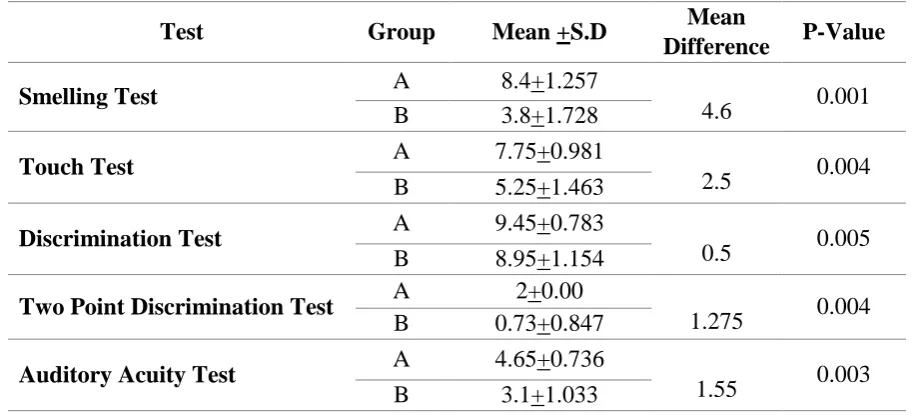
The results revealed the mean age group for group A to be 21.3+2.9 while for group B, it is found to be 23.6+3.0. Additionally, the test results showed that group A has heightened senses than group B as all the tests showed a significant mean difference with p-value<0.05. In Foot Tap Test, group A and group B showed a difference of 7.12+0.9 between their mean scores while in Smelling Test, there was a difference of 4.6+1.48. In Touch Test (Graphesthesia), the mean scores showed a difference of 2.5+1.22. Furthermore, there was also a significant difference between the mean scores of Group A and Group B for Discrimination Test, Two Point Discrimination Test, Auditory Acuity Test and Sixth Sense Test.
Table 1: Mean scores of various tests between blinds and sighted individuals
Test Group Mean +S.D Mean
Difference P-Value
Smelling Test A 8.4+1.257
4.6 0.001 B 3.8+1.728
Touch Test A 7.75+0.981
2.5 0.004 B 5.25+1.463
Discrimination Test A 9.45+0.783
0.5 0.005 B 8.95+1.154
Two Point Discrimination Test A 2+0.00
1.275 0.004 B 0.73+0.847
Auditory Acuity Test A 4.65+0.736
Sonya Arshad 50
Foot Tap Test A 7.75+1.316
7.125 0.001 B 0.63+0.49
Sixth Sense Test A 6+0.385 5.175 0.001 B 0.83+1.519
*Group A=blinds; Group B=sighted individuals *SD= Standard Deviation
Discussion
There is growing evidence that sensory indigence is confederate with cross-modal neuroplastic changes in the brain. Neuroplasticity is basically the brain's adaptation after a sensory loss (Merabet & Pascual-Leone, 2010). This study aimed to corroborate experimentally that early blinds have heightened senses including olfaction, tactile, audition and extrasensory perception. Many research studies support this phenomenon by neuroimaging studies (Voss et al., 2014&Théoret et al., 2004). Patrice Voss and Bruce evidenced this reorganizational compensatory mechanism in contrast to disuse atrophy using magnetization transfer ratio and found higher magnetization ratios in the occipital regions of early blinds as compared to the sighted ones with a p-value <0.005 (Voss et al., 2014). In a systemic review, T. Kujala et al., concluded that the occipital cortex of the blind is activated by auditory stimulus when the task is to detect the change of sound, which highlights the neuroplasticity with attentive processing of stimuli (Kujala, 2000). These findings also support the results of this study as in foot tap test, blinds outperformed the sighted ones with a difference of 7.125 points. Moreover, they also found some evidence regarding neuroplasticity in the healthy human brain, these findings form the basis for advancements in rehabilitation sector (Doucet et al., 2006).
Our brain is adaptable in nature by some practice or experience. We can mold or adjust our brain according to our need or
environment (Voss et al., 2014; Merabet & Pascual-Leone, 2010 & Jones, 2000). In an annual review by Centre of Neuroscience, conclusive remarks were made regarding this which states that although visual information is necessary for activity but in any case visual sense is lost, the neurons from surrounding sensory areas sprouts in occipital region along with divergence of preexisting circuits and expression of latent synapses (Jones, 2000).
In the current study, different tests were performed to experimentally validate the phenomena of neuroplasticity by comparing the intact senses. The first test was Smelling Identification Test, done to assess the olfactory sense, in which the results showed the potential difference between the olfactory capabilities of both the groups with a difference of 4.6 out of 10 (Table 1). The results contradicted to the findings of a meta-analysis conducted by Agnieszka Sorokowska et al. which concluded blindness does not seem to affect odor identification, discrimination or odor thresholds (Sorokowska et al., 2018). However Cuevas and Renier et al., supported functional modulation of occipital cortex in early blinds and reported favored results for early blinds in odor discrimination (p < 0.0002), free-identification (p < 0.0001) and categorization (p < 0.0004) (Renier et al., 2013 & Cuevas et al., 2009).
Sonya Arshad 51
is the tactile localization of our brain in which stronger mechanoreceptors functions as a tactile sensor, as evident in a study by Jones B which records increased cutaneous localization by the blind than the sighted (t = 2.50, df = 236, p < .01) (Jones, 1972). Stereognosis was also found to be well developed in non-sighted individuals which depend on memory, experience and practice including an intact somatosensory system. Heller concludes congruent results to the current findings regarding tactile enhancement in blinds and states that visual experience is not necessary for picture perception (Heller, 2002). This enhancement may be a result of practicing tactile discrimination while Braille reading (Burton et al., 2002). 2PD Test was performed to measure it and similar outweighed results were found in favor of the blind population as shown in (Table 1).
In a study in 2016, Bhavana G. Bhirud stated that adaptation to environment depends upon quickness of response as he found mean auditory reaction time for blinds to be 0.21+0.03s while 0.32+0.06 for the sighted group which was statistically significant with a p-value 0.000 (Bhirud & Chandan, 2017). The current study also assessed auditory localization and response time by Auditory Acuity Test and Foot Tap Recognition. In auditory acuity test, blinds were found to have a value of 4.65+0.736s while 3.1+1.033s for the sighted group. The increase in value was expected as the reaction time was also added in the recorded results. In both the tests, blinds outperformed the sighted individuals. Results also validate the findings of the studies which favor the recruitment of occipital cortex by auditory modalities (Gougoux et al., 2004 & Lessard et al., 1998).
Conclusively, it is evident that despite the lack of vision, congenitally blind subjects are able to build and manipulate neuronal circuit for spatial navigation. This neuronal substitution is more evident in early blinds as compared to late blinds (Lessard et al., 1998 & Wanet-Defalque et al., 1988).
Conclusion
It can be concluded from the results that following sensory deprivation, neuro-compensatory mechanisms generate new axonal pathways which makes the brain proficient in other functions. This mechanism should be considered as the basis for the management regimen of the sensory deficient population so that their productivity could be increased. Also, healthy people can ameliorate their normal functions by repetitive trials to enhance their specific activity.
Conflicts of Interests None.
Acknowledgment
We acknowledge the administration and staff of Dar-ul-sukun and Ida Rieu School for Blind and Deaf for extending their immense support. We are also thankful to Muhammad Nisar, Sr. lecturer at Department of Physiology, University of Karachi for his guidance.
Funding None.
References
Sonya Arshad 52
changes in early and late blind: A fMRI study of Braille reading. J. Neurophysiol., 87(1), 589-607.
Courtright, P., Hutchinson, A. K., & Lewallen, S. (2011). Visual impairment in children in middle-and lower-income countries. Arch. Dis. Child, 96(12), 1129-1134.
Cuevas, I., Plaza, P., Rombaux, P., De Volder, A. G., & Renier, L. (2009). Odour discrimination and identification are improved in early blindness. Neuropsychologia, 47(14), 3079-3083.
Doucet, M. E., Bergeron, F., Lassonde, M., Ferron, P., & Lepore, F. (2006). Cross-modal reorganization and speech perception in cochlear implant users. Brain, 129(12), 3376-3383. Gilbert, C., & Awan, H. (2003).
Blindness in children: Half of it is avoidable, and suitable cost effective interventions are available. BMJ: Br Med J, 327(7418), 760.
Gougoux, F., Lepore, F., Lassonde, M., Voss, P., Zatorre, R. J., & Belin, P. (2004). Neuropsychology: pitch discrimination in the early blind. Nature, 430(6997), 309.
Heller, M. A. (2002). Tactile picture perception in sighted and blind people. Behav Brain Res, 135(1-2), 65-68.J Can Acad Child Adolesc Psychiatry, 20(4), 265–276.
Johnston, M. V. (2009). Plasticity in the developing brain: implications for rehabilitation. Dev Disabil Res Rev., 15(2), 94-101.
Jones, B. (1972). Development of cutaneous and kinesthetic localization by blind and sighted children. Dev Psychol., 6(2), 349-352.
Jones, E. G. (2000). Cortical and subcortical contributions to activity-dependent plasticity in primate somatosensory cortex. Annu Rev Neurosci., 23(1), 1-37.
Kolb, B., & Gibb, R. (2011). Brain Plasticity and Behaviour in the Developing Brain.
Kolb, B., & Whishaw, I. Q. (1998). Brain plasticity and behavior. Annu Rev Psychol., 49(1), 43-64.
Kujala, T., Alho, K., & Näätänen, R. (2000). Cross-modal reorganization of human cortical functions. Trends Neurosci, 23(3), 115-120.
Lessard, N., Paré, M., Lepore, F., &Lassonde, M. (1998). Early-blind human subjects localize sound sources
better than sighted
subjects. Nature, 395(6699), 278-280. Merabet, L. B., & Pascual-Leone, A.
(2010). Neural reorganization following sensory loss: the opportunity of change. Nature Rev Neurosci, 11(1), 44-52.
Pascual-Leone, A., Amedi, A., Fregni, F., & Merabet, L. B. (2005). The plastic human brain cortex. Annu. Rev. Neurosci., 28, 377-401.
Renier, L., Cuevas, I., Grandin, C. B., Dricot, L., Plaza, P., Lerens, E., & De Volder, A. G. (2013). Right occipital cortex activation correlates with superior odor processing performance in the early blind. PLoS One, 8(8), e71907.
Sonya Arshad 53 Sorokowska, A., Sorokowski, P.,
Karwowski, M., Larsson, M., & Hummel, T. (2018). Olfactory perception and blindness: a systematic review and meta-analysis. Psychol Res, 1-17.
Sterr, A., Müller, M. M., Elbert, T., Rockstroh, B., Pantev, C., & Taub, E. (1998). Perceptual correlates of changes in cortical representation of fingers in blind multifinger Braille readers. J Neurosci, 18(11), 4417-4423.
Théoret, H., Merabet, L., & Pascual-Leone, A. (2004). Behavioral and neuroplastic changes in the blind: evidence for functionally relevant cross-modal interactions. J. Physiol. Paris, 98(1-3), 221-233.
Voss, P., Pike, B. G., & Zatorre, R. J. (2014). Evidence for both compensatory plastic and disuse atrophy-related neuroanatomical changes in the blind. Brain, 137(4), 1224-1240.