keeping their teeth longer. As a result, patients are more likely to have complex restorative and endodontic procedures leaving teeth more susceptible to cracks. Stressful lifestyle resulting in parafunctional habits such as clenching and bruxism and consumption of paan, betel nut etc. are important contributory factors. Additionally, in recent years, practitioners have been more aware of the existence of cracks and, therefore, more cracks are being diagnosed. Thus the diagnosis and management of cracked teeth is an integral part of dental practise today. This review article aims to highlight the different types of cracks, their diagnosis, associated signs and symptoms and treatment.
Key Words
Cracked tooth syndrome; bruxism; bite test; staining; craze line
INTRODUCTION
Cameron coined the term cracked tooth syndrome (CTS) to define the condition as “an incomplete fracture of a vital posterior tooth that involves the dentin and occasionally extends to the pulp”.[1] In recent times, the definition has been modified as follows: “A fracture plane of unknown depth and direction passing through tooth structure that, if not already involving, may progress to communicate with the pulp and or periodontal ligament”.[2] Cracked or incompletely fractured teeth can become symptomatic. Patients often present with a protracted history of pain of varying intensity; the origin of which may be difficult to locate. While intermittent pain on biting is the most consistent complaint associated with these teeth, cracks in teeth may result in a wide range of symptoms ranging from occasional discomfort to severe and prolonged pain. Symptoms are often dependent on the depth and direction of the crack and the tissuesinvolved.[3] Cracks in teeth may occur in both horizontal and vertical directions involving the crown and/or root. The etiology is generally a result of occlusal forces and iatrogenic procedures.[4] Crown and crown-root fractures are usually
incomplete fractures commencing in the crown of posterior teeth from an internal line angle at the floor of a restoration, and often involving a marginal ridge with the fracture extending in a mesiodistal direction. The fracture commences in the crown and may terminate in the vicinity of the cemento-enamel junction or extend apically into the root.[5] Vertical root fractures are longitudinally orientated fractures of the root that extend from the root canal to the periodontium.[6] These fractures are usually complete and extend a variable length along the root generally in a bucco-lingual direction and may extend into the crown.[7] This article reviews the classification, diagnosis and management of cracked teeth in dental practise.
Classification
The American Association of Endodontists, in a document titled “Cracking the Cracked Tooth Code”[8] identified five types of cracks in teeth. Craze lines are found in the majority of adult teeth and only involve enamel. In posterior teeth, craze lines are usually evident crossing marginal ridges and/or extending along buccal and lingual surfaces. Long vertical craze lines are often found in anterior teeth (Fig. 1). Fractured cusps usually result from
Kartik Pendharkar3, Amritaksha Bhattacharyya4, Harmeeta Budhiraja5, Madhurjya Chakraborty6
1MDS, Conservative Dentistry & Endodontics,
Registrar, FAA Medical College & Hospital, Barpeta, Assam, India
2Senior Lecturer, Department of
Prosthodontics, Daswani Dental College & Research Centre, Kota, Rajasthan, India
3MDS, Conservative Dentistry & Endodontics,
Private Practitioner, Mumbai, India
4MDS, Oral and Maxillofacial Pathology,
Private Practitioner, Guwahati, Assam, India
5Reader, Department of Periodontics,
Maharana Pratap Dental College, Gwalior, Madhya Pradesh, India
6
insufficient cusp support when the marginal ridge is weakened by an intra-coronal restoration (Fig. 2). The crack often extends in mesio-distal and bucco-lingual directions commonly involving one or both marginal ridges as well as a buccal or lingual groove and terminates in the cervical region either parallel to the gingival margin or slightly subgingival. A cracked tooth is indicative of a crack extending from the occlusal surface of the tooth apically without separation of the two segments. The crack is generally located centrally in a mesio-distal direction and may involve one or both marginal ridges (Fig. 3). A split tooth is indicative of a crack extending through both marginal ridges usually in a mesio-distal direction splitting the tooth completely into two separate segments (Figs. 4a-b). The crack is generally
located centrally in the tooth and this entity is the result of crack propagation of a cracked tooth. Vertical root fractures commence in the root generally in a bucco-lingual direction (Fig. 5). The crack is generally complete though may be incomplete and involve only one surface. The crack may involve either the entire root or only a portion of the root.[3]
Incidence
The presence of a cracked tooth occurs primarily in adulthood. Cameron5 reported that 80% of 102 cracked teeth occurred with patients over 40 years of age. Other reports[9,10] about the incidence and prevalence of cracked teeth were commonly associated with intracoronal restorations and most prevalent in mandibular molars. The wedging effect of the prominent mesio-palatal cusp of the maxillary first molar may account for this observation.[9] The disto-lingual cusp of mandibular molars is the most susceptible cusp for fracture. Nonfunctional cusps may be more susceptible to fracture than functional cusps.11 This observation may be a result of cuspal dimension as functional cusps are significantly larger in a bucco-lingual dimension and are covered with a thicker layer of enamel.[12] While functional cusps are supported on the inner and outer inclines by the opposing tooth, non-functional cusps may be more susceptible to fracture from lateral excursive occlusal forces due to the lack of support from the outer incline.[11]
Fig. 1: Craze lines in enamel Fig. 2: Occlusal, lingual and distal/proximal
views
Fig. 3: Craze lines in enamel Fig. 4a: Craze lines in enamel
Clinical Symptoms
Classically, the symptoms related to these teeth are pain on biting and sensitivity to thermal changes, particularly cold.[5] Pain associated with the release of pressure, ‘rebound pain’ is also a consistent finding.[13] Occasionally, there is sensitivity to sweets.[9] A chronic pulpitis with no clinical symptoms can exist as a result of microleakage of bacterial by-products and toxins. Pulpal and periodontal symptoms may occur when the fracture extends to involve the pulp.[14] The character, duration and the stimuli of pain has important implications for both diagnosis and treatment.15 An understanding of the mechanism of pain will often aid in assessment of the extent and direction of the crack. Luebke16 suggested the following terms to diagnose pain from a cracked tooth:
1. Dentin pain - A brief, sharp twinge.
2. Pulpal pain - The deep, demanding, radiating pain precipitated by thermal shock to an inflamed pulp. The pain at times may be spontaneous.
3. Periodontal pain - The aggravating throbbing of a sore tooth.
The pain associated with an incomplete fracture of a cusp is generally accepted to be due to the rapid movement of dentin fluid in the dentin tubules according to the “Hydrodynamic theory of dentin sensitivity” as proposed and investigated by Brännström.[17] The clinical presentation of a vertical root fracture is variable. Teeth with vertical root fractures often present with a history of
discomfort and localized chronic inflammation. Patients may complain of a bad taste and pain on biting. If swelling is present it is generally broad-based and any sinus tract is located in or close to the attached gingiva rather than in the apical area. Double or multiple sinus tracts are common.[3] DIAGNOSIS
Basic steps in crack detection includes, 1. History
Patient’s history should be assessed very carefully. History of any masticatory accidents, Para functional habits like bruxism, past dental treatment (iatrogenic factors),[18] dietary habits, beetle nut chewing, trauma or accidents may give a clue to our diagnosis. History of previous cracked teeth due to many anatomical[12] and developmental factors[18] may predispose teeth to crack. History of periodontal disease with extensive bone loss in the site can cause increased stresses on dentin predisposing the tooth to crack.[19] Cracks in root canal treated teeth are often due to size, design and placement of posts.[8]
2.Visual examination
Extra oral examination, shows enlarged masseter muscles due excessive stress on mastication or Para functional habits like bruxism.[20] Intraoral examination should be done using rubber dam in a clear and dry field. Visualisation of cracks becomes easier in Contrasting and brighter colour of rubber dam. Check for the wear facets. Brown studied that teeth subjected to thermal cycling between 90 to 140 degrees had severe cracking, check for
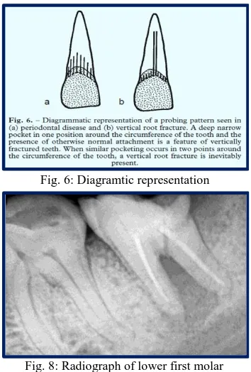
Fig. 5: Vertical root fracture around the mesial root of the mandibular molar (arrow)
Fig. 6: Diagramtic representation
abfraction lesions. Abfraction lesions are found to be associated with 94.5% of occlusal wear facets. Excessive occlusal loading has been suggested to be a possible cause of fracture in some unrestored teeth with abfraction lesions.[5] Steep cusps and wedging effect of cusp fossae relationship create hammer and anvil “chewing effect” that can propagate cracks. Wide and deep restorations also predispose to cracks. Magnifying loupes with illumination enhances our vision in crack detection.[21]
3.Tactile examination
Palpate the gingiva around the tooth for any dehiscence or fenestration typical of vertical root fracture. Palpation may show tenderness and swelling over the root and in Periapical region in cases of vertical root fracture. Running the tip of a sharp probe along the tooth surface produces a clicking sound when it passes over the fracture line.[22]
4.Bite tests
Rubber wheel, wooden stick or tooth sloth fracture detector can be placed on the cusp of the suspected tooth and ask the patient to bite down with moderate pressure and then release. Pain during biting or chewing is a classic symptom.[10]
5. Periodontal probing
Localised bone loss can result in excessive stresses on the exposed dentin predisposing crack formation.[19] Careful periodontal probing in small
increments will restrict the movement of the probe thus helps to locate a crack.[8] A deep narrow isolated periodontal pocket is present in cases of vertical root fracture (Fig. 6).[23]
6. Staining
2% Methylene blue, 0.25% sodium fluorescein ophthalmic solution, snoop caries detecting dye can be used to disclose a crack on surgically exposed roots or crack in the cavity after removal of the restoration. Wright, in his study proved that methylene blue combined with Transilluminaton was the best in crack detection.[21]
7. Transilluminaton
A fibre optic light source combined with magnification helps in visualisation of cracks on tooth surface.[16] The light beam is directed in a horizontal direction perpendicular to the plane of the suspected crack. Cracks will block the light beam from reaching the part of the tooth beyond the fracture, whereas sound teeth will transmit light through the crown.[21]
8. Radiographs
Radiographs are not conclusive in detecting cracks[24] in the earlier stages unless there is separation of segments of the tooth. Typical radiographic features for vertical root fracture includes
1. When a well condensed root canal filling appears very close to one wall of the root.[25]
6. Pitts and Natkin described halo like radiolucency running around the tooth (Fig. 8).[6] 7. Step like bone defect may be seen when fracture
runs obliquely across the root.[6]
8. V shaped radiolucency which is widest at the crestal bone and narrowest towards the apex.[6] 9. Thickened periodontal ligament space or a
diffuse radiolucency with J-shaped appearance in the apical region.[25]
Ultrasound imaging system is capable of imaging cracks in simulated human tooth and would be a possible diagnostic tool in the future.[27]
9. Restoration removal
Bulky restorations or restorations with pin head designs[28] should be cautiously examined for a crack. Removal of restorations allows for visual examination of the crack. Marginal ridges should be closely examined for a crack.[23] Thin marginal ridges predispose to cracks. Magnifying loupes and staining the tooth can aid in crack detection. 10. Surgical assessment
Surgical exploration remains the last option when all the other diagnostic means had failed to detect a crack. This consists of elevation of a full thickness
linguo-pulpal line angle of a cusp to the cemento-enamel junction or slightly below. (b) Horizontal fracture of a cusp not involving the pulp.
2. Good: A coronal vertical fracture that runs mesio-distally into the dentin but not into the pulp.
3. Poor: A coronal vertical fracture that runs mesio-distally into the dentin and pulp but is confined to the crown.
4. Hopeless: A coronal vertical fracture that runs mesio-distally through the pulp and extends into the root.
Treatment
A rough guideline for therapy of fractured teeth is given in Fig. 9.
Immediate Therapy (Table 1)
To avoid irreversible damage, it is imperative that a cracked tooth be treated as soon as possible. Occlusal adjustment of affected teeth must be done immediately to reduce the stress on the tooth and prevent further damage to the tooth. If the tooth in question presented with a preexisting restoration, it should be removed. This may cause the affected cusp to “splinter off” and the further treatment protocol can then be decided.[30] In case there is no splintering, immediate immobilization should be employed, using an “immediate extra-coronal circumferential splint.” A copper ring or a stainless steel orthodontic band can be used for this purpose.[31] The use of full coverage acrylic provisional crowns has been advocated for “immediate splinting”.[32] Another available option is the use of bonded composite resin to splint the teeth, which is known as a “direct composite splint” (DCS).[33]
Definitive therapy
In case of non splintering teeth, intra coronal restorations, without cuspal coverage may also be used. Dental amalgam, composite resin and glass-ionomer cements are most commonly used. The strength of the tooth can be restored by using these materials. Amalgam overlays have been found to increase the fracture energies of cracked teeth to
levels equivalent to that of intact teeth. Hence direct restorations with cuspal coverage have been advocated as a primary mode of treatment.[30] In cases where aesthetics is not a matter of great concern, cast metal inlays with cusp coverage or partial crowns with circumferential external splinting are applied or else, adhesive ceramic restorations can also be used. Full coverage crowns are the most appropriate form of restoration for cases where the crack extends from the occlusal incline to the cervical third of the clinical crown.[32] It has been reported by a number of authors that the loss of vitality following the application of full coverage single unit crowns is in the range of 15-19%. Loss of pulpal vitality is an obvious problem following the preparation of teeth to receive a full coverage crown. In CTS, reversible pulpitis would already be a pre- existing condition so the problem appears to be further compounded.[34]
CONCLUSION
A cracked tooth is a complex condition and can present with a wide variety of symptoms. Early diagnosis is of paramount importance in preventing further propagation of the crack. It is important to diagnose and differentiate between dentin, pulpal and periodontal pain before treatment is commenced. Though various treatment options have ben outlined for the management of cracked teeth very little evidence is available in literature to substantiate their use. The extent of the fracture, the time of intervention and the type of restoration are crucial parameters in determining the outcome of the treatment. So we as dentists must take all these factors into account and help the patients decide and initiate a proper treatment plan.
REFERENCES
1. Ehrmann EH, Tyas MJ. Cracked tooth syndrome: diagnosis, treatment and correlation between symptoms and post-extraction findings. Aust Dent J 1990;35(2):105-12. 2. Ellis SG. Incomplete tooth fracture-proposal
for a new definition. Br Dent J
2001;190(8):424-8.
3. Kahler W. The cracked tooth conundrum: terminology, classification, diagnosis, and management. Am J Dent 2008;21(5):275-82. 4. Bender IB, Freedland JB. Adult root fracture. J
Am Dent Assoc 1983;107(3):413-9.
5. Cameron CE. The cracked tooth syndrome. J Am Dent Assoc 1964;68:405-11.
6. Pitts DL, Natkin E. Diagnosis and treatment of vertical root fractures. J Endod 1983;9(8):338-46.
7. Walton RE, Michelich RJ, Smith GN. The histopathogenesis of vertical root fractures. J Endod 1984;10(2):48-56.
8. American association of endodontics; colleagues of excellence; “cracking the cracked tooth Code detection and treatment of various longitudinal tooth fractures” summer
2008 available at
http://www.aae.org/dentalpro/colleaguenews.h tm; bonus material C.
9. Hiatt WH. Incomplete crown-root fracture in pulpal-periodontal disease. J Periodontol 1973;44(6):369-79.
10. Geurtsen W. The cracked-tooth syndrome: clinical features and case reports. Int J Periodontics Restorative Dent 1992;12(5):395-405.
11. Cavel WT, Kelsey WP, Blankenau RJ. An in vivo study of cuspal fracture. J Prosthet Dent 1985;53(1):38-41.
12. Khers SC, Carpenter CW, Vetter JD, Staley RN. Anatomy of cusps of posterior teeth and their fracture potential. J Prosthet Dent 1990;64(2):139-47.
13. Cameron CE. The cracked tooth syndrome: additional findings. J Am Dent Assoc 1976;93(5):971-5.
14. Bergenholtz G. Pathogenic mechanisms in pulpal disease. J Endod 1990;16(2):98-101. 15. Figdor D. Aspects of dentinal and pulpal pain.
Pain of dentinal and pulpal origin--a review for the clinician. Ann R Australas Coll Dent Surg 1994;12:131-42.
16. Luebke RG. Vertical crown-root fractures in posterior teeth. Dent Clin North Am 1984;28(4):883-94.
17. Brännström M. Dentin and pulp in restorative dentistry. London: Wolfe Medical Publications Ltd 1982:47-63.
18. Lynch CD, McConnell RJ. The cracked tooth syndrome. J of Canadian Dent Assoc 2002;68(8):470-5.
19. American association of endodontics; colleagues of excellence; “cracking the crack tooth Code detection and treatment of various longitudinal tooth fractures” winter 1997. 20. Sonnomiya EK, Goncalves M, Cavalcanti MP.
Journal of Am Dent Assoc 2012;134:434-41. 24. White SC, Pharoah MJ. Oral radiology,
principles and interpretation (5th edn). St. Louis, MO: Mosby, 2004: p. 623.
25. Mathew S, Boopathithangavel, das A. Diagnosis of cracked tooth syndrome. JOf pharm and Bioallied Sciences 2012;4(2);242-5.
26. Moule AJ, Kahler B. Diagnosis and management of teeth with vertical root fractures. Aust Dent J 1999;44(2);75-87. 27. Culjat MO, Singh RS, Brown ER. Ultrasound
crack detection in a simulated human tooth. DentoMaxilloFacial Radiology 2005;34:80-5. 28. Standler JP, Collard EW, Caputo AA. Dentinal
defects caused by some twist drills and retentive pins. J Prosthet Dent 1970;24(2);185-92.
29. Clark LL, Caughman WF. Restorative treatment for the cracked tooth. Oper Dent 1984;9:136-42.
30. Shetty R. Cracked tooth syndrome. Journal of Dental Research and Scientific Development 2014;1(2):51-6.
31. Geurtsen W, Schwarze T, Gunay H.
Diagnosis, therapy and prevention of the cracked tooth syndrome. Quintessence Int 2003;34:409 17.
32. Gutherie RC, Difiore PM. Treating the cracked tooth with a full crown. J Am Dent Assoc 1991;122:71 3.
33. Banerji S, Mehta SB, Millar BJ. Br Dent J 2010;208:459-63.
34. Cheung GS, Lia SC, Ng RP. Fate of vital pulps beneath a metal ceramic crown or a bridge retainer. Int Endod J 2005;38:521-30.