REVIEW
Epigenetic regulation of prostate cancer
Suyin P. Chin&Joanne L. Dickinson&Adele F. Holloway
Received: 1 February 2011 / Accepted: 23 May 2011 / Published online: 14 June 2011 #Springer-Verlag 2011
Abstract Prostate cancer is a commonly diagnosed cancer in men and a leading cause of cancer deaths. Whilst the underlying mechanisms leading to prostate cancer are still to be determined, it is evident that both genetic and epigenetic changes contribute to the development and progression of this disease. Epigenetic changes involving DNA hypo- and hypermethylation, altered histone modifi-cations and more recently changes in microRNA expression have been detected at a range of genes associated with prostate cancer. Furthermore, there is evidence that particular epigenetic changes are associated with different stages of the disease. Whilst early detection can lead to effective treatment, and androgen deprivation therapy has a high response rate, many tumours develop towards hormone-refractory prostate cancer, for which there is no successful treatment. Reliable markers for early detection and more effective treatment strategies are, therefore, needed. Consequently, there is a considerable interest in the potential of epigenetic changes as markers or targets for therapy in prostate cancer. Epigenetic modifiers that demethylate DNA and inhibit histone deacety-lases have recently been explored to reactivate silenced gene expression in cancer. However, further understanding of the mechanisms and the effects of chromatin modulation in prostate cancer are required. In this review, we examine the current literature on epigenetic changes associated with prostate cancer and discuss the potential use of epigenetic modifiers for treatment of this disease.
Keywords Prostate cancer. Epigenetics . DNA methylation . Histone acetylation . MicroRNA
Introduction
Prostate cancer is the most commonly diagnosed cancer for men living in developed countries (other than non-melanoma skin cancer). According to Cancer Research UK, an estimated 913,000 men worldwide were diagnosed in developed countries in 2008. According to the US National Cancer Institute, it was estimated that almost 217,730 men would be diagnosed in the USA alone in 2010 and more than 32,050 would die as a direct result of the disease. The use of prostate-specific antigen (PSA) as a screening tool has allowed the detection of prostate cancer in the early stages whilst it is still locally confined. Whilst more than 70% of diagnosed cases now survive beyond 5 years, this cancer is still associated with significant mortality and morbidity. Metastatic prostate tumours are responsible for the majority of deaths associated with this cancer. The most frequent site of prostate cancer metastasis is to bone; over 80% of men who die of prostate cancer have metastatic boney lesions (Bubendorf et al. 2000). In terms of current treatments for prostate cancer, we are still unable to identify with certainty those tumours requiring aggressive and immediate intervention (associated with considerable morbidity) and those where a“watchful-waiting” approach may be more appropriate. Thus, identification of markers predicting tumour behaviour has become of intense interest to researchers working to discover new prognostic and diagnostic markers and new targets for treatment.
Prostatic intraepithelial neoplasia (PIN) and, in particular, high-grade PIN has been identified as precancerous lesions most likely leading to prostatic carcinoma. On the prostate morphological spectrum, PIN refers to precancerous lesions involving cell proliferation within prostatic ducts, ductules and acini (De Marzo et al.2004). PIN is believed to pre-date carcinoma by 10 or more years. High-grade PIN is considered clinically significant as men with high-grade
S. P. Chin:J. L. Dickinson
:
A. F. Holloway (*)Menzies Research Institute Tasmania, University of Tasmania, Private Bag 23,
PIN have up to 50% chance of subsequently developing prostate cancer (Lee et al. 2011). A second morphological abnormality termed atypical small acinar proliferation has also been associated with increased risk of diagnosis with prostate cancer in subsequent biopsies. Prostate tumours are most commonly graded using the Gleason score, determined by the histological characteristics of the glandular architecture within the tumour.
It is clear that epigenetic changes within a cell play a significant role in the development and progression of cancer (Esteller2008; Jones and Baylin 2007) and, as in most other human cancers, prostate cancer development and progression appears to involve an interplay between both genetic and epigenetic changes. There is now considerable evidence that changes in gene expression which involve epigenetic alterations may be an important factor in prostate cancer progression, and development of panels of epigenetically modified genes as markers of disease progression is of considerable topical interest.
Epigenetic mechanisms
Epigenetic alterations are heritable changes in gene expression that occur without changes in DNA sequence, with the broadest definition including all factors other than DNA sequence changes that heritably influence gene expression (Berger et al. 2009). Whilst the best described of these mechanisms is DNA methylation, other epigenetic mechanisms include physical and chemical changes to chromatin and regulation of gene expression by microRNAs (miRNAs).
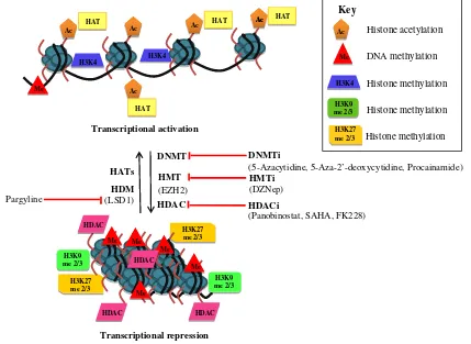
DNA methylation plays an important role in DNA repair, recombination and replication, as well as regulating gene activity (see Fig.1). DNA methylation involves the addition of a methyl group to the 5′-carbon of cytosine in CpG dinucleotide sequences, catalysed by a family DNA methyl-transferases (DNMTs). CpG-rich regions, known as CpG islands are commonly found associated with the 5′-region of vertebrate genes (Gardiner-Garden and Frommer1987) and are generally protected from methylation (Bird 2002). For many years, CpG islands have been implicated in gene regulation with their methylation strongly correlated with gene silencing (Illingworth and Bird2009). DNA methyla-tion can regulate gene activity via two mechanisms. Firstly, methylation of CpG dinucleotides within transcription factor binding sites can inhibit transcription factor binding and, therefore, directly influence gene activity (Hark et al.2000). Secondly, methylated CpG dinucleotides act as binding sites for methyl CpG binding proteins, which are associated with other factors such as histone deacetylases, involved in establishing repressive chromatin structures (Jones et al.
1998; Nan et al.1998).
Changes in DNA methylation patterns have been linked with cancer for many years now (Jones and Baylin 2007). However, the methylation changes are complex, with both hypomethylation and hypermethylation occurring in cancer cells. Aberrant DNA hypermethylation occurs when there is a gain of DNA methylation at regions that are normally unmethylated and when this occurs at gene promoters can lead to gene inactivation. Localised hypermethylation of gene promoters has been reported in virtually all types of cancers, including prostate cancer. On the other hand, DNA hypomethylation is the demethylation of normally methyl-ated DNA and can lead to chromosomal instability and activation of proto-oncogenes (Dunn 2003; Eden et al.
2003; Sharma et al. 2010). Both global and gene-specific hypomethylation events have also been implicated in prostate cancer.
Whilst alterations in DNA methylation have long been linked to cancer, there is also mounting evidence that other epigenetic changes, such as changes to chromatin composition or structure, contribute to cancer. Within the eukaryotic nucleus, DNA is assembled into chromatin, the basic unit of which is the nucleosome. Nucleosomes are composed of approximately 147 base pairs of DNA wrapped around an octamer of core histone proteins, containing two each of histones H2A, H2B, H3 and H4 (Kornberg and Lorch 1999). The N termini of the core histones protrude from the nucleosome and are subjected to a range of covalent modifications, catalysed by various histone-modifying enzymes. At least ten different histone modifications have now been reported, including acetyla-tion, methylaacetyla-tion, phosphorylation and ubiquitination (Kouzarides 2007; Gardner et al. 2011). Each of these modifications affects the chromatin structure and function in a different way by either disrupting chromatin contacts or affecting the recruitment of other proteins to the chromatin (Kouzarides 2007). Acetylation of lysine resi-dues in histone H3 and H4 (H3Ac and H4Ac) is in general associated with transcriptional activity, whereas histone methylation is associated with transcriptional activation or repression depending on the site of modification and the number of methyl groups added (see Fig.1). For example, histone H3 lysine 4 (H3K4) methylation is generally associated with transcriptional activation, whereas histone H3 lysine 9 (H3K9) and histone H3 lysine 27 (H3K27) di- and tri-methylation is generally associated with transcriptional repression (Kouzarides 2007). As with DNA methylation, there is an increasing body of evidence that changes in histone modifications due to aberrant activity or mis-targeting of chromatin-modifying enzymes is involved in carcinogenesis (Hake et al.2004).
long, which regulate gene expression by affecting the stability or the translation efficiency of target mRNAs (Pang et al. 2010). The miRNA binds to complementary sequences within the 3′mRNA tails, although usually only with partial base pair complementarity. When miRNAs bind to mRNA with partial complementarity, they function as translational repressors whereas perfect complementarity induces degradation of the target mRNA (He and Hannon
2004; Porkka et al. 2007). Recent studies have found that aberrations in miRNA expression are associated with various human cancers including prostate cancer. About half of the known miRNA genes are located at cancer-related genomic regions (Calin et al.2004), and in cancer, miRNAs can function as either oncogenes or tumour suppressor genes (Hayashita et al. 2005; He et al. 2005). One miRNA can target hundreds of mRNAs and, therefore, alterations in a single miRNA can have dramatic effects on cell biology. Our understanding of the function and regulatory mechanisms of miRNAs has expanded enor-mously over recent years; however, we still have limited capacity to identify targets of miRNAs and have much to learn about their involvement in cellular processes and disease.
Epigenetic alterations in prostate cancer progression
DNA hypomethylation
There is considerable evidence that changes in methylation patterns occur in prostate cancer with DNA hypomethyla-tion in tumour samples first documented more than 20 years ago. In 1987, Bedford and van Helden analysed DNA 5′ -methylcytosine content in human prostate samples and reported a correlation between global hypomethylation and development of benign prostatic hyperplasia (BPH) and metastatic tumours (Bedford and van Helden 1987). Comparison of DNA methylation levels in tumour and normal prostate tissue by immunohistochemistry similarly detected global hypomethylation in prostate cancer (Brothman et al.
2005). In keeping with this, Santourlidis et al. (1999) examined methylation of LINE-1 repetitive sequences in prostate adenocarcinomas and found that LINE-1 methyla-tion tended to decrease with tumour stage. A further study analysing tumour samples similarly demonstrated an associ-ation between DNA hypomethylassoci-ation, tumour state and metastasis, with extensive hypomethylation of LINE-1 observed in 64% of cases with lymph node or distant
Key
Histone acetylation
DNA methylation
Histone methylation
Histone methylation
Histone methylation Ac
Me
H3K9 me 2/3
H3K27 me 2/3 H3K4
H3K4
H3K4
HDM (LSD1) Pargyline
HDACi
(Panobinostat, SAHA, FK228) DNMTi
(5-Azacytidine, 5-Aza-2’-deoxycytidine, Procainamide) HMTi
(DZNep) HMT
(EZH2) HATs
H3K9 me 2/3 H3K27
me 2/3
H3K27 me 2/3
H3K9 me 2/3
HDAC DNMT Transcriptional activation
Transcriptional repression
Me Ac
Ac
Ac Ac
Ac Ac HAT
HAT
HAT HAT
Me
Me
Me
Me
HDAC HDAC
HDAC HDAC Me
metastases but only in 21% of cases without detectable metastases (Schulz et al. 2002). All cases of hormone-refractory locally recurrent tumours displayed extensive LINE-1 hypomethylation. The study by Schulz et al. (2002) also found a strong association between LINE-1 DNA hypomethylation and chromosomal alterations, in support of the notion that DNA hypomethylation increases genomic instability (Schulz et al. 2002). Most prostate cancer samples with prominent DNA hypomethylation also exhibited a large number of chromosomal alterations and vice versa. These findings were supported by a further study which found increased prevalence of LINE-1 hypomethyla-tion in later-stage prostate cancers and lymph-node-positive prostate cancers (Florl et al. 2004), in contrast to gene-specific hypermethylation events which did not correlate with tumour stage, being present in both early and late-stage prostate cancer. These data, therefore, suggest that hypome-thylation is a later event in prostate cancer progression compared with gene-specific hypermethylation. Consistent with this, a recent study examined LINE-1 methylation in primary prostate cancers compared with normal prostate tissues and found a significantly higher level of LINE-1 hypomethylation in metastatic prostate cancer tissues, sug-gesting that global hypomethylation occurs late in prostate cancer progression, particularly at the metastatic disease stage (Yegnasubramanian et al.2008).
Whilst global DNA hypomethylation can contribute to cancer by promoting genomic instability, the hypomethyla-tion of individual gene promoters can also contribute to cancer development and progression by directing aberrant gene expression. Hypomethylation of a number of genes has been linked to prostate cancer. For example, a study by Yegnasubramanian et al. (2008), found that CpG islands associated with a class of cancer testis antigen genes were hypomethylated in prostate cancer, correlating with their overexpression in primary prostate cancers, and more so in metastatic prostate cancer. Similarly, hypomethylation of the promoter of the heparanase gene has been reported in prostate cancer, compared with BPH (Ogishima et al.
2005). This correlates with increased mRNA expression of the heparanase gene, which is associated with tumour invasion and metastasis (Vlodavsky et al.1988; Hulett et al.
1999). Further, upregulation of urokinase expression in invasive prostate cancer cell lines correlates with hypome-thylation of the promoter (Pakneshan et al.2003). Upregu-lation of cytochrome P450 1B1 (CYP1B1) mRNA and protein levels in prostate cancer compared with BPH tissue was also found to correlate with hypomethylation of the regulatory regions of this gene (Tokizane et al.2005). Other genes such as wingless-related MMTV integration site 5A (WNT5A), S100 calcium-binding protein P and cysteine-rich protein 1 were also found to be hypomethylated in primary prostate cancer tissues. These genes are similarly
associated with tumourigenesis and metastasis (Dissanayake et al. 2007; Wang et al. 2006). Further investigation of the WNT5A gene revealed three CpG sites in the promoter region which were consistently methylated in a normal prostate cell line and normal prostate tissues but not in a prostate cancer cell line and primary prostate cancer tissues (Wang et al. 2007). Therefore, hypomethylation and conse-quent upregulation of genes involved in metastasis and cell invasion may be an important factor in prostate cancer progression. In support of this, treatment of the PC3 prostate cancer cell line with reagents that prevent DNA hypomethylation has an inhibitory effect on cell invasion in vitro and tumour growth in vivo (Shukeir et al.2006).
DNA hypermethylation
DNA hypermethylation is the most commonly reported epigenetic alteration observed in prostate cancer. Many genes have been identified as aberrantly hypermethylated in prostate cancer and these genes include tumour suppressor genes, DNA damage repair genes, hormonal response genes and genes involved in cell cycle control, tumour cell invasion and metastasis (Phé et al.2010). Hypermethylation of DNA can lead to inappropriate gene silencing, disrupting gene function and, thus, contributing to tumour initiation, progression and metastasis (Li et al. 2004). This review highlights some of the best described and most frequently hypermethylated genes in prostate cancer.
The most frequently reported hypermethylated gene in prostate cancer is the π-class glutathione-S-transferase (GSTP1) gene. GSTP1 is an enzyme involved in the metabolism, detoxification and elimination of reactive chemical compounds and in this way protects cells from DNA damage and cancer initiation (Lee 2007). Hyper-methylation of the GSTP1 gene in prostate cancer was first reported by Lee et al. (1994) who found hypermethylation of the gene in all 20 human prostate cancer tissue samples examined, but not in normal tissues or BPH. Following this, GSTP1 gene expression was analysed in 60 high-grade PIN samples, with all samples showing a loss of GSTP1 gene expression (Brooks et al. 1998). Further investigation found that this loss of GSTP1 expression was due to hypermethylation of the gene promoter. A large number of studies have now reported GSTP1 hypermethy-lation in prostate cancer samples, with methyhypermethy-lation detected in up to 90% of samples, suggesting GSTP1 hyper-methylation is a common epigenetic alteration in prostate cancer (Goessl et al.2001; Chu et al.2002; Jeronimo et al.
2002; Harden et al.2003; Nakayama et al.2003; Bastian et al. 2004).
the cadherin-catenin system. Loss of E-cadherin (CDH1) gene expression is associated with the transition from adenoma to carcinoma and the acquisition of metastatic potential (Perl et al. 1998). CDH1 hypermethylation was observed in prostate cancer cell lines and treatment with the demethylating agent, 5-aza-2′-deoxycytidine restored CDH1 mRNA and protein expression in the cell lines, suggesting that promoter hypermethylation was responsible for CDH1 silencing in these cells (Graff et al. 1995). Furthermore, Kallakury et al. (2001) reported that the CDH1 gene promoter was methylated in eight out of ten prostate cancer tissues examined. Interestingly, the degree of CDH1 promoter methylation correlated with the patho-logical stage of the prostate tumour tissues, with CDH1 promoter methylation occurring in 30% of low-grade prostate cancer tissues, but increasing to 70% in high-grade tumours (Li et al. 2001). These results suggest that methylation of the CDH1 promoter is associated with prostate tumour progression.
CD44, which is a cell surface glycoprotein involved in cell matrix adhesion and signal transduction is also silenced in prostate cancer by methylation of the gene promoter. Lou et al. (1999) examined methylation levels of the CD44 gene in 84 matched normal and prostate cancer samples and found hypermethylation of CD44 in 31 out of 40 of the primary prostate cancer samples. Further investigation by Verkaik et al. (1999) showed that in CD44-negative prostate cancer cell lines (LNCaP and PC346C) the CD44 promoter was hypermethylated compared with CD44-positive prostate cancer cell lines (Du145, PC3 and TSU). A further study of CD44 silencing was conducted on human tissue samples and demonstrated that nine out of 11 lymph node metastases of prostate cancer displayed CD44 gene promoter methylation. These data, therefore, suggest that hypermethylation of the CD44 promoter resulting in downregulation of CD44 gene expression may be involved in prostate cancer progression and metastasis (Verkaik et al.2000).
DNA methylation is also involved in regulation of the androgen receptor (AR). AR is activated by androgen, which plays a critical role in the development, growth and maintenance of the prostate (Jenster 1999). In the initial stages, prostate cancer is androgen dependent, but eventu-ally becomes androgen independent, due to the loss of AR expression (Jarrard et al.1998; Tekur et al.2001; Takahashi et al. 2002; Suzuki et al. 2003). Data from a number of studies suggest that this loss of AR expression is at least partly due to hypermethylation of the AR gene promoter. Jarrard et al. (1998) found AR promoter hypermethylation in AR-negative prostate cancer cell lines (DU145, DuPro, TSU-PR1 and PPC1) whereas the promoter was unmethy-lated in AR-positive cell lines (LNCaP and PC3). Expression of the AR gene was restored in the AR-negative cell lines by treatment with the demethylating agent 5-aza-2′deoxycitidine,
suggesting that promoter methylation was responsible for AR gene silencing in the AR-negative cell lines. Further, Suzuki et al. (2003) reported that promoter hypermethylation of AR leading to loss of AR expression occurs in 30% of hormone-refractory prostate cancers, suggesting that DNA hyper-methylation contributes to loss of AR expression in at least some prostate cancers.
The silencing of cell cycle regulation genes by DNA hypermethylation has also been observed in prostate cancer. The Ras-associated domain family 1A gene (RASSF1A) is highly methylated in several human cancers, including prostate cancer (Liu et al. 2002; Kang et al. 2004; Pfeifer and Dammann2005; Dammann et al.2005; Aitchison et al.
2007; Hesson et al. 2007). Liu et al. (2002) examined methylation of the RASSF1A promoter in primary prostate tumours and reported methylation of the RASSF1A promoter in over 70% of the tumours. Further investigation found a correlation between the frequency of methylation and the Gleason score of the tumour, with highly aggressive tumours displaying more frequent DNA methylation com-pared with less aggressive tumours. Similarly, Kang et al. (2004) reported methylation of a number of genes, including RASSF1A in prostate cancer and PIN samples, with more frequent methylation correlating with higher PSA levels and Gleason score.
Silencing of tumour suppressor genes by DNA methyl-ation is also often observed in prostate cancer. DNA hypermethylation of the adenomatous polyposis coli (APC) gene in prostate cancer individuals was observed in a study by Rosenbaum et al. (2005), which examined promoter methylation of a number of genes. Hypermethy-lation of APC alone, and hypermethyHypermethy-lation of APC and the cell cycle regulation gene cyclin D2 in combination were found to be significant predictors of prostate cancer progression. In keeping with this, Henrique et al. (2007) analysed a small panel of gene promoters in prostate biopsy samples and similarly found that hypermethylation of APC is an independent predictor of poor prognosis in prostate cancer. Subsequent studies have similarly found APC hypermethylation to be a predictor of prostate cancer progression (Liu et al. 2011b; Richiardi et al.2009).
Histone modifications
di-methylated H4 arginine 3 (H4R3me2) and di-methylated H3 lysine 4 (H3K4me2) in 183 primary prostate cancer tissue samples. In the individuals with low-grade tumours, the study found two subgroups with different risks of tumour recurrence based on the presence of similar combinations of global histone modifications. Individuals with a lower risk of tumour recurrence were those who were above the 60th percentile staining for H3K4me2 or above the 35th percentile staining for H3K18Ac and H3K4me2. However, these histone modification patterns did not correlate with the Gleason score. In contrast, a more recent study of primary and metastatic prostate cancer samples found that high global levels of H3K18Ac and H3K4me2 correlated with a threefold increased risk of prostate cancer recurrence (Bianco-Miotto et al.2010). A further study showed that global levels of H3K4me1, H3K9me2, H3K9me3, H3Ac and H4Ac were significantly reduced in prostate cancer compared with BPH and normal prostate tissue (Ellinger et al. 2010), with H3Ac and H3K9me2 in particular discriminating between the malignant and non-malignant samples. They also found that individuals with high H3K4me1 levels were more likely to experience recurrence of the prostate cancer and thus suggested that analysis of H3K4me1 may provide predictive information regarding likelihood of tumour recurrence.
In addition to the studies outlined above that have documented changes in histone modifications associated with prostate cancer, there is accumulating evidence that expression of histone-modifying enzymes is altered in prostate cancer. Bianco-Miotto et al. (2010) identified a candidate gene signature consisting of six genes encoding epigenetic modifiers, including both DNA methyltrans-ferases and histone methyltransmethyltrans-ferases (HMTs) that was associated with prostate cancer progression. In addition, a number of studies have documented alterations in the HMT EZH2, which is responsible for the repressive H3K27me3 modification, in prostate cancer. EZH2 is upregulated in hormone-refractory metastatic prostate cancer (Varambally et al.2002). Overexpression of EZH2 in prostate cancer cell lines increases the invasive characteristics of the cells, whilst knockdown of EZH2 decreases the proliferative capacity of the cells, and more so in hormone-independent cell lines (Varambally et al.2002; Karanikolas et al.2010). Furthermore, microarray analysis of metastatic prostate cancer tissue identified a group of EZH2 repressed genes that were associated with prostate cancer progression (Yu et al.2007).
A number of studies have also implicated lysine-specific demethylase 1 (LSD1) in prostate cancer. LSD1 was originally identified as a H3K4 demethylating enzyme (HDM). This enzyme was thought to function as a transcriptional co-repressor by reversing the H3K4me modifications associated with transcriptional activation
(Shi et al. 2004). However, genome-wide studies of LSD1 homologues in yeast suggest that these enzymes can act as both co-activators and co-repressors by targeting H3K4 or H3K9, respectively (Opel et al.2007). In keeping with this, LSD1 has been demonstrated to form ligand-dependent, chromatin-associated complexes with AR, stimulating AR-dependent transcription (Metzger et al. 2005). In this case, LSD1 acts by demethylating histone H3K9, thus activating AR target genes by removing repressive H3K9me modifica-tions. This study found that LSD1 and AR were co-localised in both normal prostate and prostate cancer. A further study by Kahl et al. (2006) correlated the expression patterns of AR, LSD1 and the AR co-activator FHL2 (four and a half LIM-domain protein 2) with Gleason score, Gleason grade and p53 expression in 153 prostate tumour samples with relapse after radical prostatectomy. The study found that increased LSD1, nuclear expression of FHL2, high Gleason score and grade and high levels of p53 were strongly associated with relapse during follow-up. In addition, upregulation of both LSD1 mRNA and protein levels was associated with high risk of relapse. Whilst LSD1 demethylates H3K9me1 and H3K9me2, a further study found that the Jumonji C domain containing protein JMJD2C can demethylate H3K9me3 (Wissmann et al. 2007). JMJD2C associates with AR and LSD1 in prostate cells, and this complex acts to demethylate H3K9me3 and increase AR-dependent gene transcription. A second Jumonji protein, JHDM2A, which also demethylates H3K9me1/2 has similarly been found to stimulate transcrip-tion of AR dependent genes (Yamane et al. 2006). Furthermore, Gaughan et al. (2011) showed that AR interacts with and is methylated by the HMT enzyme SET9. SET9 was originally identified as an enzyme responsible for catalysing the activating H3K4me1 modification, and, therefore, was associated with transcriptional activation (Nishioka et al. 2002). However, Gaughan et al. (2011) found that by methylating AR SET9 increased the transcrip-tional activation of AR itself. In doing so, SET9 was found to have pro-proliferative and anti-apoptotic activity in the AR-dependent LNCaP prostate cancer cell line.
Whilst the studies outlined above have highlighted differences in histone modifications and histone modifiers correlating with prostate cancer stage or recurrence, it should be noted that many of these studies have described global changes and further studies are, therefore, needed to investigate how these global changes relate to gene-specific loci, particularly at genes that have already been implicated in prostate cancer.
miRNA regulation
samples and prostate cancer cell lines (Volinia et al.2006; Tang et al.2011; Wach et al. 2011). A miRNA expression profiling study using six prostate cancer cell lines (PC3, DU145, LNCaP, 22Rv1, VCaP and LAPC4), nine prostate xenograft samples and 12 clinical prostate tissue samples revealed a miRNA signature of prostate cancer (Porkka et al. 2007). A total of 51 miRNAs were differentially expressed in benign tumours and carcinoma tumours, of which 37 miRNAs were downregulated whilst 14 miRNAs were upregulated. Based on miRNA expression, the authors were able to accurately separate the carcinomas from the BPH samples and also classify the tumours according to their androgen dependence. This suggests that androgens might regulate, or be regulated by, some miRNAs. A further study investigating both miRNA and mRNA profiles in prostate cancer, similarly found evidence for androgen regulation of miRNAs (Ambs et al. 2008). This study reported deregulation of both miRNAs and miRNA processing pathways in prostate cancer as well as demon-strating regulation of a set of cancer-associated genes by miRNAs. Similarly, analysis of miRNA profiles in androgen-dependent and androgen-inandrogen-dependent cell lines by next generation sequencing revealed a cohort of differentially expressed miRNAs, many of which target cell communication and signal transduction pathways (Xu et al.2010).
Cao et al. (2010) identified miRNA-101 (miR-101) as an androgen responsive miRNA in prostate cancer cell lines, with the androgen agonist R1881 stimulating miR-101 expression. They also demonstrated regulation of EZH2 by miR-101. As mentioned earlier, EZH2 is overexpressed in prostate cancer and, therefore, acts as an oncogene. miR-101 was found to inhibit endogenous EZH2 in PC3, DU145 and LNCaP prostate cancer cell lines and decrease invasion and migration of DU145 and LNCaP cells, respectively. These findings are in keeping with another study which analysed miR-101 expression in human prostate tumour samples and found that miR-101 expression decreases during cancer progression, correlating with the increase in EZH2 expression (Varambally et al.2008). Therefore, these studies strongly support the notion that miR-101 functions as a tumour suppressor by targeting EZH2.
Whilst some miRNAs are clearly regulated by andro-gens, recently, a study identified a number of miRNAs which target the 3′UTR of the AR mRNA and thus, regulate AR levels (Ostling et al.2011). The study screened 1,129 miRNAs in a panel of prostate cancer cell lines quantifying changes in AR levels using protein lysate microarrays. The assays identified 13 miRNAs that inhibit AR expression. Further analysis on 47 clinical prostate tumour samples confirmed a negative correlation between expression of two of these, miR-34a and miR-34c and AR expression levels. miR-34a was also found to positively correlate with expression of the tumour suppressor p53,
which inhibits expression of genes involved in cell cycle and apoptosis (Fujita et al.2008). miR-34a expression was found to be higher in p53 positive cell lines (PrEC and LNCaP) than in p53-defective cell lines (PC3 and DU145). This study further identified the histone deacetylase SIRT1, which had been implicated in diverse cellular processes including apoptosis as a direct target of miR-34a. Down-regulation of SIRT1 was observed in PC3 and DU145 cells compared with LNCaP cells, and SIRT1 mRNA levels also decreased in response to ectopic expression of miR-34a. Ectopic miR-34a expression also resulted in cell cycle arrest at G1 phase and inhibited growth of PC3 cells. In addition, ectopic miR-34a expression attenuated chemoresistance to the anticancer drug camptothecin (a topoisomerase I inhibitor) by inducing apoptosis. These data suggest that modulation of miR-34a activity may represent a novel approach for treating malignant prostate cancers with aberrant p53 function.
Interestingly, CD44, which has also been implicated in prostate cancer, as noted above, has also recently been identified as a target of miR-34a (Liu et al.2011a). CD44+ prostate cancer cell populations have previously been shown to possess cancer stem cell (CSC) properties, which includes an enhanced tumour initiating capacity, self-renewal capabilities, and resistance to conventional anticancer thera-peutics. Thus, CSCs have been implicated in tumour recurrence and metastasis (Visvader and Lindeman 2008; Patrawala et al.2006,2007). A comparison of 137 miRNAs in CD44+ versus CD44-prostate cancer cells purified from xenograft and primary tumours, suggest that miR-34a is underexpressed in the CD44+ populations. Furthermore, overexpression of miR-34a in CD44+ prostate cells and CD44 knockdown in CD44− prostate cells inhibited clono-genic expansion, tumour regeneration, and metastasis. In a prostate-tumour-bearing mouse model, systemically delivered miR-34a-inhibited prostate cancer metastasis and extended survival of tumour bearing mice. This emerging role of miR-34a in regulating CSCs further supports the idea that miR-miR-34a represents a potential therapeutic target in prostate cancer.
Loda 1998; Tsihlias et al. 1998; Yang et al. 1998). Furthermore in the study, knockdown of miR-221 and miR-222 increased p27 expression in PC3 cells and reduced their clonogenicity in vitro. Following this, a study was conducted to determine the role of miR-221and miR-222 in vivo (Mercatelli et al.2008). LNCaP cells stably trans-fected with miR-221 were subcutaneously injected into SCID mice. A significant increase in growth was observed in tumours overexpressing miR-221 correlating with a reduction in p27 expression. Furthermore, SCID mice injected with PC3 cells in which miR-221/222 were knocked down, displayed significantly reduced tumour growth asso-ciated with increased p27 expression.
There is currently intense interest in the action of miRNAs and their important role in cancer progression is increasingly recognised. It is clear that there are global changes to miRNAs in prostate cancer, and a number of these miRNAs, including those highlighted above, are already being recognised as potential biomarkers or therapeutic targets. However, we still have much to learn about the mechanism of action and target selection of miRNAs, and further research is clearly needed in order to understand the role of these molecules in prostate and other cancers.
Epigenetic modifications as potential prostate cancer biomarkers
The current challenge for prostate cancer is its early detection, as this provides the best chance of successful treatment. Since the introduction of PSA testing, there has been a significant increase in the diagnosis of prostate cancer. Although PSA is the best tumour marker available today, PSA screening has limited accuracy. Whilst a PSA level of more than 4.0 ng/mL has predictive value for the diagnosis of prostate cancer (Thompson et al. 2004), the Prostate Cancer Prevention Trial conducted by Thompson et al. (2004) showed that 15% of men with a serum PSA value less than 4.0 ng/mL have prostate cancer, with 25% of these men having cancer of Gleason score 7 or above. Further, the majority of those with elevated PSA do not have prostate cancer (Villers et al.1997). Therefore, there is an urgent need for more accurate biomarkers that can identify men at high prostate cancer risk and therefore improve current treatments.
Recently, increased understanding of the involvement of epigenetic alterations in the early stages of cancer has led to the investigation of epigenetic changes, particularly DNA hypermethylation as potential biomarkers. DNA hypermethy-lation is a relatively stable modification and has therefore been investigated as a potential biomarker, as reviewed in Phé et al. (2010). Furthermore, DNA methylation can be easily
detected using highly sensitive PCR-based methods and bisulphite sequencing. In addition, as outlined above, many studies suggest that DNA hypermethylation is an important mechanism leading to gene loss of function in prostate cancer (Li et al. 2004; Whang et al. 1998; Nakayama et al.
2003). Further, recent studies suggest that DNA hyper-methylation is involved in an early event in carcinogenesis (Feinberg et al.2006) and, therefore, DNA hypermethylation may prove to be a useful tumour biomarker.
As described earlier, GSTP1 hypermethylation is a common occurrence in prostate cancer and has therefore been investigated as a potential biomarker. Jeronimo et al. (2004) examined promoter hypermethylation at GSTP1 and several other gene promoters in 118 prostate cancer samples, 38 paired high-grade PIN and 30 BPH, in order to determine the sensitivity of using GSTP1 hypermethy-lation as a biomarker to detect prostate cancer. Interestingly, the results showed that the frequency of methylation was different between the three groups with no GSTP1 promoter hypermethylation observed in BPH. This suggests that GSTP1 promoter hypermethylation is a sensitive prostate cancer marker with the ability to distinguish between the different stages of prostate cancer. In addition, the study found that using a combination of GSTP1 and APC methylation levels increased the detection rate of prostate cancer to almost 94%. Similarly, another study of promoter hypermethylation of GSTP1, APC and prostaglandin-endoperoxide synthase 2 (or cyclo-oxygenase 2 (COX-2)) reported that GSTP1 methylation analysis in combination with either or both of the other genes provided good correlation with tumour stage and Gleason score but not PSA levels (Bastian et al. 2007). Therefore, analysis of a combination of hypermethylated genes may provide increased sensitivity and specificity of prostate cancer detection and therefore may have potential as a biomarker in prostate cancer diagnosis.
methylation of a panel of genes in post-prostate massage-voided urine (Roupret et al.2007). The study involved 95 individuals with early prostate cancer and found that a four-gene combination of GSTP1, RASSF1a, retinoic acid receptorβ2 (RARβ2) and APC was the best discriminator of malignant compared with non-malignant samples with a sensitivity of 86% and an accuracy of 89%. These studies, therefore, suggest that post-prostate massage-voided urine may be a realistic alternative as a non-invasive method of prostate cancer screening, with multiple gene panels providing a better discrimination of malignant from non-malignant cells.
Another potential cancer biomarker is the cell-free circulating DNA in serum/plasma. Recent studies have shown that individuals with prostate cancer have higher levels of cell-free DNA and that the DNA fragmentation pattern from healthy individuals is different to that from individuals with benign prostate cancer (Ellinger et al.
2008b; Jung et al.2004; Papadopoulou et al.2004; Altimari et al. 2008). The cell-free circulating DNA seems to be released from solid malignant tumours that undergo cellular necrosis and apoptosis (Jahr et al.2001). Allen et al. (2004) showed that individuals with prostate cancer and PIN had significantly higher levels of cell-free circulating DNA compared with those with BPH. However, this increase in cell-free DNA appears to be a common feature of cancer, as it is also observed in breast (Huang et al. 2006), cervical (Trejo-Becerril et al. 2003), lung (Sozzi et al. 2001) and bladder (Ellinger et al.2008a) cancer. Therefore, detecting methylation of a panel of prostate cancer-specific genes in the cell-free circulating DNA may be useful to allow a more specific diagnosis. Cell-free DNA hypermethylation at GSTP1 detected in individuals with prostate cancer, in a number of studies have shown a diagnostic specificity of nearly 100% (Jeronimo et al. 2002; Bastian et al. 2005; Papadopoulou et al.2004; Reibenwein et al.2007; Chuang et al. 2007; Altimari et al. 2008). However, detection of methylation at a panel of prostate-cancer-specific genes (GSTP1, TIGI, COX-2 and RPRM) increased the sensitivity of diagnosis and the ability to predict prognosis of prostate cancer even further (Ellinger et al.2008c). Therefore, cell-free DNA may be a promising non-invasive biomarker for prostate cancer.
Finally, very recently miRNAs have been suggested as potential biomarkers in prostate cancer. An expression profile study consisting of six prostate cancer-related miRNAs (miR-141, miR-16, miR-101, miR-34c, miR-21 and miR-125b) was conducted on human prostate samples and prostate cell lines of both benign and malignant tumours (Hao et al. 2011). These miRNAs were selected on the basis of their association with prostate cancer-related proteins, PSA, Bcl-2, COX-2, and p53. However, expression levels of all six miRNAs were different across cell lines,
corresponding xenograft tumours and human specimens. Interestingly, all the miRNAs showed a significantly different expression pattern when compared between the benign cell line, BPH1, and the tumourigenic subline, BPH1CAFTD, with miR-21 and miR-125b highly expressed in BPH1 CAFTD, and lower expression of miR-16, miR-34c, miR-101 and miR-141 detected in BPH1 cells. Therefore, profiles of these miRNAs clearly distinguished the benign BPH1 from the tumourigenic BPH1CAFTD cells. Analysis of human tissue samples showed a twofold upregulation of 21 and miR-141 in tumour samples compared with BPH samples, suggesting that miR-21 and miR-141 might be a potential biomarker useful for distinguishing tumours from benign samples. In addition, miRNAs have been detected in human plasma in a stable form and, thus, may be an attractive non-invasive biomarker (Mitchell et al. 2008). A panel of six miRNAs (miR-100, miR-125b, miR-141, miR-143, miR-205 and miR-296) were also analysed in serum samples from 25 individuals with metastatic prostate cancer and 25 healthy individuals as controls. Of all the miRNAs examined, miR-141 showed a significantly higher expression in individuals diagnosed with prostate cancer compared with individuals without prostate cancer, correlating well with the previous study by Hao et al. (2011). Therefore, miR-141 appears to be a potential non-invasive biomarker that can distinguish prostate cancer from benign or healthy controls.
Pharmacological reversal of epigenetic modifications in prostate cancer
As highlighted above, changes in epigenetic modifications are increasingly being associated with prostate cancer. Unlike genetic mutations, epigenetic alterations are potentially reversible and a range of pharmacological reagents that reverse epigenetic modifications have been characterised in recent years (see Fig. 1). This review highlights several of these agents and their potential use in the treatment of prostate cancer.
Pharmacological reversal of DNA hypermethylation in prostate cancer
within the cell and thus, genomic DNA is demethylated following DNA replication. However, 5-azacytidine is a ribose nucleoside which must be converted to a deoxyribo-nucleoside triphosphate before incorporation into DNA, and therefore, some of the 5-azacytidine is incorporated into RNA, and thus, it has cellular effects unrelated to DNA demethylation (Cihak 1974). Consequently, the analogue 5-aza-2′-deoxycytidine (decitabine) was developed, which is directly incorporated into DNA.
There have been a plethora of studies demonstrating the ability of the nucleoside inhibitors to re-express genes silenced by DNA methylation in prostate cancer cell lines, and due to space limitations only a few of these studies will be highlighted here. In 1997, two independent studies examined the effect of DNMTi on cyclin-dependent kinase 2a (CDKN2) gene expression (Chi et al.1997; Jarrard et al.
1997). CDKN2 is a tumour suppressor gene which is suggested to play an important role in prostate cancer progression (Cairns et al. 1995). Re-expression of the CDKN2 gene in non-expressing prostate cancer cell lines (PC3, PPC1 and TSU-PR1) was observed following treatment with 5-azacytidine and 5-aza-2′-deoxycytidine. In both studies, morphological changes were observed in the cell lines following treatment with the demethylating agents. Further, both studies reported a decrease in growth rate in the PC3 cell lines treated with the DNMTi. Subsequently, Whang et al. (1998) examined the candidate tumour suppressor gene PTEN/MMAC1. This gene is implicated in multiple tumours, including prostate cancer. In this study, a LuCaP-35 xenograft was explanted from an animal, then propagated in culture and treated with 5-aza-2′ -deoxycytidine. The treatment successfully restored PTEN/ MMAC1 gene expression in the LuCAP-35 prostate cancer xenograft cells. Additionally, given the prevalence of GSTP1 hypermethylation in prostate cancer, as discussed earlier, demethylation of the GSTP1 gene following DNMTi treat-ment has been studied extensively. For example, studies conducted by Lin et al. (2001b) and Singal et al. (2001) demonstrated rescue of GSTP1 expression in LNCaP cells using 5-azacytidine and 5-aza-2′-deoxycytidine, respectively. Similarly, Vidanes et al. (2002) successfully restored expres-sion of GSTP1 in the MDA PCa 2a and MDA PCa 2b prostate cancer cell lines by treatment with 5-azacytidine.
The effects of DNMTi have also been studied using in vivo models of prostate cancer, including the transgenic adenocarcinoma of the mouse prostate (TRAMP) mouse model. TRAMP is a prostate cancer mouse model, which closely mirrors the pathogenesis of human prostate cancer. Zorn et al. (2007) conducted a study to determine the effect of 5-azacytidine on pre-existing TRAMP mouse prostate cancers and its ability to prevent androgen-independent prostate cancer. In this study, 5-azacytidine treatment prevented prostate enlargement and prolonged the time
taken to develop androgen-independent status. Further, a recent study by Gravina et al. (2010) suggests 5-azacytidine in combination with anti-androgen therapy may have therapeutic potential in some individuals. This study, in which cells were treated with 5-azacytidine along with the anti-androgen bicalutamide (BCLT), was conducted using both cell lines and an in vivo model in which PC3 and 22RV1 cells where subcutaneously injected into the flank of CD1 nude mice. 5-Azacytidine acted synergistically with BCLT in both cases, upregulating cell cycle proteins and increasing the balance of pro-apoptotic compared with anti-apoptotic proteins. In addition, treatment with 5-azacytidine and BCLT in combination significantly repressed tumour growth in vivo, suggesting that the combined treatment has an anti-tumour effect. Further investigation of the in vivo model also detected upregulation of caspases, which suggests this effect may be mediated by the induction of apoptosis.
5-Azacytidine and 5-aza-2′-deoxycytidine are the most clinically advanced DNMTi and have been examined exten-sively in clinical trials in a range of cancers, with 5-aza-2′ -deoxycytidine receiving US Food and Drug Administration (FDA) approval for the treatment of myelodysplastic syn-drome (Kaminskas et al.2005). However, a small number of clinical trials conducted in individuals diagnosed with prostate cancer suggest these inhibitors may have more limited use in prostate cancer. Although the in vitro studies of nucleoside inhibitors have been successful in restoring silenced gene expression in prostate cancer cell lines, as described above, a phase II study of 5-aza-2′-deoxycytidine conducted in 14 men with androgen-independent metastatic prostate cancer had limited success with only two of the 12 individuals displaying stable disease with delayed time to progression (Thibault et al.1998). This study concluded that whilst 5-aza-2′-deoxycytidine is well tolerated, it has only moderate effects on hormone-independent prostate cancer. Further, although these inhibitors may reverse aberrant DNA hypermethylation, they have previously been found to have side effects such as myelosuppression (Kantarjian et al.
2003; Schrump et al.2006; Appleton et al.2007; Cashen et al. 2008; Batty et al. 2010; Chuang et al. 2010), tumouri-genesis (Walker and Nettesheim1986; Schnekenburger et al.
2011; Hamm et al.2009) and mutagenesis (Jackson-Grusby et al. 1997; Perry et al. 1992; Saunthararajah et al. 2003; Lavelle et al.2007), which may limit their usefulness as a therapeutic drug in prostate cancer.
Procainamide was also tested in an in vivo model in which LNCaP prostate cancer cells were inoculated into the subcutaneous region of the flanks of athymic mice. Following treatment with procainamide for 2 weeks, the xenograft tumours were excised and found to display increased GSTP1 expression, correlating with a reduction in GSTP1 CpG island methylation. Whilst this study suggests that non-nucleoside DNMTi may effectively reverse methylation-dependent gene silencing in prostate cancer cells, a study by Chuang et al. (2005) which examined the effectiveness of procainamide and two other non-nucleoside demethylating agents; (−)-epigallocatechin-3-gallate and hydralazine, compared with the nucleoside inhibitor 5-aza-2′-deoxycytidine in a range of prostate cancer cell lines, found that 5-aza-2′-deoxycytidine is considerably more effective in demethylating DNA and reactivating genes compared with the non-nucleoside inhibitors.
Whilst agents that inhibit DNA methylation are therefore being actively investigated as potential therapeutic agents in prostate cancer, one issue that also needs to be considered is their potential to hypomethylate and therefore upregulate genes involved in metastasis. As outlined above, hypomethy-lation of a number of genes has been linked to prostate cancer progression and metastasis (Shukeir et al.2006) and the effect of DNMTi on these genes also needs to be considered.
Use of histone deacetylase inhibitors in prostate cancer
Two types of enzymes are involved in determining the acetylation and deacetylation of the histone tails: histone acetyltransferases and histone deacetylases (HDACs). Studies on prostate cancer samples, cell lines and prostate cancer mouse models have shown that HDAC activity is upregulated in prostate cancer (Halkidou et al. 2004; Waltregny et al.
2004). HDAC complexes can be inhibited by a variety of agents known as HDAC inhibitors (HDACi), as indicated in Fig. 1. These fall into a number of categories, namely, hydroxamic acids, aliphatic acids, cyclic peptides, and benzamides. A range of these HDACi have been examined for their potential use in prostate cancer, and the most extensively examined and those with the most promising therapeutic potential will be highlighted here.
Trichostatin A (TSA) was one of the first natural compounds found to be a potent HDACi. A study by Huang et al. (1999) treated the prostate cancer cell line PC3 with the HDACi sodium butyrate and TSA. They found that PC3 cells undergo differentiation and apoptosis following sodium butyrate treatment. Another study con-ducted by Rashid et al. (2001), also treated a range of prostate cancer cell lines with sodium butyrate and TSA. This study investigated whether HDACi treatment modu-lated responses of prostate cancer cells to 1 alpha, 25-dihydroxyvitamin D(3). 1 Alpha, 25-25-dihydroxyvitamin D
(3) is involved in regulating the growth and differentiation of the prostate (Schwartz and Hulka 1990; Konety et al.
1996), but prostate cancer cells are less sensitive to this hormone (Miller et al.1995; Krill et al.1999). They found that sodium butyrate and TSA had little effect on their own at low doses but synergized with 1 alpha, 25-dihydroxyvitamin D(3) to inhibit the growth of the prostate cancer cell lines. These studies suggest that TSA and sodium butyrate inhibit prostate cancer cell growth by inducing apoptosis. However, the anti-proliferative effects of these HDACi are likely to be complicated as Suenaga et al. (2002), have also shown that TSA and sodium butyrate downregulate telomerase activity in both PC3 and LNCaP cell lines.
Suberoylanilide hydroxamic acid (SAHA; Vorinostat) was the first HDACi approved by the US FDA for the treatment of cancer, in particular the treatment of cutaneous T-cell lymphoma. This compound has been found to inhibit proliferation of the prostate cancer cell lines LNCaP, DU-145 and PC3 (Lakshmikanthan et al. 2006; Chinnaiyan et al.
2005; Gediya et al.2008; Kulp et al.2006; Marrocco et al.
2007; Schmudde et al.2008). SAHA has also been tested in animal models of prostate cancer and found to have potential as an anti-tumour drug. SAHA was used to treat mice transplanted with CWR22 human prostate tumours (Butler et al. 2000). At a dose without detectable toxicity, a 97% reduction in tumour growth was detected. These results suggest that SAHA is relatively non-toxic and may be useful in treating prostate cancer. As most metastatic prostate cancer cells tend to be oxidatively stressed (Halliwell
2006), a recent study by Basu et al. (2011), using LNCaP and PC3 prostate cancer cell lines, showed that the response to SAHA was enhanced by combination treatment with an anti-oxidant like vitamin E and SAHA. Qian et al. (2006) also investigated the effect of another HDACi, panobinostat, in a prostate cancer mouse model. A PC3 xenograft mouse model was examined following a daily treatment with panobinostat and this treatment was found to inhibit tumour growth and angiogenesis.
The cyclic peptide HDACi, FK228 (depsipeptide; romidepsin) has also been extensively examined in exper-imental studies of prostate cancer. Lai et al. (2008) conducted a study to determine the effects FK228 has on prostate cancer cells in vivo. 22RV1 cells were subcutane-ously inoculated into male non-obese diabetic-severe combined immune-deficient mice and were either left untreated or treated with FK228, given orally three times a week. All the untreated mice had died at 98 days whereas, 61% of the treated mice remained alive. In addition, in untreated mice the human prostate cancer cells metastasized to the lung, whereas FK228-treated mice showed normal lung morphology. These data, therefore, suggest that FK228 is efficient at suppressing prostate cancer metastases. Recently, Zhang et al. (2007) also conducted a study to determine whether FK228 can augment the effect of other chemotherapeutic agents against androgen-independent prostate cancer. They tested the combination of FK228 with docetaxel (a microtubule stabilizer and conventional chemotherapeutic agent), 5-aza-2′-deoxycytidine (the nu-cleoside DNMTi described above) and genistein (a protein kinase inhibitor) on three androgen-independent prostate cancer cell lines, PC3, DU-145 and C4-2. They found that FK228/docetaxel had greater cytotoxic effects than the other combinations. Furthermore, in vivo treatment of nude mice with PC3 tumours with FK228/docetaxel had greater inhibitory effects than the other compounds. Thus, FK228 augments the effect of docetaxel against androgen-independent prostate cancer, providing a promising treatment option.
MS-275 is an orally active synthetic benzamide deriva-tive with HDACi activity. Qian et al. (2005) conducted a study to evaluate the effects of combination therapy of MS-275 with 13-cis retinoic acid to reactivate RARβ2 gene expression in the human prostate cancer cell lines PC3 and DU-145. Retinoids have been used in the treatment of epithelial tumours but their use has been hindered by the development of resistance and is associated with the silencing of RARβ2 gene expression due to DNA methylation and histone deacetylation. The effects of the combination therapy to reactivate the RARβ2 gene expres-sion was also examined in nude mice, which were injected with PC3 cells stably transfected with a RARβ2 promoter-luciferase gene. The combination therapy was able to reactivate endogenous RARβ2 gene expression by acetyla-tion of the RARβ2 promoter both in vitro and in vivo. The combination therapy also resulted in synergistic activation of the luciferase reporter gene both in vitro and in vivo. In addition, combination therapy restored retinoid sensitivity in prostate cancer cell lines and had a greater inhibitory effect on tumour cell growth compared with treatment with the agents by themselves. Therefore, this study suggests that HDACi and retinoid combination therapy may be a
promising treatment option for retinoid-resistant prostate cancer. Subsequently, Qian et al. (2007) conducted another study to examine the biological effects of MS-275 both in vitro (PC3, LNCaP and DU-145) and in vivo (PC3, LNCaP and DU-145 injected mice and TRAMP mice). MS-275 significantly upregulated histone H3 acetylation and p21 (a potent cyclin-dependent kinase inhibitor) gene expression in the PC3, LNCaP and DU-145 cell lines. Upregulation of p21 corresponded with growth arrest in PC3 and LNCaP cells and MS-275 treatment induced cell death in DU-145 cells. In the in vivo models, MS-275 successfully inhibited the growth of PC3, LNCaP and DU-145 in subcutaneous xenografts. Tumour samples from MS-275 treated mice had increased histone acetylation levels and increased expression of p21. Long-term treatment of MS-275 in TRAMP mice slowed progression of prostate carcinomas with a significant reduction in cell proliferation. Therefore, these data strongly suggest that MS-275 is a promising HDACi for the treatment of prostate cancer.
Clinical trials
There have been a number of clinical trials on HDACi conducted in individuals diagnosed with prostate cancer either alone or in combination with other anti-cancer agents. A phase I clinical study with oral panobinostat alone or in combination with docetaxel in castration-resistant prostate cancer was conducted in 16 individuals (Rathkopf et al.
2010). In this trial, a total of eight individuals were treated with oral panobinostat alone and another eight individuals were treated with oral panobinostat in combination with intravenous docetaxel. All individuals treated with panobi-nostat alone developed progressive disease. However, five individuals treated with the combination therapy achieved a partial response and this included an individual that had previously progressed on docetaxel alone. A phase I trial of the HDACi SAHA in combination with doxorubicin has also been conducted (Munster et al. 2009). This study involved 32 individuals with a range of solid tumours, including two with prostate cancer. Doxorubicin is a topoisomerase II inhibitor and a commonly used chemotherapy agent for solid tumours. The individuals with prostate cancer showed partial responses to the treatment as determined by PSA assessment and were found to have higher levels of histone acetylation in their tumour tissue samples. These studies suggest that HDACi in combination with other conventional anti-cancer agents may have potential for the treatment of prostate cancer.
Recently, a phase II clinical trial of FK228 was undertaken involving individuals with progressing, meta-static, castration-resistant prostate cancer (Molife et al.
lasting more than 6 months and with a decline in PSA levels of more than 50%. However, 11 individuals discontinued the treatment due to toxicity and reported side effects such as nausea, fatigue, vomiting and anorexia.
Use of histone methylation modulators in prostate cancer
The development of inhibitors of HMTs and HDMs is less advanced than for DNMTs and HDACs; however, there are a number of inhibitors of these molecules that have now been investigated in cancer models, including prostate cancer. Recently, a carbocyclic adenosine analogue, 3-dezaneplanocin-A (DZNep), was found to inhibit EZH2 HMT activity and, thus, inhibit H3K27 methylation (Tan et al. 2007; Chase and Cross 2011). Although subsequent studies suggest that this agent is a global HMT inhibitor rather than a specific EZH2 inhibitor (Miranda et al.2009), several studies had demonstrated anti-tumourigenic activi-ties in prostate cancer models. Crea et al. (2011) investi-gated the effects of DZNep on prostate CSCs. Treatment of two prostate cancer cell lines; the less aggressive LNCaP and the tumourigenic DU145 with 1μM DZNep inhibited EZH2 expression and reduced H3K27 methylation by 33%. Treatment of both cell lines with DZNep also inhibited formation of spheres, which are associated with CSC formation, by 95%. In addition, DZNep decreased the CSC enriched cell population (CD44+/24−) in LNCaP cells containing 0.1% CSC fraction and caused apoptosis in DU145 cells which are almost 100% CD44+and more stem cell like. The effect of DZNep on in vivo tumour growth was examined by pre-treatment of LNCaP and Du145 with DZNep and then injection of these cells into NOD/SCID mice. In LNCaP, DZNep significantly reduced tumour formation in NOD/SCID mice but this effect was not observed in DU145. This suggests that DZNep is more effective in the early stage of prostate cancer by preventing prostate cancer metastasis rather than in the advanced stage of prostate cancer.
As noted above the HDM, LSD1 is upregulated in high-risk tumours and, therefore, inhibition of LSD1 is likely to be an attractive therapeutic target (Kahl et al.
2006). Since LSD1 is an amine oxidase, its activity can be inhibited by monoamine oxidase inhibitors, such as pargyline (Metzger et al. 2005). Pargyline was found to block demethylation of H3K9me1 and H3K9me2 during androgen-induced transcription in LNCaP cells. In addition, Kahl et al. (2006) showed that pargyline treatment of the LNCaP prostate cancer cell line conferred dose-dependent growth inhibition.
Currently, there are no studies of pargyline, DZNep or other modulators of histone methylation in clinical trials. However, modulation of histone methylation represents a promising therapeutic target, and therefore further
investi-gation of these, or other inhibitors, as they become available is clearly warranted.
Conclusions
Early and accurate diagnosis of prostate cancer is essential, and available studies suggest that gene-specific DNA methylation analysis may prove to be useful as a prostate cancer biomarker. Hypermethylation of the GSTP1 promoter, particularly in combination with other genes implicated in tumourigenesis, is proving to be a highly specific and sensitive measure of prostate cancer. Hypermethylation of particular genes also appears to be an early event in prostate cancer development. Further studies are, therefore, required to identify panels of genes that are aberrantly methylated in prostate cancer and may be useful in early detection of the disease.
In prostate cancer, as in other cancers, epigenetic changes are proving to be as common and important in tumour development and progression as genetic changes. Given the potential to reverse aberrant epigenetic modifi-cations, and the promising results that have been reported for some inhibitors of epigenetic modifiers in prostate cancer animal models and clinical trials, further investiga-tion of the mechanisms of acinvestiga-tion of these agents on prostate cancer cells is warranted. As recent studies suggest, these agents are most likely to be of use in the clinic when used in combination with conventional therapies, and further studies are therefore required to investigate their action in combination with other chemotherapeutic agents.
Acknowledgements Work in the authors’laboratories is supported by The Cancer Council Tasmania, The Australian Cancer Research Foundation and Cancer Australia.
Conflicts of interest The authors declare that they have no conflict of interest.
References
Aitchison A, Warren A, Neal D, Rabbitts P (2007) RASSF1A promoter methylation is frequently detected in both pre-malignant and non-pre-malignant microdissected prostatic epithelial tissues. Prostate 67(6):638–644
Allen D, Butt A, Cahill D, Wheeler M, Popert R, Swaminathan R (2004) Role of cell-free plasma DNA as a diagnostic marker for prostate cancer. Ann N Y Acad Sci 1022:76–80
Altimari A, Grigioni AD, Benedettini E, Gabusi E, Schiavina R, Martinelli A, Morselli-Labate AM, Martorana G, Grigioni WF, Fiorentino M (2008) Diagnostic role of circulating free plasma DNA detection in patients with localized prostate cancer. Am J Clin Pathol 129(5):756–762
messenger RNA reveals deregulated microRNA expression in prostate cancer. Cancer Res 68(15):6162–6170
Appleton K, Mackay HJ, Judson I, Plumb JA, McCormick C, Strathdee G, Lee C, Barrett S, Reade S, Jadayel D, Tang A, Bellenger K, Mackay L, Setanoians A, Schatzlein A, Twelves C, Kaye SB, Brown R (2007) Phase I and pharmacodynamic trial of the DNA methyltransferase inhibitor decitabine and carboplatin in solid tumors. J Clin Oncol 25(29):4603–4609
Bastian PJ, Nakayama M, De Marzo AM, Nelson WG (2004) GSTP1 CpG island hypermethylation as a molecular marker of prostate cancer. Urologe A 43(5):573–579
Bastian PJ, Palapattu GS, Lin X, Yegnasubramanian S, Mangold LA, Trock B, Eisenberger MA, Partin AW, Nelson WG (2005) Preoperative serum DNA GSTP1 CpG island hypermethylation and the risk of early prostate-specific antigen recurrence following radical prostatectomy. Clin Cancer Res 11(11):4037– 4043
Bastian PJ, Ellinger J, Heukamp LC, Kahl P, Muller SC, von Rucker A (2007) Prognostic value of CpG island hypermethylation at PTGS2, RAR-beta, EDNRB, and other gene loci in patients undergoing radical prostatectomy. Eur Urol 51(3):665–674 Basu HS, Mahlum A, Mehraein-Ghomi F, Kegel SJ, Guo S, Peters
NR, Wilding G (2011) Pretreatment with anti-oxidants sensitizes oxidatively stressed human cancer cells to growth inhibitory effect of suberoylanilide hydroxamic acid (SAHA). Cancer Chemother Pharmacol 67(3):705–715
Batty GN, Kantarjian H, Issa JP, Jabbour E, Santos FP, McCue D, Garcia-Manero G, Pierce S, O’Brien S, Cortes JE, Ravandi F (2010) Feasibility of therapy with hypomethylating agents in patients with renal insufficiency. Clin Lymphoma Myeloma Leuk 10(3):205–210
Bedford MT, van Helden PD (1987) Hypomethylation of DNA in pathological conditions of the human prostate. Cancer Res 47 (20):5274–5276
Berger S, Kouzarides T, Shiekhattar R, Shilatifard A (2009) An operational definition of epigenetics. Genes Dev 23(7):781–783 Bianco-Miotto T, Chiam K, Buchanan G, Jindal S, Day TK, Thomas
M, Pickering MA, O’Loughlin MA, Ryan NK, Raymond WA, Horvath LG, Kench JG, Stricker PD, Marshall VR, Sutherland RL, Henshall SM, Gerald WL, Scher HI, Risbridger GP, Clements JA, Butler LM, Tilley WD, Horsfall DJ, Ricciardelli C (2010) Global levels of specific histone modifications and an epigenetic gene signature predict prostate cancer progression and development. Cancer Epidemiol Biomarkers Prev 19(10):2611– 2622
Bird A (2002) DNA methylation patterns and epigenetic memory. Genes Dev 16(1):6–21
Brooks JD, Weinstein M, Lin X, Sun Y, Pin SS, Bova GS, Epstein JI, Isaacs WB, Nelson WG (1998) CG island methylation changes near the GSTP1 gene in prostatic intraepithelial neoplasia. Cancer Epidemiol Biomarkers Prev 7(6):531–536
Brothman AR, Swanson G, Maxwell TM, Cui J, Murphy KJ, Herrick J, Speights VO, Isaac J, Rohr LR (2005) Global hypomethylation is common in prostate cancer cells: a quantitative predictor for clinical outcome? Cancer Genet Cytogenet 156(1):31–36 Bubendorf L, Schopfer A, Wagner U, Sauter G, Moch H, Willi N,
Gasser TC, Mihatsch MJ (2000) Metastatic patterns of prostate cancer: an autopsy study of 1,589 patients. Hum Pathol 31 (5):578–583
Butler LM, Agus DB, Scher HI, Higgins B, Rose A, Cordon-Cardo C, Thaler HT, Rifkind RA, Marks PA, Richon VM (2000) Suberoylanilide hydroxamic acid, an inhibitor of histone deace-tylase, suppresses the growth of prostate cancer cells in vitro and in vivo. Cancer Res 60(18):5165–5170
Cairns P, Polascik TJ, Eby Y, Tokino K, Califano J, Merlo A, Mao L, Herath J, Jenkins R, Westra W et al (1995) Frequency of
homozygous deletion at p16/CDKN2 in primary human tumours. Nat Genet 11(2):210–212
Cairns P, Esteller M, Herman JG, Schoenberg M, Jeronimo C, Sanchez-Cespedes M, Chow NH, Grasso M, Wu L, Westra WB, Sidransky D (2001) Molecular detection of prostate cancer in urine by GSTP1 hypermethylation. Clin Cancer Res 7 (9):2727–2730
Calin GA, Sevignani C, Dumitru CD, Hyslop T, Noch E, Yendamuri S, Shimizu M, Rattan S, Bullrich F, Negrini M, Croce CM (2004) Human microRNA genes are frequently located at fragile sites and genomic regions involved in cancers. Proc Natl Acad Sci USA 101(9):2999–3004
Cao P, Deng Z, Wan M, Huang W, Cramer SD, Xu J, Lei M, Sui G (2010) MicroRNA-101 negatively regulates Ezh2 and its expression is modulated by androgen receptor and HIF-1alpha/HIF-1beta. Mol Cancer 9:108–120
Cashen AF, Shah AK, Todt L, Fisher N, DiPersio J (2008) Pharmacokinetics of decitabine administered as a 3-h infusion to patients with acute myeloid leukemia (AML) or myelodys-plastic syndrome (MDS). Cancer Chemother Pharmacol 61 (5):759–766
Chase A, Cross NC (2011) Aberrations of EZH2 in Cancer. Clin Cancer Res 17(9):2613–2618
Chi SG, deVere White RW, Muenzer JT, Gumerlock PH (1997) Frequent alteration of CDKN2 (p16(INK4A)/MTS1) expression in human primary prostate carcinomas. Clin Cancer Res 3 (10):1889–1897
Chinnaiyan P, Vallabhaneni G, Armstrong E, Huang SM, Harari PM (2005) Modulation of radiation response by histone deacetylase inhibition. Int J Radiat Oncol Biol Phys 62(1):223–229 Chu DC, Chuang CK, Fu JB, Huang HS, Tseng CP, Sun CF (2002)
The use of real-time quantitative polymerase chain reaction to detect hypermethylation of the CpG islands in the promoter region flanking the GSTP1 gene to diagnose prostate carcinoma. J Urol 167(4):1854–1858
Chuang JC, Yoo CB, Kwan JM, Li TW, Liang G, Yang AS, Jones PA (2005) Comparison of biological effects of non-nucleoside DNA methylation inhibitors versus 5-aza-2′-deoxycytidine. Mol Can-cer Ther 4(10):1515–1520
Chuang CK, Chu DC, Tzou RD, Liou SI, Chia JH, Sun CF (2007) Hypermethylation of the CpG islands in the promoter region flanking GSTP1 gene is a potential plasma DNA biomarker for detecting prostate carcinoma. Cancer Detect Prev 31(1):59–63 Chuang JC, Warner SL, Vollmer D, Vankayalapati H, Redkar S, Bearss
DJ, Qiu X, Yoo CB, Jones PA (2010) S110, a 5-Aza-2′ -deoxycytidine-containing dinucleotide, is an effective DNA methylation inhibitor in vivo and can reduce tumor growth. Mol Cancer Ther 9(5):1443–1450
Cihak A (1974) Biological effects of 5-azacytidine in eukaryotes. Oncology 30(5):405–422
Crea F, Hurt EM, Mathews LA, Cabarcas SM, Sun L, Marquez VE, Danesi R, Farrar WL (2011) Pharmacologic disruption of polycomb repressive complex 2 inhibits tumorigenicity and tumor progression in prostate cancer. Mol Cancer 10(1):40. doi:10.1186/1476-4598-10-40
Dammann R, Schagdarsurengin U, Seidel C, Strunnikova M, Rastetter M, Baier K, Pfeifer GP (2005) The tumor suppressor RASSF1A in human carcinogenesis: an update. Histol Histopathol 20 (2):645–663
De Marzo AM, DeWeese TL, Platz EA, Meeker AK, Nakayama M, Epstein JI, Isaacs WB, Nelson WG (2004) Pathological and molecular mechanisms of prostate carcinogenesis: implications for diagnosis, detection, prevention, and treatment. J Cell Biochem 91(3):459–477