R E S E A R C H
Open Access
Altered epigenetic features in circulating
nucleosomes in idiopathic pulmonary
fibrosis
J. Guiot
1*, I. Struman
2, V. Chavez
3, M. Henket
1, M. Herzog
4, K. Scoubeau
4, N. Hardat
4, B. Bondue
5, JL. Corhay
1,
C. Moermans
1and R. Louis
1Abstract
Background:Idiopathic pulmonary fibrosis (IPF) is a progressive, fatal lung disorder of unknown origin with a highly variable and unpredictable clinical course. Polymorphisms and environmentally induced epigenetic variations seem to determine individual susceptibility to the development of lung fibrosis.
Methods:We have studied circulating epitopes on cell-free nucleosomes (cfnucleosomes) in 50 IPF patients. We have compared untreated IPF (n= 23) with IPF receiving antifibrotic therapy (n= 27) and healthy subjects (HS) (n= 27). We analyzed serum levels of five cfnucleosomes including bound HMGB1 (nucleosomes adducted to high-mobility growth protein B1), mH2A1.1 (nucleosomes containing the histone variant mH2A1.1), 5mC (nucleosomes associated with methylated DNA), and H3K9Ac and H3K27Ac (nucleosomes associated with histone H3 acetylated at lysine 9 or 27 residue).
Results: Our findings showed that serum levels of bound HMGB1, mH2A1.1, 5mC, H3K9Ac, and H3K27Ac
were significantly lower in IPF patients than in HS (p < 0.001, p < 0.001, p < 0.01,p < 0.001, and p < 0. 0001, respectively). Moreover, we found differences in epigenetic profiles between untreated IPF patients and those receiving anti-fibrotic therapy with mH2A1.1 and 5mC being significantly lower in untreated than in treated patients (p< 0.01 and p < 0.05, respectively). Combination of four cfnucleosomes (HMGB1, 5mC, H3K9Ac, and H3K27Ac) allow to discriminate IPF vs HS with a good coefficient of determination (R2 = 0.681). The AUC for the ROC curve computed by this logistic regression was 0.93 (p < 0.001) with 91% sensitivity at 80% specificity.
Conclusion: Our observations showed that cfnucleosomes (bound HMGB1, mH2A1.1, 5mC, H3K9Ac, and H3K27Ac)
might have potential as biomarkers for diagnosis and treatment response. These results deserve further validation in longitudinal cohorts.
Keywords:Idiopathic pulmonary fibrosis, Epigenetic, Biomarkers, Nucleosome modifications, Interstitial lung disease
Background
Idiopathic pulmonary fibrosis (IPF) is a progressive, fatal lung disorder of unknown origin with a highly vari-able and unpredictvari-able clinical course [1–3]. Post-transcriptional modifications and gene-environment interactions (e.g., smoking, gastro-oesophageal reflux, and viral infections) appear to be key factors in IPF
pathophysiology [4–7]. Polymorphisms and environmen-tally induced epigenetic variations seem to be implicated in individual susceptibility to fibrosis development and disease progression [8]. Profound phenotype changes occur in the lung; hence, alveolar epithelial cells become hyperplasic and potentially mesenchymal cells while fibro-blasts present myofibroblast characteristics. Differential expression patterns of mRNA and microRNA (correlated with epigenetic modifications) have been observed in IPF compared to healthy subjects (HS) [9]. This suggests that epigenetic marks may be mediators of environmental
* Correspondence:J.Guiot@chu.ulg.ac.be
1Pneumology Department, CHU Liège, Domaine universitaire du Sart-Tilman B35, 4000 Liège, Belgium
Full list of author information is available at the end of the article
exposure effects in genetically predisposed individuals and may lead to the transcriptional changes that occur in IPF [8]. Although little is known about epigenetic alterations in IPF [4, 10], experimental evidence has shifted the pathophysiological paradigm from chronic inflammation towards abnormal epithelial wound repair with repeated epigenetic mutational challenge [4].
Nucleosomes are the basic unit of chromatin consist-ing in a 147-bp DNA strand wrapped around a core of eight histone protein cores. During cellular damage such as apoptosis or necrosis, chromatin is fragmented into oligo- or mono-nucleosomes which are released into the blood stream [11, 12]. During the last decade, cfnucleo-somes have appeared as novel non-invasive biomarkers that could inform on disease potential progression. However, the level of cfnucleosomes itself may not have optimal diagnostic value, due to their non-specific release as it may occur in cancer and acute and chronic non-malignant inflammatory conditions [13]. Nonethe-less, specific epigenetic changes could be more inform-ative. Histone and DNA are both subjected to epigenetic modifications. Epigenetic nucleosome modifications in-cluding histone acetylation, histone methylation, or DNA methylation play fundamental roles in gene regula-tion and expression [14]. Histone acetylaregula-tion reduces the affinity between histones and DNA, thereby facilitating gene transcription. Histone methylation or DNA methy-lation has been mostly associated with a repression of gene transcription even if activation has also been de-scribed. In addition, nucleosome can be associated with adduct proteins such as high-mobility group protein 1, a protein involved in inflammation and in pulmonary fi-brosis [15]. As a new approach for diagnosis and monitoring of IPF, we have studied the relevance of some nucleosome epigenetic features including his-tone and DNA modifications as well as hishis-tone vari-ants or nucleosome adducts in serum samples of treated and untreated IPF patients.
Methods
Subject characteristics
Patients were recruited from the ambulatory care poly-clinics of both Liège University Hospital and Erasme University Hospital (Brussels). Patients were divided into two groups: patients with untreated IPF (n = 23) and patients with treated IPF (n= 27: 18 treated with pirfeni-done: 9.9±7.4 months; 9 treated with nintedanib: 14.3 ±15.8 months (mean ± SD)). The diagnosis of (definite) IPF was made according to the international recommen-dations of the ATS [1] using the respiratory function test, HRCT scan (probable UIP pattern), bronchoalveolar lavage (when available), as well as the clinical history of the patient. All other causes of interstitial lung disease (such pneumoconiosis, hypersensitivity pneumonitis,
pneumonia associated with connective tissue disease, or drug-induced interstitial lung disease) were excluded. Diagnosis was based on the combination of the above results. All cases were discussed by an interstitial lung disease multidisciplinary group of pulmonologists, rheu-matologists, radiologists, pathologists, and occupational medicine and pulmonary rehabilitation specialists. Six-teen patients underwent a surgical biopsy and seven patients trans-bronchial cryobiopsies. Twenty-seven patients were treated with a specific treatment of IPF (pirfenidone (n = 18) or nintedanib (n = 9)). HS were recruited from Liège university hospital by advertising in the waiting room. None showed any symptoms of a respiratory disease, and all had normal spirometric values with predicted forced expired volume in one second (FEV1) > 80% and FEV1/FVC ratio > 70%. Pa-tients treated with histone deacetylase activators like theophylline were also excluded.
The protocol was approved by the ethics committee of CHU of Liège, and all subjects gave written consent be-fore their enrolment (Belgian number: B707201422832; ref.: 2014/302).
Cell-free circulating nucleosome measurements
Venous blood was collected at enrolment in Vacutainer tubes (for serum sampling) from an antecubital site. Ten nucleosome epitopes, corresponding to nucleosomes as-sociated methylated DNA (5-methylcytosine (5mC)); nu-cleosomes associated with histone modifications H3K9Ac, H3K27Ac, H4PanAc, H3K9me3, H4K20me3, H3K4Me2, or pH2AX; nucleosomes containing the histone variant mH2A1.1; and nucleosomes adducted to high-mobility growth protein B1 (HMGB1), were analyzed in the serum of IPF patients and controls.
Statistical analysis
All the statistical analyses were performed with the SPSS Statistics 24 software (IBM). For the different clinical groups, the mean value of duplicate measure-ments was used. Samples with a coefficient of vari-ation (%CV) above 20% between its duplicates were repeated. The mean values were taken into consider-ation for the statistical analysis only if it reached the criteria: CV < 20. The distribution of the marker levels is given as median, interquartile, and total ranges. Comparisons of cfnucleosomes between the groups were performed by Kruskall-Wallis followed by Mann-Whitney U test in case of statistical signifi-cances. Area under the receiver operating characteristic (ROC) curves and sensitivities at defined specificities were calculated to test the performance of the biomarkers for differential diagnosis. Binary multiple logistic regressions were used to validate the statistical significance of the cfnucleosome biomarkers and to evaluate their capacity to predict disease.pvalues < 0.05 were considered as statisti-cally significant.
Results
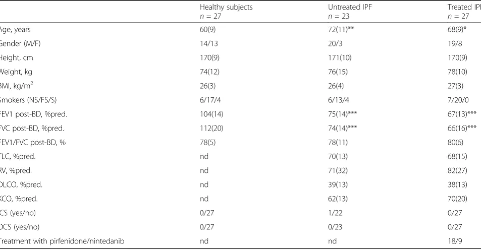
Subject demographic and functional characteristics The demographic functional and treatment characteris-tics are given in Table 1. The average age of IPF patients was 72 ± 11 years for untreated patients vs 68 ± 9 years for treated patients. Spirometric values were moderately
lower and comparable in both the treated and untreated IPF while diffusion lung capacity for CO (DLCO) was sharply reduced in both IPF groups.
Circulating cell-free nucleosomes
Epigenetic profiles of cfnucleosomes in subjects with un-treated IPF, un-treated IPF, or HS were investigated using ELISA-based Nu.Q™ assays. Ten epigenetic features of serum cfnucleosomes were analyzed. The receiver oper-ator characteristic (ROC) curves for each nucleosome assay in the untreated IPF group vs HS were established (data not shown). The area under the curve (AUC) for the individual ROC curves varied from 0.54 to 0.85 (Table 2).
Of note, diagnostic sensitivity at 80% specificity for the five best nucleosome biomarkers ranged from 61 to 86% (Table 2). Remarkably, the level of these five cfnucleo-some features, namely, HMGB1, mH2A1.1, 5mC, H3K9Ac, and H3K27Ac were significantly lower in untreated IPF than in HS (Fig. 1). Box plots derived from the best five individual assays based on the AUC are shown in Fig. 1.
Interestingly, 5mC and mH2A1 levels allowed to dis-criminate between untreated and treated IPF patients (p< 0.05 andp< 0.01, respectively) (Fig. 1). Remarkably, in our study, specific anti-fibrotic therapies significantly modified these two markers in treated IPF patients bringing them close to HS values (Fig. 1).
Table 1Subject characteristics
Healthy subjects
n= 27
Untreated IPF
n= 23
Treated IPF
n= 27
Age, years 60(9) 72(11)** 68(9)*
Gender (M/F) 14/13 20/3 19/8
Height, cm 170(9) 171(10) 170(9)
Weight, kg 74(12) 76(15) 78(10)
BMI, kg/m2 26(3) 26(4) 27(3)
Smokers (NS/FS/S) 6/17/4 6/13/4 7/20/0
FEV1 post-BD, %pred. 104(14) 75(14)*** 67(13)***
FVC post-BD, %pred. 112(20) 74(14)*** 66(16)***
FEV1/FVC post-BD, % 78(5) 78(11) 80(6)
TLC, %pred. nd 70(13) 68(15)
RV, %pred. nd 71(32) 82(27)
DLCO, %pred. nd 39(13) 38(13)
KCO, %pred. nd 62(13) 70(20)
ICS (yes/no) 0/27 1/22 0/27
OCS (yes/no) 0/27 0/23 0/27
Treatment with pirfenidone/nintedanib nd nd 18/9
Data are expressed as mean (SD)
ndnot determined,NSnon-smoker,FSformer smoker,Ssmoker,FEV1forced expired volume in one second,FVCforced vital capacity,TLCtotal lung capacity,
Correlation studies between lung function indices and cfnucleosomes showed a positive correlation between DLCO/VA (%pred) and H3K27Ac or H3K9Ac (r= 0.313, p= 0.049;r= 0.341,p= 0.032, respectively).
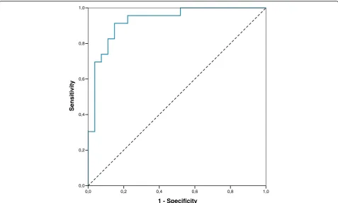
In addition, we assessed if an increase in the diagnostic value could be observed by combining levels of cfnucleo-some modifications. Statistical significance was calculated for several combinations using binary logistic regressions and the best combination was selected. This study revealed a highly statistically significant (p< 0.001) combination of four cfnucleosomes: HMGB1, 5mC, H3K9Ac, and
H3K27Ac with good coefficient of determination (R2 = 0.681). In this combination, each individual predictor was already highly statistically significant. The AUC for the ROC curve computed by this logistic re-gression was 0.93 (p < 0.001) with 91% sensitivity at 80% specificity for the discrimination of IPF patients vs HS (Fig. 2). We also notice that this analysis was gender independent (all p values > 0.1). Pearson correlation analysis between age and levels of cfnucleosome modifications showed only a marginal correlation between the two parameters (r coefficient of 0.299, 0.330, 0.210, 0.265, and 0.281 for HMGB1, 5mC, H3K9Ac, H3K27Ac, and mH2A1.1, respectively).
Discussion
We have investigated the clinical diagnostic accuracy for IPF using ELISA tests for epigenetic epitope variation in cfnucleosomes in small volumes of serum as a readily available and non-invasive sample type.
Our finding showed a reduced level of cfnucleosomes associated with HMGB1. HMGB1 is loosely bound to the chromatin during interphase and mitosis and has a rapid turnover between its chromatin-bound and soluble states. On the contrary, hyperacetylation occurring in apoptosis favors its chromatin-bound state, thereby pre-venting HMGB1 release [17]. Although HMGB1 impli-cation in inflammation and tissue repair has been well described, its role in IPF seems to be complex and need further elucidation [18].
Our results showed lower circulating chromatin-bound HMGB1 in IPF than in normal subjects. This suggests increase HMGB1 tendency towards its soluble Table 2Single cfnucleosome AUC and sensitivity at 80%
specificity Nu.Q™ assay
IPF (untreated) vs healthy
AUC % sensitivity at 80% specificity
mH2A1.1 0.85 86
5mC 0.83 78
HMGB1 0.81 74
H3K27Ac 0.79 74
H3K9Ac 0.77 61
H4K20Me3 0.70 50
pH2AX 0.66 39
H4Pan(Ac) 0.57 13
H3K4Me2 0.57 14
H3K9Me3 0.54 13
cfnucleosome features between healthy subject (n= 27) and untreated (n= 23) idiopathic pulmonary fibrosis patients. The receiver operator characteristic (ROC) curve analysis is calculated at 80% specificity. All serum nucleosome modifications were analyzed by ELISA Nu.Q™assays
AUCarea under the curve
Fig. 1Serum cfnucleosome modifications in IPF. Discrimination of five Nu.Q™assays for untreated IPF (n= 23), treated IPF (n= 27), and healthy
form in IPF as it has been previously described [15]. Therefore, extracellular HMGB1 in IPF could play a pro-tective role in preventing abundant release of nucleo-somes as it was previously described during apoptosis and repair process in lung remodeling [19].
Recent data have shown high histone deacytelase activ-ity in fibroblasts and myofibroblasts from patients with IPF [20, 21]. Our results are in agreement with that find-ing since we found lower levels of histone H3 acetylated markers (H3K9Ac and H3K27Ac) in untreated IPF pa-tients. Interestingly, in the current study, patients treated with anti-fibrotic drugs display cfnucleosome epitope profile comparable to HS, which suggests that these drugs may partially restore normal levels of histone H3 acetylation. Moreover, there is a positive correlation be-tween hypoacetylation and the severity of the disease assessed by the DLCO/VA quotient, which is known to be a good marker of the alveolo-capillar membrane function. However, this correlation is not very strong and has to be confirmed in a larger prospective study.
Acetylation homeostasis in nucleosomes is the result of a fine equilibrium between histone acetyl transferases (HAT) and histone deacetylases (HDAC). Disruption of this balance either by an increase of HDAC or a de-crease of HAT or both has been claimed to be involved
in the pathophysiologic process of IPF [15]. Given that increased acetylation by HDAC inhibition reduces colla-gen production in different fibroblast types, new anti-fibrotic drug (suberoylanilide hydroxamic acid) acting as a histone deacetylase inhibitor (HDACi) appeared to be a promising therapeutic option in IPF by reducing colla-gen deposition [22, 23].
Methylation is known to be deregulated in IPF [9]. IPF-associated differentially methylated regions occurred primarily in promoter regions of genes involved in biological processes such as cellular assembly and organization, cellular growth and proliferation, gene expression, and cell death. All of these processes are re-ported to be implicated in IPF pathogenesis [9]. Our study also highlighted that the level of cfnucleosome associated methylated DNA (5-methylcytosine) is significantly reduced in untreated IPF compared to HS. Moreover, specific anti-fibrotic therapies restore 5mC to levels closed to those seen in HS. Therefore, nucleosome-associated 5mC could potentially be useful for therapeutic monitoring or prognostic factor to treatment response.
Likewise, we identified a significant reduction in the level of the cfnucleosome carrying the histone variant mH2A1.1 in untreated IPF while those patients receiving anti-fibrotic drugs had levels similar to HS.
1 - Specificity
1,0 0,8
0,6 0,4
0,2 0,0
Sensitivity
1,0
0,8
0,6
0,4
0,2
0,0
Fig. 2A model with four cfnucleosome biomarkers discriminates IPF vs healthy patients. ROC curve for the discrimination of untreated IPF vs
Thinking beyond a single biomarker, we have assessed the value of combining several cfnucleosome modifica-tions. After performing specific logistic regression com-paring HS to untreated IPF, we found that a model of four Nu.Q™ cfnucleosome biomarkers (5mC, HMGB1, H3K27ac, and H3K9ac), reached an AUC of 0.93 in a ROC curve analysis (80% specificity and 91% sensitivity), which holds promise for use in clinical practice.
Conclusions
In conclusion, we have identified new blood-based cfnu-cleosome markers in untreated IPF patients. Intriguingly, some of those biomarkers tend to normalize in the pa-tients receiving anti-fibrotic treatment. We believe these cfnucleosomes may have potential as diagnostic and treatment response tools. This observation warrants fur-ther validation in larger longitudinal cohorts to confirm their value as biomarkers in IPF.
Abbreviations
5mC:5-Methylcytosine; ATS: American Thoracic Society; AUC: Area under the curve; CV: Coefficient of variation; DLCO: Diffusion lung capacity for CO; FEV1: Forced expired volume in one second; FVC: Forced vital capacity; HMGB1: High-mobility group protein 1a; HRCT: High-resolution computed tomography; HRP: Horseradish peroxidase; HS: Healthy subjects; IPF: Idiopathic pulmonary fibrosis; KCO: Diffusion lung capacity for CO/ alveolar ventilation; ROC: Receiver operating characteristic; SD: Standard deviation; UIP: Usual interstitial pneumonia
Acknowledgements
Thank you to all co-authors for their contributions.
Funding
There was no funding for this article.
Availability of data and materials
Data contains information that could be used to identify study participants and is available upon request from the corresponding author at J.Guiot@chu.ulg.ac.be.
Authors’contributions
IS, VC, JLC, and RL involved in the conception, hypothesis, and design of the study. MH contributed to the acquisition and analysis of samples. Belgian Volition SPRL contributed to the analysis of cfnucleosome modification, and in particular, MH contributed to the connection between the groups. BB helped in the recruitment of the study. JG, JLC, and RL guaranty the integrity of the work as a whole, from inception to published article. All authors read and approved the final manuscript.
Ethics approval and consent to participate
The protocol was approved by the ethics committee“hospitalo-facultaire”of Liège, and all subjects gave written consent before their enrolment (Belgian number: B707201422832; ref.: 2014/302).
Consent for publication Not applicable
Competing interests
MH, KS, and NH are employees of Belgian Volition SPRL. The authors declare that they have no competing interests.
Publisher’s Note
Springer Nature remains neutral with regard to jurisdictional claims in published maps and institutional affiliations.
Author details
1Pneumology Department, CHU Liège, Domaine universitaire du Sart-Tilman B35, 4000 Liège, Belgium.2Molecular Angiogenesis Laboratory, GIGA R, University of Liège, B34, 1 avenue de l hospital Sart-Tilman, Liège, Belgium. 3Department of Clinical Hematology, CHU Sart Tilman, Liège, Belgium. 4Belgian Volition SPRL, Rue du Seminaire 20A, 5000 Namur, Belgium. 5Pneumology Department, Erasme hospital, université libre de bruxelles, Belgium Route de Lennik, 808, 1070 Brussels, Belgium.
Received: 27 February 2017 Accepted: 3 August 2017
References
1. Raghu G, Collard HR, Egan JJ, Martinez FJ, Behr J, Brown KK, et al. An official ATS/ERS/JRS/ALAT statement: idiopathic pulmonary fibrosis: evidence-based guidelines for diagnosis and management. Am J Respir Crit Care Med. 2011 Mar 15;183(6):788–824.
2. Guiot J, Corhay JL, Louis R. Idiopathic pulmonary fibrosis. Rev Med Liege. 2014 Nov;69(11):605–10.
3. Guiot J, Bondue B, Henket M, Corhay JL, Louis R. Raised serum levels of IGFBP-1 and IGFBP-2 in idiopathic pulmonary fibrosis. BMC Pulm Med. 2016 May 23;16(1):86.
4. Tzouvelekis A, Kaminski N. Epigenetics in idiopathic pulmonary fibrosis. Biochem Cell Biol. 2015 Apr;93(2):159–70.
5. Kropski JA, Lawson WE, Young LR, Blackwell TS. Genetic studies provide clues on the pathogenesis of idiopathic pulmonary fibrosis. Dis Model Mech. 2013 Jan 1;6(1):9–17.
6. Raghu G. Idiopathic pulmonary fibrosis: increased survival with
‘gastroesophageal reflux therapy’. Am J Respir Crit Care Med. 2011 Dec 15; 184(12):1330–2.
7. Ye Q, Huang K, Ding Y, Lou B, Hou Z, Dai H, et al. Cigarette smoking contributes to idiopathic pulmonary fibrosis associated with emphysema. Chin Med J. 2014;127(3):469–74.
8. Yang IV, Schwartz DA. Epigenetics of idiopathic pulmonary fibrosis. Transl Res. 2015 Jan;165(1):48–60.
9. Rabinovich EI, Kapetanaki MG, Steinfeld I, Gibson KF, Pandit K V., Yu G, et al. Global methylation patterns in idiopathic pulmonary fibrosis. Eickelberg O, editor. PLoS One. 2012 Apr 10;7(4):e33770.
10. Robinson CM, Watson CJ, Baugh JA. Epigenetics within the matrix: a neo-regulator of fibrotic disease. Epigenetics. 2012 Sep;7(9):987–93. 11. Silva MT. Secondary necrosis: the natural outcome of the complete
apoptotic program. FEBS Lett. 2010 Nov 19;584(22):4491–9.
12. Brinkmann V. Neutrophil extracellular traps kill bacteria. Science (80- ). 2004 Mar 5;303(5663):1532–5.
13. Holdenrieder S, Nagel D, Schalhorn A, Heinemann V, Wilkowski R, von Pawel J, et al. Clinical relevance of circulating nucleosomes in cancer. Ann N Y Acad Sci. 2008 Aug;1137(1):180–9.
14. Kouzarides T. Chromatin modifications and their function. Cell. 2007 Feb; 128(4):693–705.
15. Hamada N, Maeyama T, Kawaguchi T, Yoshimi M, Fukumoto J, Yamada M, et al. The role of high mobility group box1 in pulmonary fibrosis. Am J Respir Cell Mol Biol. 2008 Oct;39(4):440–7.
16. Holdenrieder S, Dharuman Y, Standop J, Trimpop N, Herzog M, Hettwer K, et al. Novel serum nucleosomics biomarkers for the detection of colorectal cancer. Anticancer Res. 2014 May;34(5):2357–62.
17. Bauden M, Pamart D, Ansari D, Herzog M, Eccleston M, Micallef J, et al. Circulating nucleosomes as epigenetic biomarkers in pancreatic cancer. Clin Epigenetics BioMed Central. 2015 Dec 7;7(1):106.
18. Kang R, Zhang Q, Hou W, Yan Z, Chen R, Bonaroti J, et al. Intracellular Hmgb1 inhibits inflammatory nucleosome release and limits acute pancreatitis in mice. Gastroenterology. 2014 Apr;146(4):1097–1107.e8. 19. Scaffidi P, Misteli T, Bianchi ME. Release of chromatin protein HMGB1 by
necrotic cells triggers inflammation. Nature. 2002 Jul 11;418(6894):191–5. 20. Korfei M, Skwarna S, Henneke I, MacKenzie B, Klymenko O, Saito S, et al.
Aberrant expression and activity of histone deacetylases in sporadic idiopathic pulmonary fibrosis. Thorax. 2015 Nov;70(11):1022–32. 21. Coward WR, Watts K, Feghali-Bostwick CA, Knox A, Pang L. Defective
22. Wang Z, Chen C, Finger SN, Kwajah S, Jung M, Schwarz H, et al. Suberoylanilide hydroxamic acid: a potential epigenetic therapeutic agent for lung fibrosis? Eur Respir J. 2009;34(1):145–55.
23. Wang W, Yan M, Ji Q, Lu J, Ji Y, Ji J. Suberoylanilide hydroxamic acid suppresses hepatic stellate cells activation by HMGB1 dependent reduction of NF-κB1. PeerJ. 2015 Nov 3;3:e1362.
• We accept pre-submission inquiries
• Our selector tool helps you to find the most relevant journal • We provide round the clock customer support
• Convenient online submission • Thorough peer review
• Inclusion in PubMed and all major indexing services • Maximum visibility for your research
Submit your manuscript at www.biomedcentral.com/submit