R E S E A R C H
Open Access
Sensor-based postural feedback is more
effective than conventional feedback to
improve lumbopelvic movement control in
patients with chronic low back pain: a
randomised controlled trial
Thomas Matheve
1*, Simon Brumagne
2, Christophe Demoulin
3and Annick Timmermans
1Abstract
Background:Improving movement control can be an important treatment goal for patients with chronic low back pain (CLBP). Although external feedback is essential when learning new movement skills, many aspects of feedback provision in patients with CLBP remain currently unexplored. New rehabilitation technologies, such as movement sensors, are able to provide reliable and accurate feedback. As such, they might be more effective than conventional feedback for improving movement control. The aims of this study were (1) to assess whether sensor-based feedback is more effective to improve lumbopelvic movement control compared to feedback from a mirror or no feedback in patients with chronic low back pain (CLBP), and (2) to evaluate whether patients with CLBP are equally capable of improving lumbopelvic movement control compared to healthy persons.
Methods:Fifty-four healthy participants and 54 patients with chronic non-specific LBP were recruited. Both participant groups were randomised into three subgroups. During a single exercise session, subgroups practised a lumbopelvic movement control task while receiving a different type of feedback, i.e. feedback from movement sensors, from a mirror or no feedback (=control group). Kinematic measurements of the lumbar spine and hip were obtained at baseline, during and immediately after the intervention to evaluate the improvements in movement control on the practised task (assessment of performance) and on a transfer task (assessment of motor learning).
Results:Sensor-based feedback was more effective than feedback from a mirror (p< 0.0001) and no feedback (p < 0.0001) to improve lumbopelvic movement control performance (Sensor vs. Mirror estimated difference 9.9° (95% CI 6.1°-13.7°), Sensor vs. Control estimated difference 10.6° (95% CI 6.8°-14.3°)) and motor learning (Sensor vs. Mirror estimated difference 7.2° (95% CI 3.8°-10.6°), Sensor vs. Control estimated difference 6.9° (95% CI 3.5°-10.2°)). Patients with CLBP were equally capable of improving lumbopelvic movement control compared to healthy persons. Conclusions:Sensor-based feedback is an effective means to improve lumbopelvic movement control in patients with CLBP. Future research should focus on the long-term retention effects of sensor-based feedback.
Trial registration:clinicaltrials.govNCT02773160, (retrospectively registered on May 16th, 2016).
Keywords:Low back pain, Feedback, Movement control, Motor learning, Sensors, Technology
* Correspondence:Thomas.Matheve@uhasselt.be
1Rehabilitation Research Center - Biomed, Faculty of Medicine and Life Sciences, Hasselt University, Hasselt, Belgium
Full list of author information is available at the end of the article
Background
The lifetime prevalence of low back pain (LBP) is reported to be as high as 84%, whereas the estimated prevalence of chronic LBP (CLBP) is approximately 23% [1]. Globally, it is the leading cause of disability [2] and one of the most important reasons for work absenteeism, resulting in a high socioeconomic burden [3].
Patients with CLBP form a heterogeneous group, which is exemplified by the differences in movement patterns within this population. While some patients with CLBP stiffen their spine and avoid spinal movements, others show the opposite pattern and adopt end range postures or move excessively into their painful direction [4]. For the latter type of patients, movement control exercises are often prescribed [5]. The aim of these exercises is to learn how to control movements into the painful direction, thereby reducing the mechanical load on the painful structures and decreasing peripheral nociceptive input [6]. Changing movement patterns requires motor learning. The importance of external feedback (i.e. feedback com-ing from a source external to the person performcom-ing the task [7]) in motor learning has been well established, and optimizing the way feedback is provided is therefore essential [8,9]. While there is an abundance of literature on the role of extrinsic feedback to improve motor learning in a healthy population, many aspects of feed-back provision in patients with LBP remain currently unexplored [9]. When patients with LBP perform lumbar movement control exercises in the absence of a therap-ist, they typically have to rely on visual feedback (e.g. from a mirror) or palpation [10]. However, the reliability and accuracy of these types of feedback can be ques-tioned [11,12], which may lead to a suboptimal learning process [7]. With the development of rehabilitation technologies, new opportunities for providing external feedback have emerged [13]. For example, wireless inertial motion sensors can be used to provide easy to understand and accurate feedback to the patient (e.g. via an avatar) [13,14]. As such, sensor-based postural feedback might be more effective than conventional feedback for improving movement control, which in turn may enhance treatment effects.
Although movement control exercises are widely used in a variety of chronic pain populations, little is known about the influence of chronic pain on the capacity to learn new movement skills. From a theoretical perspective, it has been suggested that patients with CLBP might have a reduced motor learning capacity [15]. One of the reasons for this hypothesis is that LBP can negatively influence pro-prioceptive acuity, leading to impaired intrinsic feedback from the lumbar spine [16]. As a consequence, patients with LBP might have to rely more on external feedback and become more dependent on it [7]. In addition, pain demands attention and can distract patients from the
movement task [17], which might in turn interfere with the learning process [15]. However, empirical evidence for a reduced motor learning capacity in patients with CLBP is currently lacking and the scantly available research in other chronic pain populations shows equivocal results [18,19].
Therefore, the aims of this study were (1) to assess whether sensor-based feedback is more effective to im-prove lumbopelvic movement control compared to feed-back from a mirror or no feedfeed-back in patients with chronic low back pain (CLBP), and (2) to evaluate whether patients with CLBP are equally capable of improving lumbopelvic movement control compared to healthy persons.
Methods Design
A randomised controlled trial including healthy persons and patients with CLBP was conducted. Both groups of participants were randomised into three subgroups, each receiving a different type of feedback during the inter-vention, i.e. feedback from sensors, a mirror or no feed-back (= control group). Randomisation was done with a computerised random sequence generator and allocation concealment was obtained by using sequentially num-bered, sealed, opaque envelopes prepared by a person not further involved in the study.
The intervention consisted of a single exercise session during which participants practised a movement control task while receiving their assigned type of feedback. Move-ment control was assessed with lumbopelvic kinematics, which were obtained at baseline, during and immediately after the intervention.
Participants
Participants were recruited at private physiotherapy and GP practices and via social media. To be included, all participants needed to be between 18 and 65 years old and patients had to be diagnosed with chronic non-specific LBP (> 3 months, ≥3 days/week). Exclusion criteria for all participants were: spinal surgery in the past, an underlying serious disease or a physical problem interfering with daily life activities (e.g. severe knee pain), signs or symptoms of nerve root involvement, performance of lumbopelvic movement control exercises in the past year and pregnancy. Healthy subjects were also excluded if they experienced LBP in the past year.
range of motion could only be calculated after comple-tion of the full protocol. Therefore, all of the included participants completed the protocol, but only those ful-filling the abovementioned criterion were included in the final analysis.
Assessments Baseline assessments
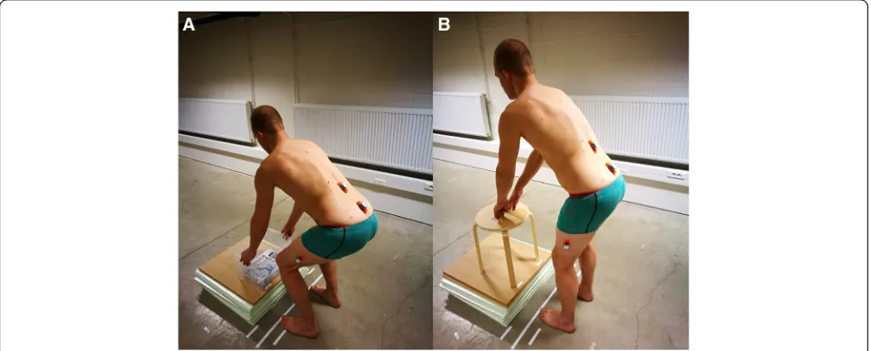
Sociodemographic data were obtained from all partici-pants. Patients with CLBP also completed the Numeric pain rating scale (NPRS) [20] to assess current pain and the average pain during the past 7 days, the Roland Morris Disability questionnaire (RMDQ) [21] to assess disability and the Tampa scale for kinesiophobia (Miller RP, Kori SH, Todd DD: The Tampa Scale, unpublished) to assess the fear of movement/re-injury due to physical activity. After completing the questionnaires, participants per-formed two movement control tasks, i.e. a lifting task followed by a waiter’s bow (Fig.1). Both tasks were stan-dardised for the participants’ height and assessed with lumbopelvic kinematic measurements in the sagittal plane. Before the baseline kinematic measurements, the tasks were explained and demonstrated in a standardised way. For the lifting task, participants started from a relaxed standing position and were asked to lift a box with handles from a platform on the floor and to put it back down, while maintaining their lumbar curvature (i.e. not to flex or extend the lumbar spine). Participants were allowed to flex their knees as far as they wanted to. The distance from the box to the hallux was 15 cm. The dimensions of the box were 40 × 30 × 23.5 cm, and it weighed 4 kg. The top of the box was positioned 10 cm below the apex of the subjects’patella. For the waiter’s bow, participants started with slightly flexed knees (±20°). Participants were instructed to keep their knees in the same position and to
bend forward in the hips while maintaining their lumbar curvature. Participants had to touch the middle of a stool, marked with a piece of tape, which was positioned 15 cm in front of the hallux, and to return to their starting pos-ition. No familiarisation was allowed, and each task was performed five times at a self-selected speed.
Assessments during and after the intervention
Kinematics were also obtained during and three minutes after the intervention. For the post-intervention kinematic assessment, participants first performed the waiter’s bow and then the lifting task as described above. Immediately after the post-intervention kinematic assessment, all participants were asked to complete the Borg-scale for perceived exertion [22], and to answer two questions on a 0 to 10 numeric rating scale:‘what was your aver-age LBP intensity during the experiment?’ (0 = no pain at all, 10 = worst imaginable pain),‘how fearful were you to damage your back?’ (0 = not fearful at all, 10 = ex-tremely fearful). If significant between group differences would be present on the post-intervention questionnaires, these would be controlled for in the data analysis, as they might influence movement patterns [23–25].
Equipment
The Valedo®motion research tool (Hocoma, Switzerland) was used to assess the lumbopelvic kinematics and to provide feedback in the sensor groups. This system con-sists of a laptop and three wireless inertial measurement sensors, which contain a magnetometer, 3D-accelerometer and a 3D-gyroscope. The sensors were placed on the spin-ous process of L1 and S1, and 20 cm above the lateral femoral condyle (Fig. 1). All three sensors were used for the kinematic assessment, while only the L1 and S1 sensors were used to provide feedback in the sensor groups. Details
on the kinematic data acquisition have been previously described [26].
Intervention
During the intervention, participants practised the waiter’s bow during three sets of six repetitions while they received their assigned form of feedback. Each set of exercises was separated by one minute of rest. The lifting task was not practised. The feedback in the different groups was pro-vided as follows:
Sensor group
The sensor-feedback was given via an avatar on a computer screen in front of the participants. The ava-tar was controlled by two movement sensors that were placed on the spinous process of L1 and S1. The upper body of the avatar corresponded with the S1-sensor and the green rectangle with the L1-sensor (Fig. 2). First, the system was calibrated when the participants assumed the starting position so that the green rectangle was placed in the middle of the ava-tar’s upper body. Participants were instructed to keep the green rectangle on the avatar during the exercises, as this meant that the lumbar curvature was
maintained (Fig. 2a). If the rectangle moved anteriorly of the avatar, this corresponded with a lumbar flexion (Fig. 2b), while a posterior displacement indicated a lumbar extension.
Mirror group
A large mirror was placed laterally to the participants so they could see the stool and their whole body, and observe their spinal curvature during the exercises.
Control group
No feedback was provided.
Before the exercise trials, participants were ex-plained how to use pelvic tilts to adjust the lumbar curvature. Hereafter, they were allowed to perform up to five pelvic tilts, during which participants in the sensor group could see how pelvic movements af-fected the position of the green rectangle relative to the avatar, participants in the mirror group could ob-serve in the mirror how the pelvic tilts changed their lumbar curvature, while the control group received no feedback.
Outcome measures for addressing the primary and secondary aims of the study
Primary aim - effectiveness of feedback
The influence of the different types of feedback on movement control performance and motor learning was of primary interest. Performance can be measured during or shortly after training, whereas motor learning can be assessed with a transfer test [27]. As the partici-pants only practised the waiter’s bow, we used the dif-ferences between baseline and post-intervention kinematics of the waiter’s bow as a measure of perform-ance, while differences in the lifting task kinematics were used as a measure for motor learning. For each repetition, the maximal range of motion in the lumbar spine and hip joint was calculated and expressed in absolute values. Lumbar spine angles were calculated from the L1 and S1 sensors, while hip joint angles were calculated from the S1 and femoral sensors. This method is highly reliable for both tasks in this study (ICCs = 0.89–0.93) [26]. The minimal detectable change between two measurements for the lifting task is 5.3° for the lumbar spine and 8.8° for the hip, while for the waiter’s bow this is 6.5° and 11.8°, respectively [26]. An improvement in movement control was defined as a de-crease in the lumbar range of motion and an inde-crease in the hip range of motion between baseline and post-intervention assessment. In addition to statistical significance, the abovementioned minimal detectable changes were used to interpret the results.
Secondary aim–Comparison between healthy persons and patients with CLBP
The differences between healthy subjects and patients with CLBP in movement control performance improvement and motor learning was evaluated. This was done by com-paring the change in lumbopelvic kinematics between baseline and post-intervention between both participant groups. In addition, the evolution of the performance on
the waiter’s bow task during the intervention was com-pared. In this way, it could be determined whether healthy participants and patients with CLBP needed the same number of repetitions to achieve an improvement on the waiter’s bow.
To investigate whether participants became dependent on the external feedback, the performance on the last exercise trial (with feedback) was compared with the
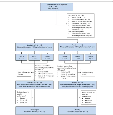
Fig. 3Design and flow of participants through the trial. FB = feedback.aParticipants were excluded after the trial, based on their performance on the
post-intervention performance (without feedback) on the waiter’s bow. A significant decline on the post-intervention performance would indicate such dependence [7].
Data analysis
The statistical analysis was performed with SAS JMP Pro (Version 12.2). To examine the effectiveness of the feed-back and the difference in movement control improve-ment between healthy participants and patients with CLBP, a multiple linear regression was performed. The following variables were entered in the initial model to pre-dict the differences between baseline and post-intervention kinematics: type of feedback (i.e. control, mirror or sensor), health status (i.e. healthy or CLBP), joint (i.e. lumbar spine or hip) and all their pairwise interactions. To control for the baseline values of the lumbar spine and hip angles, this variable was also put in the initial model. The variable
‘joint’was included in the model because the movements of the spine and hip were considered to be related to each other. The final model was obtained by stepwise backward regression. The variable with the least significant p-value was left out first, and this was repeated until all the variables reached significance (p< 0.05). A Tukey all pair-wise comparison was used as a post-hoc test.
A mixed model was used to assess the difference between healthy participants and patients with CLBP in the evolution of the performance on the waiter’s bow task. This model was also used to examine the difference between the last repetition of the intervention and the post-intervention performance on the waiter’s bow. The same variables from the linear regression were included in the mixed model, but‘repetition number’(i.e. baseline, repetitions during the intervention and post-intervention
measurements were numbered) and its pairwise inter-actions with other variables were added as fixed factors.
‘Participant’was used as a random factor to account for multiple measurements for the same participant.
Sample size calculation was based on an effect size (f2) of 0.2, power of 0.80 and α-level of 0.05. With these parameters, a total sample size of 80 participants was needed. Taking into account an attrition rate of 30% be-cause of baseline performance on the movement control tests, 54 healthy persons and 54 patients with CLBP had to be recruited.
Results
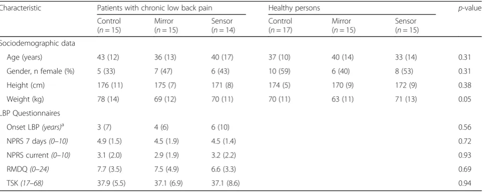
The flow of participants through the study is shown in Fig. 3. Ten (19%) patients with CLBP and seven (13%) healthy participants were excluded based on their baseline performance on the movement control tasks. No signifi-cant differences in demographics (Table 1) and baseline scores on kinematic outcome measures (Table 2, first column) were observed between groups.
Effectiveness of feedback
The results of the linear regression and post-hoc tests are presented in Table3(see Additional file1for a detailed sum of squares table). In both the healthy participants and patients with CLBP, the sensor group improved signifi-cantly more than the mirror and control group (post-hoc tests, p< 0.0001), while no differences were observed between the mirror and control group (post-hoc tests,
p> 0.91). These results were obtained for both the waiter’s bow and the lifting task, as well as for the lumbar spine and hip. The improvements in the sensor groups were also larger than the measurement error (i.e.
Table 1Baseline characteristics of the participants
Characteristic Patients with chronic low back pain Healthy persons p-value
Control (n= 15)
Mirror (n= 15)
Sensor (n= 14)
Control (n= 17)
Mirror (n= 15)
Sensor (n= 15)
Sociodemographic data
Age (years) 43 (12) 36 (13) 40 (17) 37 (10) 40 (14) 33 (14) 0.31
Gender, n female (%) 5 (33) 7 (47) 6 (43) 10 (59) 6 (40) 8 (53) 0.31
Height (cm) 176 (11) 175 (7) 171 (8) 174 (5) 170 (9) 172 (9) 0.38
Weight (kg) 78 (14) 69 (12) 70 (11) 70 (11) 63 (11) 71 (13) 0.05
LBP Questionnaires
Onset LBP(years)a 3 (7) 4 (6) 6 (10) 0.56
NPRS 7 days(0–10) 4.9 (1.5) 4.5 (1.9) 4.5 (1.4) 0.72
NPRS current(0–10) 3.1 (2.0) 2.9 (1.9) 3.2 (2.2) 0.93
RMDQ(0–24) 7.7 (3.5) 7.5 (4.9) 6.6 (3.3) 0.69
TSK(17–68) 37.9 (5.5) 37.1 (6.9) 37.1 (8.6) 0.94
Data are mean (SD), unless mentioned otherwise
LBPlow back pain,NPRSNumeric pain rating scale,NPRS 7 daysaverage pain during the past 7 days measured with a NPRS,NPRS currentcurrent pain measured with a NPRS,RMDQRoland-Morris Disability Questionnaire,TSKTampa scale for kinesiophobia
a
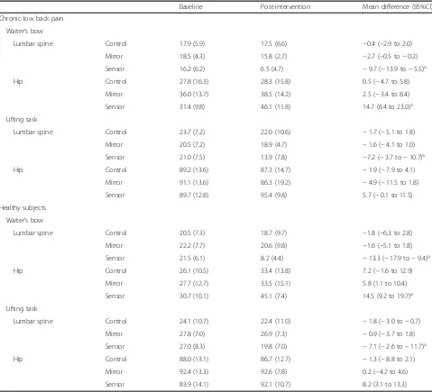
minimal detectable change), except for the hip during the lifting task (Table 2). There were no between groups differences in the post-intervention questionnaires (see Additional file2).
Based on the type III sum of squares tables (see Additional file 1), it is clear that the type of feedback is the most important factor contributing to the variance that is explained by the final regression models of the waiter’s bow and lifting task, while the factor joint only explains a small proportion. A significant part of the vari-ance that is explained by the final model of the lifting task can be attributed to the baseline scores on the kinematic assessments. Participants who had a worse performance on the baseline lifting task had a larger improvement.
Comparison between healthy persons and patients with CLBP
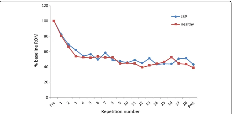
The variable health status (i.e. healthy or CLBP) and its interaction with repetition number were not retained in the final mixed model (Table 4). This indicates that patients with CLBP were equally capable of improving lumbopelvic movement control, and that the evolution of the performance on the waiter’s bow task was similar between both participant groups (see Fig. 4 for an ex-ample of the sensor groups). These results are further supported by the fact that only a small proportion of the variance that is explained by the final model can be at-tributed to each of the variables pertaining to our second research question (see Additional file 3 for a detailed Table 2Baseline and post-intervention maximal range of motion in the lumbar spine and hip joint
Baseline Post-intervention Mean difference (95%CI)
Chronic low back pain
Waiter’s bow
Lumbar spine Control 17.9 (5.9) 17.5 (6.6) −0.4 (−2.9 to 2.0)
Mirror 18.5 (4.3) 15.8 (2.7) −2.7 (−0.5 to−0.2)
Sensor 16.2 (6.2) 6.5 (4.7) −9.7 (−13.9 to−5.5)a
Hip Control 27.8 (16.3) 28.3 (15.8) 0.5 (−4.7 to 5.8)
Mirror 36.0 (13.7) 38.5 (14.2) 2.5 (−3.4 to 8.4)
Sensor 31.4 (9.8) 46.1 (11.8) 14.7 (6.4 to 23.0)a
Lifting task
Lumbar spine Control 23.7 (7.2) 22.0 (10.6) −1.7 (−5.1 to 1.8)
Mirror 20.5 (7.2) 18.9 (4.7) −1.6 (−4.1 to 1.0)
Sensor 21.0 (7.5) 13.9 (7.8) −7.2 (−3.7 to−10.7)a
Hip Control 89.2 (13.6) 87.3 (14.7) −1.9 (−7.9 to 4.1)
Mirror 91.1 (13.6) 86.3 (19.2) −4.9 (−11.5 to 1.8)
Sensor 89.7 (12.8) 95.4 (9.8) 5.7 (−0.1 to 11.5)
Healthy subjects
Waiter’s bow
Lumbar spine Control 20.5 (7.3) 18.7 (9.7) −1.8 (−6.3 to 2.8)
Mirror 22.2 (7.7) 20.6 (9.8) −1.6 (−5.1 to 1.8)
Sensor 21.5 (6.1) 8.2 (4.4) −13.3 (−17.9 to−9.4)a
Hip Control 26.1 (10.5) 33.4 (13.8) 7.2 (−1.6 to 12.9)
Mirror 27.7 (12.7) 33.5 (15.1) 5.8 (1.1 to 10.4)
Sensor 30.7 (10.1) 45.1 (7.4) 14.5 (9.2 to 19.7)a
Lifting task
Lumbar spine Control 24.1 (10.7) 22.4 (11.0) −1.8 (−3.0 to−0.7)
Mirror 27.8 (7.0) 26.9 (7.3) −0.9 (−3.7 to 1.8)
Sensor 27.0 (8.3) 19.8 (7.0) −7.1 (−2.6 to−11.7)a
Hip Control 88.0 (13.1) 86.7 (12.7) −1.3 (−8.8 to 2.1)
Mirror 92.4 (13.3) 92.6 (7.8) 0.2 (−4.2 to 4.6)
Sensor 83.9 (14.1) 92.1 (10.7) 8.2 (3.1 to 13.3)
All data are expressed as angles in degrees (°). Data for baseline and post-intervention are mean (SD). Mean difference = post-intervention minus baseline
a
sum of squares table). Post-hoc tests also showed that there were no differences between the performance on the last exercise trial and the post-intervention assess-ment of the waiter’s bow. This demonstrates that partici-pants in the mirror and sensor groups did not become dependent on the feedback.
Discussion
The primary aim of this study was to compare the effect-iveness of different types of external feedback to improve lumbopelvic movement control in healthy persons and patients with CLBP. Our results show that sensor-based postural feedback was more effective to improve lumbo-pelvic movement control than feedback from a mirror or no feedback. Furthermore, being provided with feedback from a mirror did not lead to better results than receiving no feedback at all.
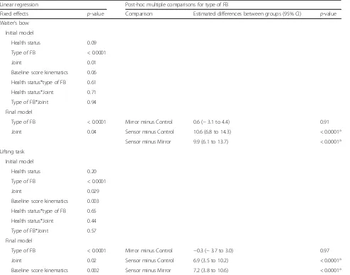
We hypothesize that the lack of improvement in the mirror group could be explained by the difficulty for unexperienced persons to visually detect changes in the lumbar curvature during the waiter’s bow. Although physiotherapists can reliably assess the waiter’s bow by observation, observer training may play an important role in this assessment [28]. Possibly, a longer teaching and familiarization period before the intervention could have enhanced the effectiveness of the mirror-feedback. In contrast, the very short introduction to the sensor-feedback was sufficient to improve lumbopelvic move-ment control. We believe that the avatar provided more accurate and easy-to-understand feedback, which required no advanced training in order to interpret it correctly. It has been shown that abstract visualisations can be more effective than very realistic feedback (e.g. via a video or mirror) because they can provide information about key features of the task only, without overwhelming the Table 3Results of the linear regression analysis and post-hoc tests for type of feedback
Linear regression Post-hoc multiple comparisons for type of FB
Fixed effects p-value Comparison Estimated differences between groups (95% CI) p-value
Waiter’s bow
Initial model
Health status 0.09
Type of FB < 0.0001
Joint 0.01
Baseline score kinematics 0.06
Health status*type of FB 0.61
Health status*Joint 0.71
Type of FB*Joint 0.94
Final model
Type of FB < 0.0001 Mirror minus Control 0.6 (−3.1 to 4.4) 0.91
Joint 0.04 Sensor minus Control 10.6 (6.8 to 14.3) < 0.0001a
Sensor minus Mirror 9.9 (6.1 to 13.7) < 0.0001a
Lifting task
Initial model
Health status 0.20
Type of FB < 0.0001
Joint 0.029
Baseline score kinematics 0.003
Health status*type of FB 0.65
Health status*Joint 0.44
Type of FB*Joint 0.57
Final model
Type of FB < 0.0001 Mirror minus Control −0.3 (−3.7 to 3.0) 0.97
Joint 0.02 Sensor minus Control 6.9 (3.5 to 10.2) < 0.0001a
Baseline score kinematics 0.002 Sensor minus Mirror 7.2 (3.8 to 10.6) < 0.0001a
FBFeedback,Health statushealthy of CLBP,Jointlumbar spine or hip,Type of FBsensor, mirror or control
a
participants with irrelevant information [8]. Participants in the sensor group only had to look at the green dot rela-tive to the avatar’s upper body, while participants in the mirror group could also see movements in other body regions that were irrelevant to the task.
In addition, the screen displaying the avatar could be placed in front of the participants, whereas the mirror had to be positioned laterally to visualise the movements in the sagittal plane. Although this difference in position could be interpreted as a confounding factor because participants in the mirror group had to turn their heads in order to view their spinal curvature, the possibility to place the computer screen in the most convenient pos-ition should rather be considered as an inherent advantage of the sensor-feedback.
Finally, the improvements on the lifting task were partially explained by the baseline kinematic scores. Partici-pants who performed worse on the lifting task at baseline assessment had a significantly larger improvement, which indicates that the motor learning effect was more pro-nounced in these participants. This might be explained by the fact that persons who performed worse at the baseline lifting task also had a larger potential for improvement.
Besides a mirror, various other types of conventional feedback, including tape or palpation [10], can be used to support patients during lumbopelvic movement control exercises. The rationale for comparing the sensor-based feedback to feedback from a mirror was twofold: First, a mirror is frequently being used or recommended to pro-vide postural feedback during lumbopelvic movement Table 4Results for the mixed model
Fixed effects p-value
Initial model
Health status 0.40
Type of FB < 0.0001
Joint < 0.0001
Baseline score kinematics < 0.0001
Repetition number < 0.0001
Health status*type of FB 0.83
Health status*Joint 0.01
Type of FB*Joint 0.08
Repetition number*type of FB < 0.0001
Repetition number*Health status 0.28
Repetition number*Joint 0.09
Final model
Health status 0.38
Type of FB < 0.0001
Baseline score kinematics < 0.0001
Joint < 0.0001
Repetition number < 0.0001
Health status*Joint 0.01
Repetition number*Type of FB < 0.0001
FBFeedback,Health statushealthy of CLBP,Jointlumbar spine or hip,Type of FBsensor, mirror or control
control exercises [10, 29–31]. Second, and more import-antly, both the mirror and sensors provided visual feed-back, whereas palpation and a tape provide tactile feedback. Because visual motion detection is processed differently than tactile motion detection [32], we chose to compare the sensor feedback to feedback from a mirror.
Healthy subjects and patients with CLBP were equally capable of improving lumbopelvic movement control. It has been suggested that pain could negatively influence skill acquisition and motor learning by distracting people from the task they are performing [15]. However, this distraction mainly occurs when the pain is more intense, unfamiliar or unexpected [17]. The patients with CLBP in our study did not report an increase in pain during the exercise trials and there is no reason to assume that the pain they felt was unexpected or unfamiliar. Therefore, it is unlikely that patients with CLBP were distracted from the movement task.
Pain can also affect proprioceptive acuity and impair the intrinsic feedback system [33]. When less reliable intrinsic feedback is available, the dependency on the extrinsic feed-back may increase [7]. Overall, patients with CLBP have de-creased lumbosacral proprioception compared to healthy persons [16, 34], so it can be argued that removing the external feedback could influence the performance on the waiter’s bow more in patients with CLBP than in healthy participants. On the other hand, these proprioceptive im-pairments may be position specific (e.g. sit versus stance) [34] and little is known about proprioception during dy-namic tasks [16], such as the ones in the present study. Our results show that omitting the external feedback had no influence on the performance on the waiter’s bow in both participant groups. This suggests that patients with CLBP also used information from the sensorimotor sys-tem to adjust their spinal curvature during the exercise trials [8], and that they did not rely more on the external feedback than healthy subjects. The improvements on the lifting task in the sensor groups further support this notion, as it indicates that both participant groups were able to transfer their newly learned skills to a different task. Therefore, it seems appropriate to use concurrent sensor-based feedback during the initial learning phase of movement control tasks in patients with CLBP.
Several limitations apply to this study. First, motor learning was only assessed with a transfer test, and not with a retention test. Both the transferability of practised skills and the long-term retention effects are important as-pects of motor learning [27]. Because it is impossible to provide movement control training during every single ac-tivity an individual needs to perform, persons should be able to implement their newly acquired skills during activ-ities that were not practised. In addition, the movement control improvements should be retained in the long term. However, because a retention test was not included
in this study, we cannot make any statements regarding the longstanding effects of the sensor-based feedback.
Second, the mobility of the lower limb joints was not eval-uated at baseline assessment. According to the concept of relative flexibility, a restriction in one joint could influence the movements in an adjacent joint [6]. Especially during the lifting task, more end range movements were necessary in the hip joint. As such, a restriction in hip joint mobility could have influenced the lumbar movements. On the other hand, participants with any physical problems other than LBP (e.g. hip or knee pain) that interfered with daily life activities were excluded from this study. Therefore, we believe it is unlikely that a (pathological) restriction of lower limb joint mobility would have significantly influenced the movement patterns in the lumbar spine.
Finally, our measurement and feedback system only contained three sensors. Due to these technical limita-tions, we could only measure the movements in the lumbar spine and hip joint. Consequently, we cannot exclude that some patients might have used compensa-tory movements in the thoracic spine while performing the movement control tasks. On the other hand, the reduction in lumbar ROM in the sensor group was accompanied by an increase in hip joint motion, indicat-ing movements in the hip joint and lumbar spine were coupled.
Conclusions
The recent development of rehabilitation technologies creates new possibilities for therapists and patients to support the rehabilitation process. As such, evaluating the effectiveness of these rapidly evolving technological systems poses an important challenge. The present study shows that sensor-based postural feedback is more ef-fective than feedback from a mirror or no feedback to improve lumbopelvic movement control in the short term. Patients with CLBP were equally capable of im-proving lumbopelvic movement control as compared to healthy participants. Future research should focus on the long-term retention effects and evaluate whether sup-porting exercises with sensor-based feedback leads to larger improvements in pain and disability compared to conventional exercise therapy.
Additional files
Additional file 1:Type III sum of squares table of the final model of the regression analyses. Table showing the Type III sum of squares table of the final model of the regression analyses. (DOCX 14 kb)
Additional file 2:Results for post-intervention questionnaires. Table showing the results for post-intervention questionnaires. (DOCX 15 kb)
Abbreviations
CLBP:Chronic low back pain; LBP: Low back pain
Acknowledgements
We would like to thank Dr. Robin Bruyndonckx and Dr. Francesca Solmi for their advice on the statistical analysis.
Availability of data and materials
The datasets used and/or analysed during the current study are available from the corresponding author on reasonable request.
Authors’contributions
TM conceptualised the study, and SB and AT refined the design. TM and CD collected the data. TM drafted the manuscript, and SB, CD and AT revised it for important intellectual content and helped interpret the data. All authors read and approved the final manuscript.
Ethics approval and consent to participate
This study was approved by The Ethics Committees of Hasselt University and Jessa Hospital, Belgium (B243201423040). All participants provided written informed consent before being included in the study.
Consent for publication
Written consent for publication was obtained from the person in the pictures.
Competing interests
The authors declare that they have no competing interests.
Publisher’s Note
Springer Nature remains neutral with regard to jurisdictional claims in published maps and institutional affiliations.
Author details 1
Rehabilitation Research Center - Biomed, Faculty of Medicine and Life Sciences, Hasselt University, Hasselt, Belgium.2Department of Rehabilitation Sciences, KU Leuven–University of Leuven, Leuven, Belgium.3Department of Sport and Rehabilitation Sciences, University of Liege, Liege, Belgium.
Received: 18 May 2018 Accepted: 29 August 2018
References
1. Airaksinen O, Brox JI, Cedraschi C, Hildebrandt J, Klaber-Moffett J, Kovacs F, Mannion AF, Reis S, Staal JB, Ursin H, Zanoli G. Chapter 4. European guidelines for the management of chronic nonspecific low back pain. Eur Spine J. 2006;15(Suppl 2):S192–300.
2. Hurwitz EL, Randhawa K, Yu H, Cote P, Haldeman S. The global spine care initiative: a summary of the global burden of low back and neck pain studies. Eur Spine J. 2018.https://doi.org/10.1007/s00586-017-5432-9. 3. Dagenais S, Caro J, Haldeman S. A systematic review of low back pain
cost of illness studies in the United States and internationally. Spine J. 2008;8:8–20.
4. O'Sullivan P. Diagnosis and classification of chronic low back pain disorders: maladaptive movement and motor control impairments as underlying mechanism. Man Ther. 2005;10:242–55.
5. Luomajoki HA, Bonet Beltran MB, Careddu S, Bauer CM. Effectiveness of movement control exercise on patients with non-specific low back pain and movement control impairment: a systematic review and meta-analysis. Musculoskeletal Science Practice. 2018;36:1–11.
6. Sahrmann SA. Movement System Impairment Syndromes of the Extremities, Cervical and Thoracic Spines. 1st ed. St. Louis: Mosby; 2010.
7. Magill RA. Motor Learning and Control. Concepts and applications. 8th ed. Boston: McGraw-Hill; 2007.
8. Sigrist R, Rauter G, Riener R, Wolf P. Augmented visual, auditory, haptic, and multimodal feedback in motor learning: a review. Psychon Bull Rev. 2013;20:21–53.
9. Ribeiro DC, Sole G, Abbott JH, Milosavljevic S. Extrinsic feedback and management of low back pain: a critical review of the literature. Man Ther. 2011;16:231–9.
10. Hodges PW, Van Dillen LR, McGill SM, Brumagne S, Hides JA, Moseley GL. Integrated clinical approach to motor control interventions in low back and pelvic pain. In: Hodges PW, Cholewicki J, Van Dieen JH, editors. Spinal Control: The rehabilitation of back pain State of the art and science. 1st ed. London: Churchill Livingstone; 2013. p. 243–309.
11. Elgueta-Cancino E, Schabrun S, Danneels L, Hodges P. A clinical test of lumbopelvic control: development and reliability of a clinical test of dissociation of lumbopelvic and thoracolumbar motion. Man Ther. 2014;19:418–24. 12. Haneline MT, Cooperstein R, Young M, Birkeland K. Spinal motion palpation:
a comparison of studies that assessed intersegmental end feel vs excursion. J Manip Physiol Ther. 2008;31:616–26.
13. Wang Q, Markopoulos P, Yu B, Chen W, Timmermans A. Interactive wearable systems for upper body rehabilitation: a systematic review. J Neuroeng Rehabil. 2017;14:20.
14. Matheve T, Claes G, Olivieri E, Timmermans A. Serious gaming to support exercise therapy for patients with chronic nonspecific low back pain: a feasibility study. Games Health J. 2018;7:262–270.
15. Boudreau SA, Farina D, Falla D. The role of motor learning and neuroplasticity in designing rehabilitation approaches for musculoskeletal pain disorders. Man Ther. 2010;15:410–4.
16. Brumagne S, Janssens L, Claeys K, Pijnenburg M. Altered variability in proprioceptive control strategy in people with recurrent low back pain. In: Hodges P, Cholewicki J, Van Dieën JH, editors. Spinal control: The rehabilitation of back pain. 1st ed. London: Churchill Livingstone; 2013. p. 135–44. 17. Eccleston C, Crombez G. Pain demands attention: a cognitive-affective
model of the interruptive function of pain. Psychol Bull. 1999;125:356–66. 18. Vallence AM, Smith A, Tabor A, Rolan PE, Ridding MC. Chronic tension-type
headache is associated with impaired motor learning. Cephalalgia. 2013;33: 1048–54.
19. Parker RS, Lewis GN, Rice DA, McNair PJ. The association between Corticomotor excitability and motor skill learning in people with painful hand arthritis. Clin J Pain. 2017;33:222–30.
20. Chapman JR, Norvell DC, Hermsmeyer JT, Bransford RJ, DeVine J, McGirt MJ, Lee MJ. Evaluating common outcomes for measuring treatment success for chronic low back pain. Spine (Phila Pa 1976). 2011;36:S54–68.
21. Roland M, Morris R. A study of the natural history of back pain. Part I: development of a reliable and sensitive measure of disability in low-back pain. Spine (Phila Pa 1976). 1983;8:141–4.
22. Borg GA. Psychophysical bases of perceived exertion. Med Sci Sports Exerc. 1982;14:377–81.
23. Vaisy M, Gizzi L, Petzke F, Consmuller T, Pfingsten M, Falla D. Measurement of lumbar spine functional movement in low back pain. Clin J Pain. 2015;31:876–85.
24. Trost Z, France CR, Thomas JS. Examination of the photograph series of daily activities (PHODA) scale in chronic low back pain patients with high and low kinesiophobia. Pain. 2009;141:276–82.
25. van Dieen JH, van der Burg P, Raaijmakers TA, Toussaint HM. Effects of repetitive lifting on kinematics: inadequate anticipatory control or adaptive changes? J Mot Behav. 1998;30:20–32.
26. Matheve T, De Baets L, Rast F, Bauer C, Timmermans A. Within/between-session reliability and agreement of lumbopelvic kinematics in the sagittal plane during functional movement control tasks in healthy persons. Musculoskelet Sci Pract. 2018;33:90–8.
27. Soderstrom NC, Bjork RA. Learning versus performance: an integrative review. Perspect Psychol Sci. 2015;10:176–99.
28. Carlsson H, Rasmussen-Barr E. Clinical screening tests for assessing movement control in non-specific low-back pain. A systematic review of intra- and inter-observer reliability studies. Man Ther. 2013;18:103–10. 29. Vibe Fersum K, O'Sullivan P, Skouen JS, Smith A, Kvale A. Efficacy of
classification-based cognitive functional therapy in patients with non-specific chronic low back pain: a randomized controlled trial. Eur J Pain. 2013;17:916–28.
30. O'Sullivan PB, Caneiro JP, O'Keeffe M, Smith A, Dankaerts W, Fersum K, O'Sullivan K. Cognitive functional therapy: an integrated behavioral approach for the targeted Management of Disabling low Back Pain. Phys Ther. 2018;98:408–23. 31. Sheeran L, van Deursen R, Caterson B, Sparkes V. Classification-guided
versus generalized postural intervention in subgroups of nonspecific chronic low back pain: a pragmatic randomized controlled study. Spine (Phila Pa 1976). 2013;38:1613–25.
33. Roijezon U, Clark NC, Treleaven J. Proprioception in musculoskeletal rehabilitation. Part 1: basic science and principles of assessment and clinical interventions. Man Ther. 2015;20:368–77.