R E S E A R C H
Open Access
JunB promotes cell invasion, migration and
distant metastasis of head and neck
squamous cell carcinoma
Hiroshi Hyakusoku
1, Daisuke Sano
1,2*, Hideaki Takahashi
1,2, Takashi Hatano
1, Yasuhiro Isono
1, Shoko Shimada
1,
Yusuke Ito
3, Jeffrey N. Myers
4and Nobuhiko Oridate
1,2Abstract
Background:While treatment failure in cases of head and neck squamous cell carcinoma (HNSCC) frequently takes the form of locoregional recurrences and distant metastasis, our understanding of the mechanisms of metastasis in HNSCC is limited. We initially performed the upstream and key nodes analysis together with whole gene microarray analysis characterized by distant metastatic potentialin vivowith HNSCC cell lines and identified JunB, a member of the activator protein-1 (AP-1) family, as a key molecule in the regulation of the pathways related to distant metastasis in HNSCC. We have therefore tested the hypothesis that JunB plays a crucial role in distant metastasis in HNSCC.
Methods:To study the role of JunB on metastatic potential of HNSCC, small interfering RNA (siRNA)-mediated knockdown and clustered regularly interspaced short palindromic repeats (CRISPR)/CRISPR-associated protein 9 (cas9) system (CRISPR/Cas9)-mediated knockout of JunB in HNSCC cells were established and the abilities of cell
invasion and migrationin vitrowere examined. The efficacy of knockout of JunB was also examined using an
experimental lung metastatic mouse model of HNSCC. In addition, to study if the role of JunB in HNSCC cell migration and invasiveness is related to epithelial-to-mesenchymal transition (EMT), cell morphology and expression of mesenchymal or epithelial marker on siRNA mediated JunB knockdown in HNSCC cells were examined with or without TGF-βstimulation.
Results:siRNA knockdown and sgRNA knockout of JunB in metastatic HNSCC cells significantly suppressed both cell invasion and migrationin vitro. In addition, the knockout of JunB in metastatic HNSCC cells significantly repressed the incidence of lung metastases and prolonged the survivalin vivo. However, we did not observe any change in cell morphology with the down-regulation of mesenchymal markers and up-regulation of epithelial markers in response to siRNA-mediated JunB knockdown in HNSCC cells.
Conclusion:These results suggested that JunB could play an important role in promoting cell invasion, migration and distant metastasis in HNSCC via pathways other than EMT and that the down-regulation of JunB may
become an effective strategy for patients with invasive HNSCC.
Keywords:JunB, AP-1, Head and neck squamous cell carcinoma (HNSCC), Distant metastasis, CRISPR/Cas9-mediated knockout
* Correspondence:dsano@yokohama-cu.ac.jp
1Department of Biology and Function in Head and Neck, Yokohama City
University Graduate School of Medicine, Yokohama, Japan
2Department of Otorhinolaryngology - Head and Neck Surgery, Yokohama
City University, School of Medicine, 3-9 Fukuura, Kanazawa-ku, Yokohama 236-0004, Japan
Full list of author information is available at the end of the article
Background
Head and neck squamous cell carcinoma (HNSCC) is the sixth most frequently diagnosed cancers in the world [1]. The survival of patients with HNSCC has not dra-matically improved over the past several decades despite advances in multidisciplinary treatment [2, 3]. This is be-cause of many newly diagnosed HNSCC patients present with advanced stage disease at diagnosis, and partly due to our inability to control and our poor understanding of the regional and distant spread of this disease. In fact, most treatment failure in cases of HNSCC is due to locoregional recurrence or distant metastatic disease [4, 5]. Thus, it is still urgent to advance our understanding of the mechanisms of the progression and metastasis of HNSCC in order to improve the survival outcome for pa-tients with HNSCC.
The transcription factor AP-1 (activator protein 1), one of the major effectors activating gene transcription, is a heterodimeric protein composed of Fos family (Fos, FosB, FosL1 and FosL2), Jun family (Jun, JunB, and JunD), Atf (activating transcription factor) and Maf (musculoapo-neurotic fibrosarcoma) proteins [6, 7]. Phosphorylation of Fos and Jun or extracellular stimuli such as cytokines, stress, infection, and growth factors inducing the expres-sion of Fos and Jun, the main AP-1 proteins in mamma-lian cells, can activate the AP-1 pathway. The activated AP-1 complex then binds to a consensus DNA sequence in the promoter region to regulate AP-1 target genes ex-pression thus playing an important role in a number of cellular processes, including proliferation, differentiation, apoptosis, cell migration, and transformation [8]. While some AP-1 proteins have been reported to have tumor suppressor activity, AP-1 is well known to have oncogenic activity [6, 9, 10]. In fact, AP-1 activation in epithelial cells has been reported to be required for SCC transformation in a transgenic mouse model [11], and to promote metas-tasis in SCC [12]. Among the AP-1 family, Jun and FosL1 have also been reported to promote invasion via epithelial-to-mesenchymal transition (EMT) [9]. Thus, the activation of AP-1 has been reported to play a critical role in the invasive growth and metastasis of human cancers, although the significance of AP-1 in metastasis in HNSCC is not yet fully understood.
In this study, we initially characterized the distant metastatic potential in vivo using 26 different HNSCC cell lines in an experimental lung metastatic mouse model with tail vein injection of HNSCC. A whole gene microarray was performed with 8 selected HNSCC cell lines, and upstream and key node analysis was then used to investigate the upstream key molecules involved in the mechanisms of distant metastasis in HNSCC. The AP-1 family was identified as the key molecules regulating the pathways related to distant metastasis in HNSCC. We therefore hypothesize that the AP-1 family plays a crucial
role in inducing cell invasion, migration and distant me-tastasis in HNSCC. In the present study, we show that the small interfering RNA (siRNA)-mediated knockdown and clustered regularly interspaced short palindromic repeats (CRISPR)/CRISPR-associated protein 9 (cas9) system (CRISPR/Cas9 [13, 14])-mediated knockout of JunB in HNSCC cells significantly inhibited both invasion and mi-grationin vitro, as well as lung metastasisin vivo.
Methods
Cell lines
Information on and appropriate growth media for the 26 HNSCC cell lines are shown in Additional file 1: Table S1. All cells were authenticated by short tandem repeat geno-typing as described previously [15, 16]. Adherent mono-layer cultures were maintained on plastic and incubated at 37 °C and 5 % CO2.
Animals and maintenance
Athymic nude mice, aged 7–8 weeks, were purchased from the animal production area of the National Cancer Institute-Frederick Cancer Research and Development Center (Frederick, MD) and Oriental Yeast (Tokyo, Japan). The mice were housed and maintained in laminar flow cabinets under specific pathogen-free conditions and used in accordance with the NIH and AERI-BBRI Animal Care and Use Guidelines under protocols approved by the Institutional Animal Care Use Committee of the University of Texas M.D. Anderson Cancer Center (Houston, TX) and Yokohama City University School of Medicine (Yokohama, Japan).
Experimental lung metastatic mouse model of HNSCC
The lung metastatic potential of the 26 HNSCC cells was examined by a tail vein metastatic assay as described previ-ously [17]. Briefly, 1.0 × 10⁶HNSCC cells in a volume of 200μL were injected into the lateral tail vein using a 27-gauge needle. Eight to 11 mice were injected with each cell line. Mice were euthanized using carbon dioxide asphyxi-ation when they lost more than 15 % of their pre-injection body weight or at 90 days after cell injection. Necropsy was performed and lungs were harvested and then weighed. Each lung was fixed in formalin and embedded in paraffin for hematoxylin and eosin (H&E) staining. The presence of lung metastases was evaluated by light microscopy.
Microarray analysis and upstream and key node analysis with ExPlain™
Microarray (Agilent Technologies, Santa Clara, CA, USA). The raw data were deposited into the Gene Expression Omnibus (GEO, accession number: GSE67275).
Principal component analysis (PCA) was then performed based on all the probe sets utilized in our microarray ana-lysis. The 8 principal components were checked for differ-ences between metastatic lines and non-metastatic lines, and statistical significance was assessed by unpairedt-test. Genes with an absolute fold change value > 5.5 and P< 0.05 were selected for further analysis. Upstream and key nodes analysis with ExPlain™ (www.biobase.de) was then performed to investigate upstream key molecules involved in the mechanisms for distant metastasis in HNSCC as de-scribed previously [19, 20]. Each key node is assigned a score based on its connectivity to the pathways. Molecules with a higher score can be considered to be key factors in the regulation of the pathways.
Western blotting analysis
Western blot analyses were performed to determine the expression of FosL1, c-Jun and JunB in HNSCC cell lines as described previously [21]. Antibodies were purchased from the following sources and used at the indicated dilutions: FosL1 (1:1000; Cell Signaling Technologies, Danvers, MA), c-Jun (1:1000; Cell Signaling Technologies), JunB (1:1000; Cell Signaling Technologies), E-cadherin (1:1000; Cell Signaling Technologies), N-cadherin (1:1000; Santa Cruz Biotechnology, Dallas, TX) Vimentin (1:1000; Santa Cruz Biotechnology), and α-Tubulin (1:1000; Cell Signaling Technologies). To study TGF-β1-mediated EMT, HNSCC cells were serum-starved overnight and then treated with or without 2 ng/mL transforming growth factor-β1 (TGF-β1; R&D Systems, Minneapolis, MN) for 24 h.
siRNA-mediated knockdown of JunB in HNSCC cells
HNSCC cell lines were transiently transfected with scram-bled control or two independent siRNAs forJUNB(siRNA IDs: 7661 and s7662) (Life Technologies, Gaithersburg, MD) using Lipofectamine RNAiMAX (Life Technologies) according to the manufacturer’s instructions. JunB protein expression levels in the JunB knockdown cells were com-pared with that of cells transfected with a negative siRNA control by Western blotting.
CRISPR/cas9-mediated knockout of JunB in HNSCC cells
The cloning of top and bottom oligonucleotides, anneal-ing and ligation were performed usanneal-ing a GeneArt CRISPR Nuclease Vector with a CD4 Enrichment Kit (Life Technologies). KCC-T871 cells were transfected with single-guide RNA (sgRNA) for two independent specific sequences in JUNB (JunB#1 and JunB#2) or nonspecific sgRNA using Lipofectamine 3000 (Life technologies) and Amaxa Nucleofector 2b (Lonza, Basel, Switzerland).
Electroporation/nucleofection was performed using a Cell Line Nucleofector kit V (Lonza) and the Nucleofector program T-030. Control and JUNB oligonucleotides are shown in Table 1. Single colonies were isolated using a Dynabeads CD4 Positive Isolation Kit (Life technologies) for further passaging.
The confirmation of the genome editing was per-formed using a GeneArt Genomic Cleavage Detection kit (Life technologies) with the primers shown in Table 1. PCR products were visualized by means of an E-Gel Safe Imager on E-GeL EX 2 % agarose (Life technologies). JunB protein expression levels in sgRNA-transfected or control cells were confirmed by Western blotting.
Invasion assay
In vitrotumor cell invasion was examined using Corning Matrigel Invasion Chambers (Corning life science, Corn-ing, NY). Briefly, 5 × 10⁴of KCC-T871 cells or 1 × 10⁵of HN30 cells infected with scramble or JUNB siRNA in serum-free medium were plated in the upper chamber and incubated with medium containing 10 % fetal bovine serum (FBS) in the bottom of the chamber for 22 h. In-vaded cells were then stained with giemsa solution (WAKO, Japan) and counted in all fields. The experi-ment was repeated three times.
Scratch assay
One million KCC-T871 or HN30 cells infected with scrambled or JUNB siRNA, or with sgControl or JUNB sgRNA were seeded in 24-well plates and incubated with medium containing 10 % FBS. Once confluent, a horizon-tal wound was made in the cell layer of each well using a 200-μL pipette tip and images were captured at 0 h and 9 h post-wound for KCC-T871 and 15 h for HN30 cells. The percentage of the wound area remaining open was measured to assess the amount of movement during wound closure. The experiment was repeated three times.
Table 1Sequences of CRISPR sgRNA and confirming primers used in this study
Name sgRNA sequence (5′–3′)
Control (forward) CATTTCTCAGTGCTATAGAGTTTT
Control (reverse) TCTATAGCACTGAGAAATGCGGTG JUNB#1 (forward) GTCTCTCAAGCTCGCCTCTTGTTTT JUNB#1 (reverse) AAGAGGCGAGCTTGAGAGACCGGTG JUNB#2 (forward) GCATCAAAGTGGAGCGCAAGGTTTT JUNB#2 (reverse) CTTGCGCTCCACTTTGATGCCGGTG
Cell viability assay
Cells were seeded on 96-well microplates at the concen-tration of 1.0 × 10³cells per well and cultured at 37 °C in 5 % CO2, and then incubated for 24, 48, 72 or 96 h. Cell
viability was evaluated by Cell Counting Kit-8 (CCK-8) assay (Dojindo Laboratories, Kumamoto, Japan), in which the absorbance at OD 450 nm was measured using a mi-croplate reader (BioRad, Model 680, USA).
Experimental lung metastatic mouse model with KCC-T871/crControl and KCC-T871/JUNB/KO#1 cells
The lung metastatic potential of KCC-T871 cells trans-fected with the sgRNA control or sgRNA JUNB#1 was examined using a tail vein metastatic assay. A total of 7 × 10⁵ cells were injected into the lateral vein as de-scribed previously [17]. Fourteen or 16 mice were pre-pared for each cell line. Mice were euthanized using carbon dioxide asphyxiation when they lost more than 15 % of their pre-injection body weight or at 120 days after cell injection. The presence of lung metastasis was confirmed by H&E staining.
The animal experiment was repeated with 6 mice for the control and 8 mice for the sgRNA-mediated JunB knockout group. In the repeated experiment, mice were euthanized at 78 days after cell injection, and lungs were then weighed to evaluate for the presence of metastasis by light microscopy. To quantify the development of lung metastasis in the animal model, we calculated the average ratio of the area displaying metastatic cells in the lung to the total area of the lung in an entire field. Three lung slides were prepared for each mouse in this analysis.
Statistical analysis
Potential correlations between the incidence of lung me-tastasis (%) and median survival time in the experimen-tal lung metastasis model were analyzed using Pearson’s correlation. Survival was analyzed by the Kaplan-Meier method and compared using log-rank tests. The results of the migration and invasion assay were compared using a paired 2-tailedttest. Fisher’s exact test was used to compare the incidences of lung metastasis. Quantita-tive data related to median lung weight and the area dis-playing metastatic cells in the lung were compared using an unpaired 2-tailed ttest. Statistical analyses were per-formed with GraphPad Prism version 6.05 (GraphPad Software, La Jolla, CA). For all comparisons, P< 0.05 was considered statistically significant.
Results
The distant metastatic potential of 26 HNSCC lines in an experimental lung metastatic mouse model
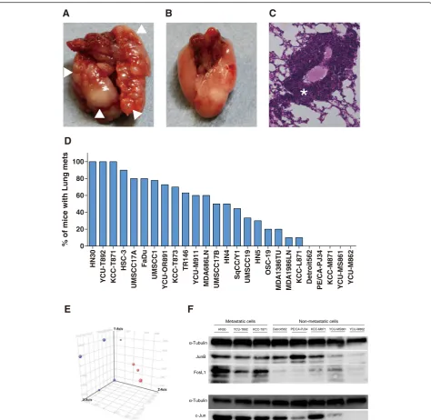
Twenty-six HNSCC cell lines showed a wide spectrum of distant metastatic potentialin vivo. We found that 21 (80.8 %) of the 26 HNSCC cell lines produced lung
metastases (Fig. 1d, Additional file 2: Table S2). Three HNSCC lines (HN30, KCC-T871, YCU-T892) estab-lished 100 % lung metastases in an experimental lung metastatic mouse model of HNSCC (Fig. 1a, c), while five HNSCC cell lines (Detroit562, PE/CA-PJ34, KCC-M871, YCU-MS861, YCU-M862) did not establish lung metastasis in any of the mice injected (Fig. 1b). Survival curves for mice injected with each of the 26 HNSCC cell lines are shown in Additional file 3: Figure S1. The median survival time ranged from 43.5 to 90 days. We found an inverse correlation between the incidence of lung metasta-sis (%) and median survival time (r=–0.5195,P= 0.0015) (Additional file 4: Figure S2).
Microarray analysis and upstream and key node analysis
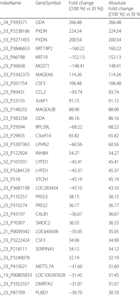
A clear separation in the 3 principal component analysis (PCA) was observed between three metastatic HNSCC cell lines (HN30, KCC-T871, YCU-T892) and five non-metastatic HNSCC cell lines (Detroit562, PE/CA-PJ34, KCC-M871, YCU-MS861, YCU-M862) (Fig. 1e). Differ-entially expressed genes showing statistically significant up- or down-regulations in expression between the metastatic and non-metastatic HNSCC cell lines are shown in Table 2.
To better understand the mechanisms underlying the gene expression findings, the microarray data were ana-lyzed in the context of complex regulatory networks. One hundred and sixty-four genes with an absolute fold change value (FC) >5.5 and P< 0.05 (unpaired t-test) were selected, and the data were then loaded into the ExPlain™ pathway search tool and key nodes were searched in the upstream pathways.
One hundred and ninety-seven genes were identified for the candidate genes as key factors in the regulation of pathways related to distant metastasis in HNSCC. A list of 20 genes with a score ≥11 according to the Ex-Plain™ tool is shown Table 3. The results, which show several AP-1 family genes such as Fos, JunB and FosL1 as having high scores, led us to hypothesize that the AP-1 family of transcription factor plays a crucial role in inducing cell invasion, migration and distant me-tastasis in HNSCC.
Expression of AP-1 family proteins in HNSCC cells
roles of JunB in regulating the pathways related to distant metastasis in HNSCC based on the high scores observed for JunB in the upstream and key node analysis for the current dataset (distant metastatic vs. non-metastatic) and the regional metastatic vs. non-metastatic data set (data not shown).
siRNA knockdown and sgRNA knockout of JunB in metastatic HNSCC cells suppresses tumor invasion and migration
To determine whether JunB promoted invasion and mi-gration in HNSCC cells, we depleted JunB in metastatic HNSCC cell lines (KCC-T871 and HN30), and performed invasion and scratch assays.
A
B
D
%o
fm
ic
ew
it
hL
un
g
m
et
s
HN30
YCU-T892 KCC-T871
HSC-3
UMSCC17A
FaDu
UMSCC1
YCU-OR891 KCC-T873
TR14
6
YCU-M91
1
MDA
686LN
UMSCC17B
HN4
SqCC/
Y
1
UMSCC1
9
HN5
OSC-19
MDA
1386TU
MDA
1986LN
KCC-L87
1
Detroit562
PE/CA
-PJ34
KCC-M87
1
YCU-MS861 YCU-M86
2
0 20 40 60 80 100
FosL1
E
C
F
*
Fig. 1The distant metastatic potential of 26 HNSCC lines in the experimental lung metastatic mouse model.aLung metastasis in the experimental lung metastatic mouse model of HNSCC. (△metastatic lesion).bNo metastasis was observed in the lungs of the mouse model.cH&E staining of lung metastasis in the experimental lung metastatic mouse model of HNSCC (* metastatic lesion).dIncidence of lung metastasis for 26 cell lines in the experimental lung metastatic mouse model of HNSCC. Three HNSCC cell lines (HN30, KCC-T871, YCU-T892) established 100 % lung metastases, while 5 cell lines (Detroit562, PE/CA-PJ34, KCC-M871, YCU-MS861, YCU-M862) did not establish lung metastasis in any of the mice injected.eThe results of principle component analysis (PCA). PCA was performed based on the expression profiles of samples. The first 3 PCAs for 8 HNSCC cells were plotted.
KCC-T871 and HN30 were transfected with siControl or two independent siRNAs for JUNB (#1 and #2) and the knockdown was then confirmed by Western blotting (Fig. 2a). The invasion assay revealed a 45.9 % reduction in invasion potential for KCC-T871/siJUNB#1 cells (39.8 ± 7.7) compared to scrambled siRNA control (73.5 ± 7.0, P= 0.0003) and an 81.2 % reduction in HN30/siJUNB#1 cells (18.4 ± 5.2) invasiveness when compared to control (98.1 ± 27.2, P= 0.011) as shown
in Fig. 2b. Thus, the siRNA-mediated knockdown of JunB in KCC-T871 and HN30 cells inhibited the inva-sive potential of these cell lines.
The scratch assay revealed significant reductions in cell motility for KCC-T871/siJUNB#1 cells (62.3 % ± 5.8 % vs. 83.6 % ± 4.4 %, P= 0.005) and KCC-T871/siJUNB#2 cells (8.25 % ± 2.9 % vs. 34.6 % ± 2.5 %,P< 0.0001) compared to control. Significant reductions in cell motility for HN30/siJUNB#1 cells (16.2 % ± 5.9 % vs. 92.2 % ± 2.4 %, P< 0.0001) and HN30/siJUNB#2 cells (60.9 % ± 2.9 % vs. 85.6 % ± 2.3 %,P< 0.0001) compared to control were also observed. Thus, the siRNA-mediated knockdown of JunB in KCC-T871 and HN30 cells also inhibited the cell migration ability as shown in Fig. 2c.
To confirm that JunB knockdown decreased cell mo-tility and invasiveness in HNSCC cells, the JunB knock-out cells were established with two independent sgRNAs (JUNB/KO#1 and #2) using the CRISPR/cas9 system and the knockout was then confirmed as shown in Fig. 3a and b. As shown in Fig. 3c, the scratch assay showed sig-nificant reductions in cell motility for KCC-T871/JUNB/ KO#1 cells (29.3 % ± 1.0 %) and KCC-T871/JUNB/KO#2 cells (34.2 % ± 2.1 %) compared to the control (58.1 % ±
Table 2Top 32 lists of expressed genes showing statistically between metastatic and non-metastatic HNSCC cells ProbeName GeneSymbol Fold change
([100 %] vs [0 %])
Absolute Fold change ([100 %] vs [0 %])
A_24_P393571 GDA 266.48 266.48
A_33_P3238166 PXDN 224.54 224.54
A_33_P3271455 PXDN 200.54 200.54
A_33_P3846653 KRT19P2 −160.22 160.22
A_23_P66798 KRT19 −152.13 152.13
A_23_P36658 MGST1 −148.41 148.41
A_33_P3342375 MAGEA6 114.26 114.26
A_23_P501754 CSF3 106.48 106.48
A_23_P89431 CCL2 −93.74 93.74
A_23_P23155 AJAP1 91.15 91.15
A_23_P148255 MAGEA2B 88.90 88.90
A_23_P383258 GDA 86.16 86.16
A_23_P29594 RPL39L −68.22 68.22
A_23_P29655 C3orf14 65.82 65.82
A_33_P3307363 LPHN2 −60.56 60.56
A_23_P122924 INHBA 54.27 54.27
A_32_P101031 LYPD1 −45.41 45.41
A_33_P3284129 LYPD1 −45.37 45.37
A_23_P518 VTCN1 −45.19 45.19
A_33_P3687198 LOC283454 −43.10 43.10
A_23_P135257 PRSS3 38.15 38.15
A_23_P310274 PRSS2 36.77 36.77
A_23_P43197 CALB1 −36.67 36.67
A_23_P70307 SMOC2 36.55 36.55
A_21_P0009342 LOC645638 −35.05 35.05
A_33_P3222424 CSF3 34.90 34.90
A_23_P218111 SERPINA1 34.12 34.12
A_33_P3240078 32.19 32.19
A_23_P415021 METTL7A −31.60 31.60
A_19_P00805833 LOC100287628 −31.45 31.45
A_33_P3352557 DMRTA2 −31.07 31.07
A_23_P87709 PLBD1 −30.70 30.70
One hundred sixty four genes with an absolute fold change value (FC) >5.5 andP< 0.05 were detected. Top 32 genes with an absolute FC > 30 were listed
Table 3Top 20 lists of upstream key molecules in lung metastatic versus non-metastatic HNSCC cells
Key molecules score
FOS 26
JUNB 23
FOSL1 21
PPARG 21
IRF8 20
IRF1 19
CBP 18
IRF4 18
ISGF3G 17
IKK-beta 16
FOXA2 14
IRF7 14
NR3C1 14
JUND 13
Src-isoform1 13
IRF2 12
beta-catenin 11
c-Myc-isoform1 11
MKK4beta 11
p38alpha 11
2.5 %, P< 0.0001 and P< 0.0001, respectively), which was consistent with our previous results. These results suggested that JunB could promote HNSCC cell migration and invasion. On the other hand, the cell proliferation ability of KCC-T871, KCC-T871/crControl, KCC-T871/ JUNB/KO#1 and KCC-T871/JUNB/KO#2 were similar in cell viability assays (Fig. 3d).
As JunB has been reported to contribute transforming growth factor-β-induced EMT [22], we next examined if the role of JunB in HNSCC cell migration and invasiveness is related to EMT. KCC-T871 cells showed mesenchymal
characteristics at baseline as well as E-cadherin expression. Their cell morphology did develop any epithelial character-istics upon siRNA-mediated JunB knockdown in KCC-T871 cells with or without TGF-βstimulation. In addition, no phenomena associated with the down-regulation of mesenchymal markers and up-regulation of epithelial markers were observed with or without TGF-βstimulation as shown in Additional file 5: Figure S3. Furthermore, we did not observe any change in cell morphology with the down-regulation of mesenchymal markers and up-regulation of epithelial markers in response to
A
C
JunBJUNB#1
**
0 20 40 60 80 100
JUNB#2
***
0 20 40 60 80 100
B
JUNB#1
0 20
***
HN30
40 60 80 100
JUNB#1
*
HN30
0 50 100 150 200
JUNB#1
**
0 50 100 150 200
JUNB#2
0 20 40 60 80
100
***
HN30 HN30
JUNB#1 JUNB#2 JUNB#1 JUNB#2
sgRNA-mediated JunB knockout in KCC-T871 cells (data not shown).
Knockout of JunB in metastatic HNSCC cells reduced the incidence of lung metastasisin vivo
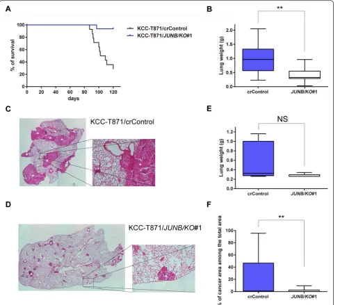
To clarify the role of JunB in cell migration and invasive-ness in HNSCC in vivo, the effect of knocking out of JunB was examined using an experimental lung meta-static mouse model. Fourteen mice for the control group with KCC-T871/crControl and 16 mice for the JunB knockout (KO) group with KCC-T871/JUNB/KO#1 were prepared for the survival study. The median survival period for the JunB KO group (120.0 days) was signifi-cantly greater than that for the control group (105.5 days, P= 0.0002, Fig. 4a). The presence of microscopic lung metastasis lesions was also impacted as 100 % of control animals had lung metastases, while 75.0 % of animals in the JunB KO group were found to have lung metastases as shown in Additional file 6: Figure S4a (P= 0.1029).
To confirm that JunB knockout reduced the incidence of lung metastasis in the animal model, thein vivostudy was repeated to measure the weight of each lung and the area occupied by metastatic HNSCC cells in the lung at 78 days after cell inoculation (6 mice were used for the control group and 8 mice for the JunB KO group). Microscopic lung metastasis was detected in 83.3 % of the control mice and 62.5 % of the JunB KO mice (P= 0.209, Additional file 6: Figure S4b). Median lung weight was 0.325 g for the control group (0.26–1.16 g) and 0.26 g for JunB KO group (0.26–0.34 g) as shown in Fig. 4e. Thus, JunB knockout markedly reduced lung weight in the animal model; however, the difference was not significant (P= 0.0727). On the other hand, we observed that JunB knockout significantly reduced area occupied by meta-static HNSCC cells in the lung in the experimental lung metastatic mouse model (Additional file 7: Figure S5). As shown in Fig. 4f, the mean percentage of metastatic HNSCC cells in the lung was 21.4 % for the control group
C
0 20 40 60 80 100
er
u
s
ol
c
d
n
u
o
w f
o
%
KCC-T871
***
crControl
JUNB/KO#1
A
crControl JUNB/KO#1 JUNB/KO#2
JunB
B
crControl
JUNB/KO#2
0 20 40 60 80 100
KCC-T871
**
% of wound closure
JUNB/KO#1 JUNB/KO#2
uncut cut
D
0 1 2 3 4
0.125 0.25 0.5 1 2
days
Lo
g2 Ab (450
nm
) KCC-T871/JUNB/KO#1
KCC-T871/JUNB/KO#2
KCC-T871 KCC-T871/crControl
Fig. 3sgRNA knockout of JunB in metastatic HNSCC cells suppresses tumor migration but not cell proliferation.aWestern blotting analysis of KCC-T871 cells transfected with control sgRNA orJUNBsgRNA (#1 and #2).bThe confirmation of genome-editing in KCC-T871 cells transfected with sgRNA-mediated JunB knockout. The efficiency of sgRNAs targeting JunB was measured using a GeneArt Genomic Cleavage Detection kit.
(0–95.6 %) and 1.6 % for the JunB KO group (0–9.4 %, P= 0.0037). Overall, we observed that knockout of JunB in metastatic HNSCC cells markedly reduced the in-cidence of lung metastasis in an experimental lung metastatic mouse model.
Discussion
In this study, we identified AP-1 family as key molecules regulating the pathways related to distant metastasis in HNSCC using the upstream and key node analysis to-gether with whole gene microarray analysis based on the
in vivometastatic potential of HNSCC cell lines. Among the AP-1 family, we determined that the knockdown and knockout of JunB in HNSCC cells significantly inhibited their invasion and migration in vitroas well as the inci-dence of lung metastasesin vivo. However, no phenom-ena related to mesenchymal-to-epithelial transition (MET), including the depletion of morphological mesen-chymal characteristics and mesenmesen-chymal markers, were observed in response to the knockdown or knockout of JunB in metastatic HNSCC cells. These results suggested that JunB could play an important role in promoting cell invasion, migration and distant metastasis in HNSCC viapathways other than EMT.
While there were several candidate genes related to the regulation of the pathways for distant metastasis in HNSCC identified by our key node analysis, as shown in Table 3, the AP-1 family genes were one of handful genes with high scores in the analysis for both the current dataset for distant metastatic vs. non-metastatic and our previous dataset for regional metastatic vs. non-metastatic. We previously characterized the regional metastatic potential in vivo using HNSCC cell lines in an orthotopic nude mouse model of HNSCC [21], and performed the upstream and key node analysis using almost the same method as that in the present study, and found that JunB and c-Fos also had high scores (data not shown). Thus, our results revealed that the AP-1 family, including JunB, might be important for regulating the pathways related not only to distant but also to regional metastasis in HNSCC.
It is well known that EMT is crucial for cancer cells not only in regard to tumor invasion and metastasis abil-ity but also in the acquisition of resistance to apoptosis and stemness properties [23]. During the EMT process, cancer cells acquire mesenchymal characteristics instead of losing epithelial features, and increased cell migration and invasiveness is induced by stimuli or cytokines in-cluding TGF-β. Recently, several studies showing the contribution of the AP-1 family to the EMT process have been reported for several malignancies [24–26]. In fact, genome-wide profiling of AP-1–regulated transcrip-tion has revealed that c-Jun and FosL1 promote cell in-vasion through the repression of E-cadherin expression by the transcriptional upregulation of ZEB2 in triple-negative breast cancer cells [24]. FosL1/AP-1 signaling has also been reported to modulate ZEB1/2 and TGF-β expression to induce EMT in triple-negative breast can-cer cells [24]. Moreover, cooperation between Twist1 and AP-1 has been reported to regulate integrin α5 expression to induce cell invasion by EMT [25]. Thus, AP-1 is closely associated with the EMT process in pro-moting the invasion and metastasis of cancer cells. How-ever, contrary to these previous reports, we did not observe any phenomena related to MET with or without
TGF-βin response to the depletion of JunB in HNSCC cells. Among the AP-1 family members, only a few stud-ies have sought to determine the contribution of JunB to EMT, suggesting that the role of JunB in regulating EMT might be less important than that of either c-Jun and/or FosL1. There is also the possibility that the con-tribution of AP-1 signaling to EMT in the metastatic process in HNSCC could be relatively low compared to those for other malignancies, as the greater number of gene mutations existing in HNSCC cells, due to a history of tobacco and/or alcohol use, could play an important role in HNSCC metastasis [27].
Other mechanisms underlying the AP-1-mediated regu-lation of tumor invasion in cancer cells have been also re-ported. Kanno et al. reported that JunB regulates several genes, such as matrix metalloproteinase-2 (MMP-2), MMP-9 and chemokine (C-C motif ) ligand-2 (CCL2), to promote tumor invasion and angiogenesis in VHL-defective renal cell carcinomas [28]. The AP-1/NFAT4 complex has also been reported to regulate the inhibition of E-cadherin expression by microRNA-23a during Fas-induced EMT in gastrointestinal cancer [29]. Ding et al. have identified KDM4A (lysine-specific demethylase 4A) as a key epigenetic factor activating JUN and FOSL1 to promote tumor invasion and cervical lymph node metas-tasis in HNSCC [12]. Thus, there are a number of mecha-nisms related to the regulation of tumor invasiveness by AP-1 in cancer cells. Further study is required to examine the details of the cellular and molecular mechanisms underlying the JunB-mediated promotion of tumor inva-sion in HNSCC.
HNSCC, since the tongue tumors in the model do not allow enough time for distant metastatic lesions to de-velop biologically from the primary tumor generated by orthotopic implantation. Moreover, Rashid et al. have reported that an experimental lung metastatic mouse model with tail vein injection could produce lung metastatic lesion with similar genomic profiles as lung metastases after orthotopic implantation [33]. An ex-perimental lung metastatic mouse model with tail vein injection of HNSCC was therefore used to elucidate the key molecules regulating the pathways related to metas-tasis in HNSCC in this study.
Conclusions
We have identified the AP-1 family as the key molecules regulating the pathways related to distant metastasis in HNSCC by use of upstream and key nodes analysis con-ducted in combination with the characterization of the in vivo distant metastatic potential of 26 different of HNSCC cell lines in an experimental lung metastatic mouse model. The knockdown and knockout of JunB re-duced tumor migration and invasion in vitro as well as lung metastasis in vivo, suggesting that the JunB path-way might be a useful a therapeutic target for inhibiting distant metastasis in patients with HNSCC. However, we did not observe any phenomena related to MET in re-sponse to JunB knockdown in HNSCC cells. Further studies are required to examine the details of the cellular and molecular mechanisms of the promotion tumor in-vasion by JunB in metastatic HNSCC in order to identify specific JunB inhibitors and demonstrate their efficacy in inhibiting tumor invasion and metastasis in HNSCC.
Availability of data and materials
The GEO accession number for the agilent gene ex-pression profiling data reported in the present study is GSE67275.
Additional files
Additional file 1: Table S1.Primary site, source, and clinical features of tumors used to derive twenty-six HNSCC cell lines used in this study. (DOCX 20 kb)
Additional file 2: Table S2.Survival, macroscopic and microscopic lung metastases, and lung weight of mice in the mouse model. (DOCX 71 kb)
Additional file 3: Figure S1.Survival curves for mice injected with each of the 26 HNSCC cell lines. Animals were asphyxiated when they had lost more than 15 % of their initial body weight or had become moribund, and the remaining mice were asphyxiated 90 days after cell injection. Survival was analyzed by the Kaplan–Meier method. (PDF 488 kb)
Additional file 4: Figure S2.Correlation of mean survival time with incidence of lung metastasis in an experimental lung metastatic mouse model of HNSCC. Inverse correlation between mean survival time and the incidence of lung metastasis in the mouse model was observed (r=–0.5192,P= 0.0015). (PDF 223 kb)
Additional file 5: Figure S3.Cell morphology and expression of mesenchymal or epithelial marker on siRNA control or siRNA mediated JunB knockdown in KCC-T871 and HN30 cells.aCell morphology of KCC-T871/siRNA control, KCC-T871/siJUNB#1 and KCC-T871/siJUNB#2.
bCell morphology of HN30/siRNA control, HN30/siJUNB#1 and HN30/ siJUNB#2.cExpression of mesenchymal or epithelial marker on siRNA control or siRNA mediated JunB knockdown in KCC-T871 and HN30 cells. (PDF 1800 kb)
Additional file 6: Figure S4.The incidence of microscopic lung metastasis in an experimental lung metastatic mouse model of HNSCC.
aThe incidence of microscopic lung metastasis of the control group (N= 14) and JunB KO group (N= 16). The incidence of lung metastasis in the JunB KO group (75.0 %) was reduced compared to that in the control group (100 %), however, the difference was not significant.P= 0.1029.
bThe incidence of microscopic lung metastasis of the control group (N= 6) and JunB KO group (N= 8) in the repeated animal study. The incidence of lung metastasis in the JunB KO group (62.5 %) was reduced compared to that in the control group (83.3 %), however, the difference was not significant.P= 0.5804.cThe incidence of total lung metastasis the control group (N= 20) and JunB KO group (N= 24) in our entire animal study. The incidence of lung metastasis in the JunB KO group (70.8 %) was remarkedly reduced compared to that in the control group (95.0 %), however, the difference was not significant.P= 0.0544. (PDF 246 kb)
Additional file 7: Figure S5.Hematoxyilin and eosin (H&E) slides of microscopic lung metastasis in an experimental lung metastatic mouse model of HNSCC. H&E slides of lung in the mouse injected with KCC-T871/ crControl or KCC-T871/JUNB/KO#1 euthanized after 78 days following cell inoculation. A. Lung sections in the mouse injected with KCC-T871/crControl cells. B. Lung sections in the mouse injected with KCC-T871/JUNB/KO#1 cells. (DOCX 905 kb)
Competing interests
We, the authors, declare that we have no competing interests.
Authors’contributions
HH was involved in the acquisition of the data, analysis and interpretation of the data, and writing, and reviewing of the manuscript. DS was involved in the design of the study, acquisition of the data, analysis and interpretation of the data, writing, reviewing, and revision of the manuscript. HT, TH, YI, SS and YI were involved in the acquisition of the data, analysis and interpretation of the data. JM was involved in the design of the study and reviewing of the manuscript. NO was involved in the design of the study, as well as review of the manuscript. All authors read and approved the final manuscript.
Acknowledgments
We thank Mari Mitsuka (Department of Biology and Function in Head and Neck, Yokohama City University Graduate School of Medicine, Yokohama, Japan) and Hideaki Mitsui (Department of Pathology) for their excellent technical assistance.
Funding
This work was supported by JSPS KAKENHI Grant Number 24791797 and 26861406 (PI: DS) from Japan Society for the Promotion of Science.
Author details
1Department of Biology and Function in Head and Neck, Yokohama City
University Graduate School of Medicine, Yokohama, Japan.2Department of
Otorhinolaryngology - Head and Neck Surgery, Yokohama City University, School of Medicine, 3-9 Fukuura, Kanazawa-ku, Yokohama 236-0004, Japan.
3Department of Urology, Yokohama City University Graduate School of
Medicine, Yokohama, Japan.4Department of Head and Neck Surgery, The
University of Texas M. D. Anderson Cancer Center, Houston, Texas, USA.
Received: 11 November 2015 Accepted: 4 January 2016
References
2. Goldberg HI, Lockwood SA, Wyatt SW, Crossett LS. Trends and differentials in mortality from cancers of the oral cavity and pharynx in the United States, 1973-1987. Cancer. 1994;74(2):565–72.
3. Edwards BK, Ward E, Kohler BA, Eheman C, Zauber AG, Anderson RN, et al. Annual report to the nation on the status of cancer, 1975-2006, featuring colorectal cancer trends and impact of interventions (risk factors, screening, and treatment) to reduce future rates. Cancer. 2010;116(3):544–73.
4. Cooper JS, Pajak TF, Forastiere AA, Jacobs J, Campbell BH, Saxman SB, et al. Postoperative concurrent radiotherapy and chemotherapy for high-risk squamous-cell carcinoma of the head and neck. N Engl J Med. 2004; 350(19):1937–44.
5. Allen CT, Law JH, Dunn GP, Uppaluri R. Emerging insights into head and neck cancer metastasis. Head Neck. 2013;35(11):1669–78.
6. Eferl R, Wagner EF. AP-1: a double-edged sword in tumorigenesis. Nat Rev Cancer. 2003;3(11):859–68.
7. Piechaczyk M, Farras R. Regulation and function of JunB in cell proliferation. Biochem Soc Trans. 2008;36(Pt 5):864–7.
8. Angel P, Karin M. The role of Jun, Fos and the AP-1 complex in cell-proliferation and transformation. Biochim Biophys Acta. 1991;1072(2-3):129–57. 9. Ozanne BW, Spence HJ, McGarry LC, Hennigan RF. Transcription factors
control invasion: AP-1 the first among equals. Oncogene. 2007;26(1):1–10. 10. Schmidt D, Textor B, Pein OT, Licht AH, Andrecht S, Sator-Schmitt M, et al. Critical role for NF-kappaB-induced JunB in VEGF regulation and tumor angiogenesis. EMBO J. 2007;26(3):710–9.
11. Young MR, Li JJ, Rincon M, Flavell RA, Sathyanarayana BK, Hunziker R, et al. Transgenic mice demonstrate AP-1 (activator protein-1) transactivation is required for tumor promotion. Proc Natl Acad Sci U S A. 1999;96(17):9827–32. 12. Ding X, Pan H, Li J, Zhong Q, Chen X, Dry SM, et al. Epigenetic
activation of AP1 promotes squamous cell carcinoma metastasis. Sci Signal. 2013;6(273):ra28. 1-13, S0-15.
13. Wang H, Yang H, Shivalila CS, Dawlaty MM, Cheng AW, Zhang F, et al. One-step generation of mice carrying mutations in multiple genes by CRISPR/Cas-mediated genome engineering. Cell. 2013;153(4):910–8. 14. Jiang W, Bikard D, Cox D, Zhang F, Marraffini LA. RNA-guided editing of
bacterial genomes using CRISPR-Cas systems. Nat Biotechnol. 2013;31(3):233–9. 15. Masters JR, Thomson JA, Daly-Burns B, Reid YA, Dirks WG, Packer P, et al.
Short tandem repeat profiling provides an international reference standard for human cell lines. Proc Natl Acad Sci U S A. 2001;98(14):8012–7. 16. Zhao M, Sano D, Pickering CR, Jasser SA, Henderson YC, Clayman GL, et al.
Assembly and initial characterization of a panel of 85 genomically validated cell lines from diverse head and neck tumor sites. Clin Cancer Res. 2011;17(23):7248–64.
17. Elkin MV, lodavsky I. Tail vein assay of cancer metastasis. Curr Protoc Cell Biol. 2001; Chapter 19:Unit 19.2.
18. Ginos MA, Page GP, Michalowicz BS, Patel KJ, Volker SE, Pambuccian SE, et al. Identification of a gene expression signature associated with recurrent disease in squamous cell carcinoma of the head and neck. Cancer Res. 2004;64(1):55–63.
19. Good DM, Zubarev RA. Drug target identification from protein dynamics using quantitative pathway analysis. J Proteome Res. 2011;10(5):2679–83. 20. Kel A, Voss N, Valeev T, Stegmaier P, Kel-Margoulis O, Wingender E. ExPlain:
finding upstream drug targets in disease gene regulatory networks. SAR QSAR Environ Res. 2008;19(5–6):481–94.
21. Sano D, Xie TX, Ow TJ, Zhao M, Pickering CR, Zhou G, et al. Disruptive TP53 mutation is associated with aggressive disease characteristics in an orthotopic murine model of oral tongue cancer. Clin Cancer Res. 2011;17(21):6658–70.
22. Gervasi M, Bianchi-Smiraglia A, Cummings M, Zheng Q, Wang D, Liu S, et al. JunB contributes to Id2 repression and the epithelial-mesenchymal transition in response to transforming growth factor-beta. J Cell Biol. 2012;196(5):589–603.
23. Lamouille S, Xu J, Derynck R. Molecular mechanisms of epithelial-mesenchymal transition. Nat Rev Mol Cell Biol. 2014;15(3):178–96. 24. Qiao Y, Shiue CN, Zhu J, Zhuang T, Jonsson P, Wright AP, et al.
AP-1-mediated chromatin looping regulates ZEB2 transcription: new insights into TNFalpha-induced epithelial-mesenchymal transition in triple-negative breast cancer. Oncotarget. 2015;6(10):7804–14.
25. Nam EH, Lee Y, Moon B, Lee JW, Kim S. Twist1 and AP-1 cooperatively upregulate integrin alpha5 expression to induce invasion and the epithelial-mesenchymal transition. Carcinogenesis. 2015;36(3):327–37.
26. Bakiri L, Macho-Maschler S, Custic I, Niemiec J, Guio-Carrion A, Hasenfuss SC, et al. Fra-1/AP-1 induces EMT in mammary epithelial cells by modulating Zeb1/2 and TGFbeta expression. Cell Death Differ. 2015;22(2):336–50. 27. Agrawal N, Frederick MJ, Pickering CR, Bettegowda C, Chang K, Li RJ, et al.
Exome sequencing of head and neck squamous cell carcinoma reveals inactivating mutations in NOTCH1. Science. 2011;333(6046):1154–7. 28. Kanno T, Kamba T, Yamasaki T, Shibasaki N, Saito R, Terada N, et al. JunB
promotes cell invasion and angiogenesis in VHL-defective renal cell carcinoma. Oncogene. 2012;31(25):3098–110.
29. Zheng H, Li W, Wang Y, Xie T, Cai Y, Wang Z, et al. miR-23a inhibits E-cadherin expression and is regulated by AP-1 and NFAT4 complex during Fas-induced EMT in gastrointestinal cancer. Carcinogenesis. 2014;35(1):173–83. 30. Talmadge JE, Singh RK, Fidler IJ, Raz A. Murine models to evaluate
novel and conventional therapeutic strategies for cancer. Am J Pathol. 2007;170(3):793–804.
31. Kim MY, Oskarsson T, Acharyya S, Nguyen DX, Zhang XH, Norton L, et al. Tumor self-seeding by circulating cancer cells. Cell. 2009;139(7):1315–26. 32. Sano D, Myers JN. Xenograft models of head and neck cancers. Head Neck
Oncol. 2009;1:32.
33. Rashid OM, Nagahashi M, Ramachandran S, Dumur CI, Schaum JC, Yamada A, et al. Is tail vein injection a relevant breast cancer lung metastasis model? J Thorac Dis. 2013;5(4):385–92.
• We accept pre-submission inquiries
• Our selector tool helps you to find the most relevant journal • We provide round the clock customer support
• Convenient online submission • Thorough peer review
• Inclusion in PubMed and all major indexing services • Maximum visibility for your research
Submit your manuscript at www.biomedcentral.com/submit