Open Access
R E S E A R C H
© 2010 Lee et al; licensee BioMed Central Ltd. This is an Open Access article distributed under the terms of the Creative Commons At-tribution License (http://creativecommons.org/licenses/by/2.0), which permits unrestricted use, disAt-tribution, and reproduction in any medium, provided the original work is properly cited.
Research
Cyclic nucleotides and mitogen-activated protein
kinases: regulation of simvastatin in platelet
activation
Ye-Ming Lee
†1,2, Wei-Fan Chen
†3, Duen-Suey Chou
2, Thanasekaran Jayakumar
3, Ssu-Yu Hou
3, Jie-Jen Lee
2,
George Hsiao
2and Joen-Rong Sheu*
2,3Abstract
Background: 3-Hydroxy-3-methyl-glutaryl coenzyme A (HMG-CoA) reductase inhibitors (statins) have been widely used to reduce cardiovascular risk. These statins (i.e., simvastatin) may exert other effects besides from their cholesterol-lowering actions, including inhibition of platelet activation. Platelet activation is relevant to a variety of coronary heart diseases. Although the inhibitory effect of simvastatin in platelet activation has been studied; the detailed signal transductions by which simvastatin inhibit platelet activation has not yet been completely resolved.
Methods: The aim of this study was to systematically examine the detailed mechanisms of simvastatin in preventing platelet activation. Platelet aggregation, flow cytometric analysis, immunoblotting, and electron spin resonance studies were used to assess the antiplatelet activity of simvastatin.
Results: Simvastatin (20-50 μM) exhibited more-potent activity of inhibiting platelet aggregation stimulated by collagen than other agonists (i.e., thrombin). Simvastatin inhibited collagen-stimulated platelet activation accompanied by [Ca2+]i mobilization, thromboxane A
2 (TxA2) formation, and phospholipase C (PLC)γ2, protein kinase C
(PKC), and mitogen-activated protein kinases (i.e., p38 MAPK, JNKs) phosphorylation in washed platelets. Simvastatin obviously increased both cyclic AMP and cyclic GMP levels. Simvastatin markedly increased NO release, vasodilator-stimulated phosphoprotein (VASP) phosphorylation, and endothelial nitric oxide synthase (eNOS) expression. SQ22536, an inhibitor of adenylate cyclase, markedly reversed the simvastatin-mediated inhibitory effects on platelet aggregation, PLCγ2 and p38 MAPK phosphorylation, and simvastatin-mediated stimulatory effects on VASP and eNOS phosphorylation.
Conclusion: The most important findings of this study demonstrate for the first time that inhibitory effect of
simvastatin in platelet activation may involve activation of the cyclic AMP-eNOS/NO-cyclic GMP pathway, resulting in inhibition of the PLCγ2-PKC-p38 MAPK-TxA2 cascade, and finally inhibition of platelet aggregation.
Background
A high incidence of atherosclerosis and thrombotic com-plications has been associated with hypercholester-olemia. Blood cholesterol levels are of fundamental importance in the pathogenesis of coronary artery dis-eases (CAD). Elevations of low-density lipoprotein (LDL) levels are not only linked to an increased risk for athero-sclerosis but may also exert prothrombotic effects via
platelet activation [1]. The effectiveness of 3-hydroxy-3-methyl-glutaryl coenzyme A (HMG-CoA) reductase inhibitors (statins) in the prevention of CAD is ascribed not only to reduced cholesterol levels [2,3], but also to a number of additional effects, including the stabilization of atherosclerotic plaque, improved endothelial function, enhanced fibrinolysis, and antithrombotic effects [3-5]. Although many studies have demonstrated that statins have antiplatelet activity in hypercholesterolemic patients and animals [6], the inhibition of platelet-dependent thrombus formation in hypercholesterolemia may not correlate with the lipid-lowering effects, suggesting that
* Correspondence: [email protected]
2 Department of Pharmacology, Taipei Medical University, Taipei, Taiwan † Contributed equally
these statins may exert another effect besides from their cholesterol-lowering actions.
Inhibition of the thromboxane B2 formation or chang-ing cholesterol content on platelet membrane by statins has been reported [7,8]. Recently, Chou et al. [6] also sug-gested that enhanced nitric oxide (NO) and cyclic GMP formation of simvastatin (20-80 μM) may be involved in the inhibitory effects on platelet aggregation. The anti-platelet activity of simvastatin in anti-platelets has been stud-ied; however, the detailed signal transduction mechanism by which simvastatin inhibits platelet activation has not yet been completely resolved. We therefore systematically examined the cellular signal events associated with sim-vastatin-inhibited platelet activation in the present study.
Methods
Materials
Collagen (type I), luciferin-luciferase, phorbol-12, 13-dibutyrate (PDBu), 5,5-dimethyl-1 pyrroline N-oxide (DMPO), SQ22536, ODQ, arachidonic acid (AA), prosta-glandin E1 (PGE1), nitroglycerin, and thrombin were pur-chased from Sigma Chem. (St Louis, MO); Fura 2-AM and fluorescein iso-thiocyanate (FITC) were from Molec-ular Probe (Eugene, OR); the thromboxane B2 enzyme immunoassay (EIA) kit was from Cayman (Ann Arbor, MI); the anti-vasodilator-stimulated phosphoprotein (VASP Ser157) monoclonal antibody (mAb) was from
Cal-biochem (San Diego, CA); the anti-phospho-p38 mito-gen-activated protein kinase (MAPK) Ser182 mAb was
from Santa Cruz (Santa Cruz, CA); the anti-p38 MAPK and anti-phospho-c-Jun N-terminal kinase (JNK) (Thr183/Tyr185) mAbs, anti-phospholipase Cγ2 (PLCγ2),
anti-phospho (Tyr759) PLCγ2 mAbs, and the
anti-phos-pho-p44/p42 extracellular signal-regulated kinase (ERK) (Thr202/Tyr204) polyclonal antibody (pAb) were from Cell
Signaling (Beverly, MA); the anti-α-tubulin mAb was from NeoMarkers (Fremont, CA); and the Hybond-P PVDF membrane, ECL Western blotting detection reagent and analysis system, horseradish peroxidase (HRP)-conjugated donkey anti-rabbit IgG, and sheep anti-mouse IgG were from Amersham (Buckinghamshire, UK). Cyclic AMP and cyclic GMP EIA kits were pur-chased from Cayman (Ann Arbor, MI). Simvastatin was dissolved in 0.5% dimethyl sulfoxide (DMSO) and stored at 4°C until used.
Platelet aggregation
Human platelet suspensions were prepared as previously described [9]. This study was approved by the Institu-tional Review Board of Taipei Medical University and conformed to the principles outlined in the Helsinki Dec-laration, and all human volunteers provided informed consent. In brief, blood was collected from healthy
human volunteers who had taken no medicine during the preceding 2 weeks, and was mixed with acid/citrate/glu-cose (9:1:1, v/v). After centrifugation, the supernatant (platelet-rich plasma; PRP) was supplemented with pros-taglandin E1 (PGE1) (0.5 μM) and heparin (6.4 IU/ml). The washed platelets were finally suspended in Tyrode's solution containing bovine serum albumin (BSA) (3.5 mg/ml). The final concentration of Ca2+ in Tyrode's
solu-tion was 1 mM.
A turbidimetric method was applied to measure plate-let aggregation [9], using a Lumi-Aggregometer (Payton, Scarborough, Ontario, Canada). Platelet suspensions (0.4 ml) were preincubated with various concentrations of simvastatin or an isovolumetric solvent control (0.5% DMSO) for 3 min before the addition of agonists. The reaction was allowed to proceed for 6 min, and the extent of aggregation was expressed in light-transmission units. When measuring ATP release, 20 μl of a luciferin/ luciferase mixture was added 1 min before the addition of agonists, and ATP release was compared to that of the control.
Measurement of cyclic AMP and cyclic GMP formations
Platelet suspensions (3.6 × 10⁸/ml) were incubated with
isovolumetric solvent control (0.5% DMSO), nitroglyc-erin (10 μM), PGE1 (10 μM), or simvastatin (30 and 50 μM) for 6 min. The incubation was stopped by the addi-tion of EDTA (5 mM), and the soluaddi-tion was immediately boiled for 5 min. Fifty microliters of the supernatant was used to determine the cyclic AMP and cyclic GMP con-tents with EIA kits following acetylation of the samples as described by the manufacturer.
Flow cytometric analysis
Triflavin, an αIIbβ3 integrin antagonist, was prepared as
previously described [10]. Fluorescence-conjugated tri-flavin was prepared as previously described [10]. Platelet suspensions (3.6 × 10⁸/ml) were preincubated with
sim-vastatin (30 and 50 μM) or a solvent control for 3 min, followed by the addition of 2 μl of FITC-triflavin (2 μg/ ml). The suspensions were then assayed for fluorescein-labeled platelets using a flow cytometer (Beckman Coulter, Miami, FL). Data were collected from 50,000 platelets per experimental group, and the platelets were identified on the basis of their characteristic forward and orthogonal light-scattering profile. All experiments were repeated at least four times to ensure reproducibility.
Measurement of platelet [Ca2+]i by Fura 2-AM fluorescence Citrated whole blood was centrifuged at 120 g for 10 min. The supernatant was incubated with Fura 2-AM (5 μM) for 1 h. Human platelets were then prepared as described above. Finally, the external Ca2+ concentration of the
rise was measured using a fluorescence spectrophotome-ter (CAF 110, Jasco, Tokyo, Japan) with excitation wave-lengths of 340 and 380 nm, and an emission wavelength of 500 nm [9].
Measurement of thromboxane B2 formation
Platelet suspensions (3.6 × 10⁸/ml) were preincubated
with simvastatin (30 and 50 μM) or solvent control for 3 min before the addition of collagen (1 μg/ml). Six minutes after the addition of agonists, 2 mM EDTA and 50 μM indomethacin were added to the suspensions. The thromboxane B2 (TxB2) levels of the supernatants were measured using an EIA kit.
Immunoblotting study
Washed platelets (1.2 × 10⁹/ml) were preincubated with
simvastatin (30 and 50 μM) or a solvent control for 3 min, followed by the addition of agonists to trigger platelet activation. The reaction was stopped, and platelets were immediately re-suspended in 200 μl of lysis buffer. Sam-ples containing 80 μg of protein were separated by SDS-PAGE (12%); the proteins were electrotransferred by semidry transfer (Bio-Rad, Hercules, CA). Blots were blocked with TBST (10 mM Tris-base, 100 mM NaCl, and 0.01% Tween 20) containing 5% BSA for 1 h and then probed with various primary antibodies. Membranes were incubated with HRP-linked mouse IgG or anti-rabbit IgG (diluted 1: 3000 in TBST) for 1 h. Immunore-active bands were detected by an enhanced chemilumi-nescence (ECL) system. The bar graph depicts the ratios of quantitative results obtained by scanning reactive bands and quantifying the optical density using vid-eodensitometry (Bio-profil; Biolight Windows Applica-tion V2000.01; Vilber Lourmat, France).
Estimation of nitrate formation
NO was assayed in platelet suspensions as previously described [10]. In brief, platelet suspensions (1.2 × 10⁹/
ml) were preincubated with PGE1 (10 μM) or simvastatin (30 and 50 μM) for 3 min, followed by centrifugation. The amount of nitrate in the platelet suspensions (10 μl) was measured by adding a reducing agent to the purge vessel to convert nitrate to NO which was stripped from the suspensions by purging with helium gas. The NO was then drawn into a Sievers Nitric Oxide Analyzer (Sievers 280 NOA, Sievers, Boulder, CO). Nitrate concentrations were calculated by comparison with standard solutions of sodium nitrate.
Measurement of hydroxyl radical by electron spin resonance (ESR) spectrometry
The ESR method used a Bruker EMX ESR spectrometer as described previously [11]. In brief, platelet suspensions (3.6 × 10⁸/ml) were preincubated with simvastatin (30
and 50 μM) or solvent control for 3 min before the addi-tion of collagen (1 μg/ml). The reacaddi-tion was allowed to proceed for 5 min, followed by the addition of DMPO (100 μM) for the ESR study. The rate of hydroxyl radical-scavenging activity is defined by the following equation: inhibition rate = 1-[signal height (simvastatin)/signal height (solvent control)] [11].
Statistical analysis
The experimental results are expressed as the means ± S.E.M. and are accompanied by the number of observa-tions. The experiments were assessed by the method of analysis of variance (ANOVA). If this analysis indicated significant differences among group means, then each group was compared using the Newman-Keuls method. P
< 0.05 was considered statistically significant.
Results
Effects of simvastatin on platelet aggregation, αIIbβ3 integrin conformational change, and [Ca2+]i mobilization in
human platelets
Simvastatin (20-70 μM) exhibited potent activity of inhibiting platelet aggregation and the ATP-release reac-tion stimulated by collagen (1 μg/ml, open circle). It also significantly inhibited platelet aggregation stimulated by thrombin (0.02 U/ml, open square), AA (60 μM, open diamond) or U46619 (1 μM, open triangle), a prostaglan-din endoperoxide at higher concentrations (70-100 μM) (Fig. 1A and 1B). The IC50 value of simvastatin for platelet aggregation induced by collagen was approximately 30 μM. The solvent control (0.5% DMSO) did not signifi-cantly affect platelet aggregation stimulated by agonists in washed platelets (Fig. 1A). When platelets were prein-cubated with a higher concentration of simvastatin (200 μM) or 0.5% DMSO for 10 min, followed by two washes with Tyrode's solution, there were no significant differ-ences between the aggregation curves of either platelet preparations stimulated by collagen (1 μg/ml), indicating that the effect of simvastatin on inhibition of platelet aggregation occurs in a reversible manner (data not shown). In subsequent experiments, we used collagen as an agonist to explore the inhibitory mechanisms of sim-vastatin in platelet activation.
Figure 1 Effects of simvastatin on the inhibition of (A and B) platelet aggregation, (C) FITC-triflavin binding to the αIIbβ3 integrin and (D)
[Ca2+]i mobilization in activated platelets. Washed platelets (3.6 × 10⁸/ml) were preincubated with simvastatin (10-100 μM) or 0.5% DMSO,
fol-lowed by the addition of collagen (1 μg/ml, open circle), U46619 (1 μM, upside down open triangle), thrombin (0.02 U/ml, open square) or arachidonic acid (60 μM, open diamond) to trigger platelet aggregation (A and B) and the ATP-release reaction (A, upper tracings) or (D) [Ca2+]i mobilization. (C)
The solid line represents the fluorescence profiles of FITC-triflavin (2 μg/ml) (a) with or (b) without EDTA (5 mM); or pretreatment of simvastatin (c) (30 μM) and (d) (50 μM), followed by the addition of FITC-triflavin (2 μg/ml). Data are presented as the means ± S.E.M. (n = 4); ***P < 0.001, compared to the control group; #P < 0.05 and ##P < 0.01, compared to the collagen group.
[Ca
2+
] i (nM)
0 20 40 60 80 100 120 140 160 180 200
Fluorescence in
tensity (folds)
0 1 2 3 4 5
0 20 40 60 80 100 120
0 20 40 60 80 100
ʳ
A
B
C
DMSO Ц
Х collagen
simvastatin 30 Ц
Х
50 Ц
Х
AT
P
r
e
le
a
se
T
(10%)
min
a b c d
D
**
*** ***
collagen + + +
simvastatin 30 50
***
#
##
0
0 0 0
85 85
85 85
100 101 102 103 100 101 102 103
100 101 102 103 100 101 102 103
Fluorescence intensity
Number
of cells
a
d c
b
Concentration (
P
M)
aggregation induced by collagen. In this study, the relative intensity of the fluorescence of FITC-triflavin (2 μg/ml) bound directly to collagen (1 μg/ml)-activated platelets was relatively higher than that of negative control (in the presence of 5 mM EDTA) (a, 1.4 ± 0.2; b, 4.8 ± 0.4) (Fig. 1C). Simvastatin (30 and 50 μM) did not significantly affect FITC-triflavin binding to the αIIbβ3 integrin in platelet suspensions (c, 4.8 ± 0.1; d, 4.9 ± 0.1) (Fig. 1C), indicating that the inhibitory effect of simvastatin on platelet aggregation does not involve binding to the plate-let αIIbβ3 integrin.
Free cytoplasmic Ca+2 concentrations in human
plate-lets were measured by the Fura 2-AM loading method. As shown in Figure 1D, collagen (1 μg/ml) evoked a marked increase in [Ca2+]i, and this increase was markedly
inhib-ited in the presence of simvastatin (30 μM, 60.9 ± 17.0%; 50 μM, 72.1 ± 7.9%).
Effects of simvastatin on TxA2, PLCγ2, and PKC activation As shown in Figure 2A, resting platelets produced rela-tively little TxB2 compared to collagen-activated platelets. Simvastatin (30 and 50 μM) concentration-dependently inhibited TxB2 formation in platelets stimulated by colla-gen (1 μg/ml). PLC hydrolyzes phosphatidylinositol 4,5-bisphosphate (PIP2) to generate two secondary messen-gers: inositol 1,4,5-trisphosphate (IP3) and diacylglycerol (DAG) [12]. DAG activates PKC, inducing protein phos-phorylation (p47) and ATP release. Phosphos-phorylation is one of the key mechanisms regulating the activity of PLC. The immunoblotting analysis revealed that treatment with simvastatin markedly abolished the phosphorylation of PLCγ2 stimulated by collagen (Fig. 2B). Stimulation of platelets with a number of different agonists induced acti-vation of PKC, which then phosphorylated p47 proteins. In this study, phosphorylation experiments were per-formed to examine the role of simvastatin in PKC activa-tion in human platelets. When collagen (1 μg/ml) (Fig. 2C) or PDBu (150 nM) (Fig. 2D) was added to human platelets, a protein with an apparent of p47 was predomi-nately phosphorylated compared to resting platelets. Simvastatin inhibited p47 phosphorylation stimulated by collagen but not by PDBu (Fig. 2C and 2D).
Effect of simvastatin on collagen-induced MAPK phosphorylation
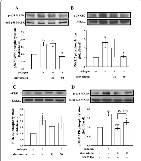
To further investigate the inhibitory mechanisms of sim-vastatin in platelet activation stimulated by collagen, we further detected MAPK signaling molecules including p38 MAPK, JNKs, and ERKs. The immunoblotting analy-sis revealed that simvastatin (50 μM) inhibited p38 MAPK (Fig. 3A) and JNKs (Fig. 3B), but not ERKs (Fig. 3C) phosphorylation stimulated by collagen. In addition, in the presence of SQ22536 (100 μM), an inhibitor of ade-nylate cyclase, significantly reversed the simvastatin-mediated inhibition of p38 MAPK phosphorylation stim-ulated by collagen (Fig. 3D).
Effects of simvastatin on cyclic nucleotides, nitrate formation and VASP phosphorylation
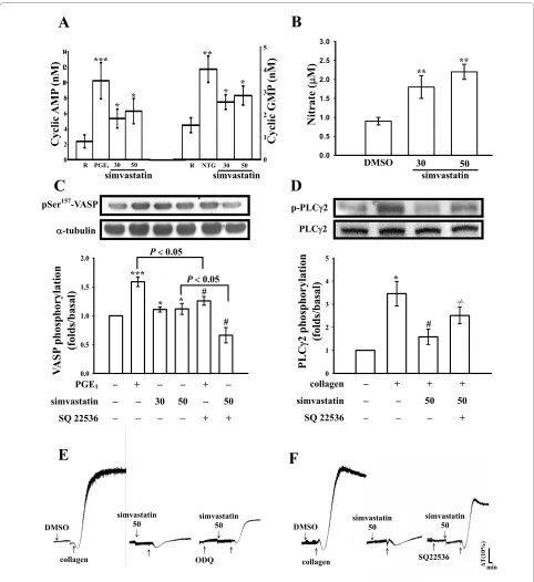
The level of cyclic AMP in unstimulated platelets was less, the addition of PGE1 (10 μM) markedly increased approximately 4.3-fold of cyclic AMP level compared with the resting group (Fig. 4A). Simvastatin (30 and 50 μM) significantly increased the cyclic AMP levels in human platelets (30 μM, 5.3 ± 1.2 nM; 50 μM, 6.3 ± 1.6 nM; n = 3) (Fig. 4A). We also performed a similar study measuring the cyclic GMP response. The level of cyclic GMP in unstimulated platelets was about 1.5 ± 0.3 nM, but when nitroglycerin (NTG, 10 μM) was added to the platelet suspensions, the cyclic GMP level markedly increased from the resting level to 4.0 ± 0.6 nM (n = 3) (Fig. 4A). The addition of simvastatin (30 and 50 μM)
Figure 2 Effects of simvastatin on (A) thromboxane B2 formation, (B) phospholipase Cγ2 and (C and D) PKC substrate (p47)
phos-phorylation in activated platelets. Washed platelets were
preincu-bated with simvastatin (30 and 50 μM) or 0.5% DMSO, followed by the addition of collagen (1 μg/ml) or PDBu (150 nM) to trigger platelet ac-tivation. Cells were collected, and subcellular extracts were analyzed for (A) thromboxane A2 formation, (B) phospholipase Cγ2 phosphory-lation, and (C and D) phospho-PKC substrate (p-p47) as described in "Methods". Data are presented as the means ± S.E.M. (n = 4); *P < 0.05 and **P < 0.01, compared to the control group; #P < 0.05 and ##P < 0.01,
compared to the collagen group. 0 1 2 3 4 5
0 1 2 3 4 0 10 20 30 40 50
0 1 2 3 4 5 6 7 8
A B
collagen + + + simvastatin 30 50
collagenʳ + + + simvastatin 30 50
PDBu + + +
simvastatin 30 50
C D
**
#
* p-PLCJ2
PLCJ2
#
p-p47 p-p47
D-tubulin D-tubulin
** **
# ##
PLC
J
2 phosphorylation (folds/basal)
Thr
omboxane B
2
(ng/ml)
p47 phosphorylation
(folds/basal)
p47 phosphorylation
(folds/basal)
resulted in significant increases in platelet cyclic GMP levels (30 μM, 2.6 ± 0.3 nM; 50 μM, 2.9 ± 0.4 nM; n = 3) (Fig. 4A). NO was quantified using a sensitive and spe-cific ozone redox-chemiluminescence detector. As shown in Figure 4B, simvastatin (30 and 50 μM) concentration-dependently increased nitrate production after incuba-tion with washed platelets (Fig. 4B). It was demonstrated that cyclic nucleotides can induce VASP Ser157
phospho-rylation in human platelets [13]. In this study, PGE1 (10 μM) and simvastatin (30 and 50 μM) markedly induced VASP Ser157 phosphorylation (Fig. 4C). SQ22536 (100
μM) significantly inhibited the phosphorylation stimu-lated by both PGE1 (10 μM) and simvastatin (50 μM) (Fig.
4C). Furthermore, SQ22536 (100 μM) obviously reversed the simvastatin (50 μM)-mediated inhibitory effect of PLCγ2 phosphorylation stimulated by collagen (Fig. 4D). On the other hand, pretreatment with SQ22536 (100 μM) or ODQ (20 μM), an inhibitor of guanylate cyclase, signif-icantly reversed the simvastatin (50 μM)-mediated inhi-bition of platelet aggregation stimulated by collagen (Fig. 4E and 4F). These results indicate that simvastatin
inhib-its platelet aggregation, al least in part, via a cyclic nucle-otides-dependent pathway.
Effects of simvastatin on eNOS phosphorylation and hydroxyl radical formation
Endothelial nitric oxide synthase (eNOS) phosphoryla-tion was markedly activated by both PGE1 (10 μM) and simvastatin (50 μM) (Fig. 5A). The simvastatin-activated eNOS phosphorylation was significantly reversed in the presence of SQ22536 (100 μM) but not by ODQ (20 μM), indicating that cyclic AMP plays an up-regulator in sim-vastatin-mediated eNOS phosphorylation in human platelets (Fig. 5A). On the other hand, a typical ESR signal of hydroxyl radical (OH•) formation was induced in
colla-gen (1 μg/ml)-activated platelets compared to resting platelets (Fig. 5B, a and 5b); pretreatment with simvasta-tin (30 and 50 μM) did not significantly reduce hydroxyl radical formation stimulated by collagen (Fig. 5B, c and 5d). The antioxidant, catalase (1000 U/ml), markedly sup-pressed hydroxyl radical formation by about 78% (data not shown).
Discussion
This study demonstrates for the first time that simvasta-tin inhibits platelet activation via a novel pathway: activa-tion of cyclic AMP-eNOS/NO-cyclic GMP and inhibiactiva-tion of MAPK phosphorylation (i.e., p38 MAPK and JNKs) in washed platelets. Simvastatin exhibited more-potent activity at inhibiting collagen-induced platelet aggrega-tion than other agonists. For the clinical therapy, the approved starting dose of simvastatin for most patients is 20 mg, and the maximal dose is 80 mg. In this study, the concentrations of simvastatin were employed at 30 and 50 μM, and the concentration of collagen was used at 1 μg/ml to trigger platelet aggregation. In general, concen-trations of collagen were employed for platelet aggrega-tion of from 0.1 to 5 μg/ml. In an attempt to elucidate the detailed mechanisms of pharmacological interest, we used a higher concentration (1 μg/ml) of collagen to induce a more-pronounced signal transduction in plate-lets (i.e., MAPKs and PKC etc.). Therefore, the pharma-cological concentrations (30-50 μM) of simvastatin employed to inhibit platelet aggregation in vitro are rea-sonable higher than that of blood concentrations obtained during a simvastatin regimen in vivo. However, the concentration employed is closely to that of other in vitro studies (20-80 μM) [6,14,15].
Stimulation of platelets by agonists (i.e., collagen) causes marked alterations in phospholipid metabolism. The activation of PLC results in the degradation of phos-phoinositides, notably, phosphatidylinositol 4,5-bisphos-phate (PI4,5-P2), resulting in the production of the second messengers, inositol 1,4,5-trisphosphate (IP3) and
Figure 3 Effects of simvastatin on (A and D) p38 MAPK, (B) JNKs,
and (C) ERKs phosphorylation in activated platelets. Washed
plate-lets (1.2 × 10⁹/ml) were preincubated with simvastatin (30 and 50 μM)
or 0.5% DMSO, followed by the addition of collagen (1 μg/ml) to trig-ger (A and D) p38 MAPK, (B) JNKs, and (C) ERKs phosphorylation. Data are presented as the means ± S.E.M. (n = 4); *P < 0.05 and **P < 0.01, compared to the control group; #P < 0.05, compared to the collagen
group.
0.0 0.5 1.0 1.5 2.0 2.5
0.0 0.5 1.0 1.5 2.0 2.5 3.0
0 1 2 3 4
0.0 0.5 1.0 1.5 2.0
collagenʳ + + +
simvastatin 30 50
collagen + + +
simvastatin 30 50
JNK1/2 p-JNK1/2 p-p38 MAPK
total-p38 MAPK
**
#
*
A B
C
p38 MAPK phosphorylation
(folds/basal)
JNK1/2 phosphorylation
(folds/basal)
collagenʳ + + +
simvastatin 30 50
p-ERK1/2 ERK1/2
ERK1/2 phosphorylation
(folds/basal)
*
#
p-p38 MAPK total-p38 MAPK
D
collagen + + +
simvastatin 50 50
SQ 22536 +
p38 MAPK phosphorylation
(folds/basal)
***
###
Figure 4 Effects of simvastatin on (A) cyclic nucleotides (B) nitrate formations, (C) Ser157-vasodilator-stimulated phosphoprotein (VASP)
and (D) phospholipase Cγ2 phosphorylation as well as (E and F) platelet aggregation in the presence of inhibitors of cyclic nucleotides in
washed platelets. Platelets were incubated with prostaglandin E1 (PGE1, 10 μM), nitroglycerin (NTG, 10 μM), simvastatin (30 and 50 μM), or 0.5%
DM-SO. Cells were collected, and subcellular extracts were analyzed for (A) cyclic nucleotides, (B) nitrate formations (C) Ser157-VASP, and (D) phospholipase
Cγ2 phosphorylations as described in "Methods". For platelet aggregation study, washed platelets were preincubated with simvastatin (50 μM) in the absence or presence of (E) ODQ (20 μM) or (F) SQ22536 (100 μM), followed by the addition of collagen (1 μg/ml). Data are presented as the means ± S.E.M. (n = 3-4); *P < 0.05, **P < 0.01, and ***P < 0.001, compared to the control group; #P < 0.05, compared to the without SQ22536 groups. ≠P < 0.05,
compared to the collagen plus simvastatin group. The profiles (E and F) are representative examples of four similar experiments.
0.0 0.5 1.0 1.5 2.0 2.5 3.0
0.0 0.5 1.0 1.5 2.0 0 2 4 6 8 10 12 14
0 1 2 3 4 5
0 1 2 3 4 5
pSer157-VASP
D-tubulin
**
**
***
* * #
B
DMSO Ц
Х collagen
simvastatin 50 Ц
Х Х
simvastatin 50 Ц
Х ODQ
DMSO Ц
Х collagen
simvastatin 50 Ц
Х
simvastatin 50 Ц Х
SQ22536 Х
min
T
(10%)
C
D
Nitrate (
P
M)
V
A
SP
phosphorylation (folds/basal)
DMSO 30 50 simvastatin
P 0.05
#
P 0.05
PGE1 + +
simvastatin 30 50 50 SQ 22536 + +
Cyclic AMP
(nM)
Cyclic GMP
(nM)
collagen + + + simvastatin 50 50
SQ 22536 + p-PLCJ2
PLCJ2
PLC
J
2 phosphorylation (folds/basal)
*
#
Ћ
A
E
F
***
* *
**
* *
R PGE1 30 50
simvastatin
NTG
R 30 50
DAG [16]. DAG activates PKC, inducing protein phos-phorylation (p47). PKC activation represents a strategy adopted by cells to allow selected responses to specific activating signals in distinct cellular compartments [17]. Phosphoinositide-specific PLC is a key enzyme in signal transduction [18]. There are six major families of PLC enzymes which consist of at least 13 PLC isoforms [18]. PLCγ2 is involved in antigen-dependent signaling in B cells and collagen-dependent signaling in platelets [19]. In this study, both PLCγ2 phosphorylation and PKC acti-vation stimulated by collagen were inhibited by simvasta-tin, suggesting that simvastatin-mediated antiplatelet activity is involved in inhibition of the PLCγ2-PKC signal pathway. Simvastatin had no direct effect on PKC activa-tion, as it did not inhibit PDBu-induced PKC activation (Fig. 2D) or platelet aggregation (data not shown). In addition, collagen-induced TxB2 formation, a stable metabolite of TxA2, was markedly inhibited by simvasta-tin. TxA2 is important for collagen-induced platelet aggregation. This may explain the more-potent activity of simvastatin in inhibiting collagen-induced platelet aggre-gation than other agonists (thrombin and U46619).
MAPKs consist of three major subgroups. Growth fac-tors preferentially activate ERKs (p44 ERK1 and p42 ERK2), which are involved in proliferation, adhesion, and cell progression [20], whereas p38 MAPK and JNKs (p46 JNK1 and p54 JNK2) are more responsive to stress, and appear to be involved in apoptosis [20]. ERKs, JNKs, and p38 MAPK have been identified in platelets [20]. The roles of JNKs and ERKs in physiopathology are unclear, but they have been suggested to be suppressors of αIIbβ3 integrin activation or negative regulators of platelet acti-vation [21]. On the other hand, p38 MAPK provides a crucial signal as a downstream effector of PKC which is necessary for aggregation caused by collagen [22]. Among the numerous downstream targets of p38 MAPK, the most physiologically relevant one in platelets is cyto-solic phospholipase A2 (cPLA2). p38 MAPK is essential
for the stimulation of cPLA2, which catalyzes AA release to produce TxA2 [23]; thus, p38 MAPK appears to pro-vide a TxA2-dependent platelet aggregation pathway. Simvastatin significantly inhibits TxA2 formation, at least in part, via inhibition of p38 MAPK phosphorylation.
Activation of human platelets is inhibited by two intra-cellular pathways regulated by either cyclic AMP or cyclic GMP. The importance of cyclic AMP and cyclic GMP in modulating platelet reactivity is well established [24]. In addition to inhibiting most platelet responses, elevated levels of cyclic AMP or/and cyclic GMP decrease intrac-ellular Ca2+ concentrations by the uptake of Ca2+ into the
dense tubular system (DTS) which negatively affects the
Figure 5 Effects of simvastatin on (A) endothelial nitric oxide
syn-thase (eNOS) phosphorylation and (B) hydroxyl radical (OH•)
for-mation in activated platelets. (A) Platelets were incubated with
prostaglandin E1 (PGE1, 10 μM), simvastatin (30 and 50 μM), or 0.5% DMSO in the absence or presence of SQ22536 (100 μM) or ODQ (20 μM) as described in "Methods". Cells were collected, and subcellular extracts were analyzed for eNOS phosphorylation. Data are presented as the means ± S.E.M. (n = 4); **P < 0.01 and ***P < 0.001, compared to the control group; #P < 0.05, compared to the PGE
1 group. (B) For the
electron spin resonance (ESR) study, platelets were preincubated with (a) Tyrode's solution (resting group), (b) a solvent control (0.5% DMSO), or simvastatin (30 and 50 μM), followed by the addition of collagen (1 μg/ml) to trigger platelet activation. Spectra are representative exam-ples of four similar experiments. Asterisk (*) indicates the formation of hydroxyl radical.
0.0 0.5 1.0 1.5 2.0 2.5 3.0 3.5
A
PGE1 +
simvastatin 50 50 50
ODQ +
SQ 22536 +
eNOS phosphorylation
(folds/basal)
p-eNOS
eNOS
***
**
B
P 0.05
#
*
* * *
* *
*
action of PLC and/or PKC [24]. Therefore, cyclic AMP and cyclic GMP act synergistically to inhibit platelet aggregation. In this study, simvastatin obviously increased the levels of both cyclic AMP and cyclic GMP in human platelets. Platelets produce NO in smaller amounts than do endothelial cells [25]. Most cellular actions of NO occur via stimulation of intracellular gua-nylate cyclase, leading to increases in cyclic GMP. Both the inducible NOS (iNOS) and eNOS isoforms have been described in platelets, but eNOS is predominant [25]. Simvastatin (80 μM) has been reported to induce NO release and stimulate eNOS activity in rabbit platelets [6]. In this study, SQ22536 markedly reversed simvastatin-mediated inhibition of platelet aggregation, PLCγ2, and p38 MAPK phosphorylation stimulated by collagen, and it also reversed the simvastatin-mediated activation of both eNOS and VASP phosphorylations. VASP is phos-phorylated by cyclic nucleotide-dependent protein kinase in platelets, which plays important role in modulating actin filament dynamics and integrin activation [13]. In this study, simvastatin was found to stimulate eNOS phosphorylation, and this effect was reversed by SQ22536 but not by ODQ. This result is in accord with that of increased cyclic AMP stimulating eNOS activity and NO biosynthesis [26].
Reactive oxygen species (i.e., hydrogen peroxide and hydroxyl radicals) derived from platelet activation might amplify platelet reactivity during thrombus formation. Free radical species act as secondary messengers that increase cytosolic Ca2+ during the initial phase of platelet
activation processes, and PKC is involved in receptor-mediated free radical production in platelets [11]. The antiplatelet effect of simvastatin did not mediate by the free radical-scavenging activity in ESR experiment.
In conclusion, the most important findings of this study demonstrate for the first time that the antiplatelet activity of simvastatin may involve an increase of the cyclic AMP-eNOS/NO-cyclic GMP pathway, followed by inhibition of the PLCγ2-PKC-p38 MAPK-TxA2 cascade, thereby leading to inhibition of platelet aggregation. Hypercho-lesterolemic patients usually associate with a high inci-dence of atherosclerosis and thrombotic complications. This study provides a new insight of antiplatelet mecha-nisms of simvastatin to explain its clinical protective effect in CAD.
Competing interests
The authors declare that they have no competing interests.
Authors' contributions
YML and WFC carried out the platelet aggregation study and drafted the man-uscript. DSC carried out the ESR study. TJ, SYH, and JJL carried out the immuno-blotting study. GH performed the statistical analysis. JRS conceived of the study, and participated in the design and coordination, and collectively pre-pared the manuscript. All authors read and approved the final manuscript.
Acknowledgements
This work was supported by grants from the National Science Council of Tai-wan (NSC97-2320-B-038-016-MY3) and Hsinchu Mackay Memorial Hospital (MMH-HB-96-02; MMH-HB-97-01).
Author Details
1Department of Surgery, Hsinchu Mackay Memorial Hospital, Hsinchu; Mackay
Medicine, Nursing and Management College, Taipei, Taiwan, 2Department of
Pharmacology, Taipei Medical University, Taipei, Taiwan and 3Graduate
Institute of Medical Sciences, Taipei Medical University, Taipei, Taiwan
References
1. Notarbartolo A, Davi G, Averna M, Barbagallo CM, Ganci A, Giammarresi C, La Placa FP, Patrono C: Inhibition of thromboxane biosynthesis and platelet function by simvastatin in type IIa hypercholesterolemia. Arterioscler Thromb Vasc Biol 1995, 15:247-251.
2. Maron DJ, Fazio S, Linton MF: Current perspectives on statins. Circulation 2000, 101:207-213.
3. Undas A, Brummel KE, Musial J, Mann KG, Szczeklik A: Simvastatin depresses blood clotting by inhibiting activation of prothrombin, factor V, and factor XIII and by enhancing factor Va inactivation. Circulation 2001, 103:2248-2253.
4. Rosenson RS, Tangney CC: Antiatherothrombotic properties of statins: implications for cardiovascular event reduction. JAMA 1998,
279:1643-1650.
5. Blumenthal RS: Statins: effective antiatherosclerotic therapy. Am Heart J 2000, 139:577-583.
6. Chou TC, Lin YF, Wu WC, Chu KM: Enhanced nitric oxide and cyclic GMP formation plays a role in the anti-platelet activity of simvastatin. Br J Pharmacol 2008, 153:1281-1287.
7. Osamah H, Mira R, Sorina S, Shlomo K, Michael A: Reduced platelet aggregation after fluvastatin therapy is associated with altered platelet lipid composition and drug binding to the platelets. Br J Clin Pharmacol 1997, 44:77-83.
8. Ma LP, Nie DN, Hsu SX, Yin SM, Xu LZ, Nunes JV: Inhibition of platelet aggregation and expression of alpha granule membrane protein 140 and thromboxane B2 with pravastatin therapy for
hypercholesterolemia. J Assoc Acad Minor Phys 2002, 13:23-26. 9. Sheu JR, Lee CR, Lin CH, Hsiao G, Ko WC, Chen YC, Yen MH: Mechanisms
involved in the antiplatelet activity of Staphylococcus aureus
lipoteichoic acid in human platelets. Thromb Haemost 2000, 72:617-621. 10. Sheu JR, Hung WC, Wu CH, Ma MC, Kan YC, Lin CH, Lin MS, Luk HN, Yen
MH: Reduction in lipopolysaccharide-induced thrombocytopenia by triflavin in a rat model of septicemia. Circulation 1999, 99:3056-3062. 11. Chou DS, Hsiao G, Shen MY, Tsai YJ, Chen TF, Sheu JR: ESR spin-trapping
of a carbon-centered free radical from agonist-stimulated human platelets. Free Rad Biol Med 2005, 39:237-248.
12. Singer WD, Brown HA, Sternweis PC: Regulation of eukaryotic phosphatidylinositol-specific phospholipase C and phospholipase D. Annu Rev Biochem 1997, 66:475-509.
13. Aszodi A, Pfeifer A, Ahmad M, Glauner M, Zhou XH, Ny L, Anderson KE, Kegrel B, Offermanns S: The vasodilator-stimulated phosphoprotein (VASP) is involved in cGMP- and cAMP-mediated inhibition of agonist-induced platelet aggregation, but is dispensable for smooth muscle function. EMBO J 1999, 18:37-48.
14. Kaesemeyer WH, Caldwell RB, Huang J, Caldwell W: Pravastatin sodium activates endothelial nitric oxide synthease independent of its cholesterol-lowering actions. J Am Coll Cardiol 1999, 33:234-241. 15. Huang KC, Chen CW, Chen JC, Lin WW: HMG-CoA reductase inhibitors
inhibit inducible nitric oxide synthase gene expression in macrophages. J Biomed Sci 2003, 10:396-405.
16. Mangin P, Yuan Y, Goncalves I, Eckly A, Freund M, Cazenave JP, Gachet C, Jackson SP, Lanza F: Signaling role for phospholipase C gamma 2 in platelet glycoprotein Ib alpha calcium flux and cytoskeletal reorganization. Involvement of a pathway distinct from FcR gamma chain and Fc gamma RIIA. J Biol Chem 2003, 278:32880-32891. 17. Pascale A, Amadio M, Govoni S, Battaini F: The aging brain, a key target
for the future: the protein kinase C involvement. Pharmacol Res 2007,
55:560-569.
Received: 18 January 2010 Accepted: 4 June 2010 Published: 4 June 2010
This article is available from: http://www.jbiomedsci.com/content/17/1/45 © 2010 Lee et al; licensee BioMed Central Ltd.
This is an Open Access article distributed under the terms of the Creative Commons Attribution License (http://creativecommons.org/licenses/by/2.0), which permits unrestricted use, distribution, and reproduction in any medium, provided the original work is properly cited.
18. Bunney TD, Katan M: Phospholipase C epsilon: linking second messengers and small GTPases. Trends Cell Biol 2006, 16:640-648. 19. Ragab A, Severin S, Gratacap MP, Aguado E, Malissen M, Jandrot-Perrus M,
Malissen B, Ragab-Thomas J, Payrastre B: Roles of the C-terminal tyrosine residues of LAT in GPVI-induced platelet activation: insights into the mechanism of PLC gamma 2 activation. Blood 2007, 110:2466-2474. 20. Bugaud F, Nadal-Wollbold F, Levy-Toledano S, Rosa JP, Bryckaert M:
Regulation of c-jun-NH2 terminal kinase and extracellular-signal regulated kinase in human platelets. Blood 1999, 94:3800-3805. 21. Hughes PE, Renshaw MW, Pfaff M, Forsyth J, Keivens VM, Schwartz MA,
Ginsberg MH: Suppression of integrin activation: a novel function of a Ras/Raf-initiated MAP kinase pathway. Cell 1997, 88:521-530. 22. Ravi D, Muniyappa H, Das KC: Caffeine inhibits UV-mediated NF-kappaB
activation in A2058 melanoma cells: an ATM-PKCdelta-p38 MAPK-dependent mechanism. Mol Cell Biochem 2008, 308:193-200. 23. Coulon L, Calzada C, Moulin P, Vericel E, Lagarde M: Activation of p38
mitogen-activated protein kinase/cytosolic phospholipase A2 cascade in hydroperoxide-stressed platelets. Free Rad Biol Med 2003, 35:616-625. 24. Walter U, Eigenthaler M, Geiger J, Reinhard M: Role of cyclic
nucleotide-dependent protein kinases and their common substrate VASP in the regulation of human platelets. Adv Exp Med Biol 1993, 344:237-249. 25. Gkaliagkousi E, Ritter J, Ferro A: Platelet-derived nitric oxide signaling
and regulation. Circ Res 2007, 101:654-662.
26. Queen LR, Xu B, Horinouchi K, Fisher I, Ferro A: β2-Adrenoceptors activate nitric oxide synthase in human platelets. Circ Res 2000,
87:39-44.
doi: 10.1186/1423-0127-17-45