OPINION
Atraumatic splenic rupture, an underrated
cause of acute abdomen
Massimo Tonolini1&Anna Maria Ierardi2&Gianpaolo Carrafiello2
Received: 31 March 2016 / Accepted: 3 May 2016 / Published online: 18 May 2016 #The Author(s) 2016. This article is published with open access at Springerlink.com
Dear Sir,
We recently read with interest the pictorial review of splenic emergencies by Unal et al., just published in Insights into Imaging. After describing the common injuries from blunt abdominal trauma, this paper comprehensively describes sev-eral more or less unusual non-traumatic acute conditions in-cluding splenic infarction, aneurysms and pseudoaneurysms, arterial and venous thrombosis, splenic torsion, sequestration in sickle-cell anaemia, infections, and abscesses, with an ap-propriate emphasis on the mainstay role of multidetector com-puted tomography (CT) [1].
On the basis of our personal experience, we strongly agree with the Authors when they state that the spleen is an underrated cause of acute abdomen, and that severe morbidity and mortality result from delayed or missed diagnosis of splenic lesions. However, upon finishing reading the article we thought that general radiologists should be well aware of the condition known as life-threatening atraumatic splenic rupture (ASR), which has not yet been presented [1–3].
ASR is uncommon but not exceptional: in fact, searching the English-language literature using PubMed yields more than a thousand publications, over 300 in the last ten years, the vast majority being case reports. The incidence, mecha-nisms, treatment guidelines, and prognosis are poorly defined due to heterogeneity and limited availability of
comprehen-sive reviews. ASR may occur in a wide age range, from teen-agers and young people (particularly from infectious causes) to the elderly. The predominant manifestations include vari-able degrees of upper or left-sided abdominal pain, tachycar-dia, and hypotension, followed at a later stage by malaise, vomiting, generalised abdominal tenderness and peritonism, and progressive haemodynamic shock [4–8].
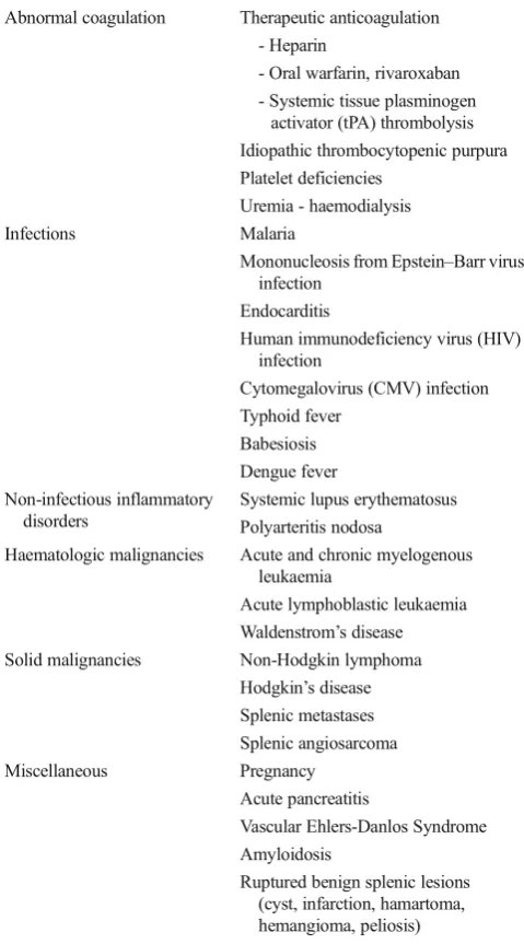
The vast majority (over 90 %) of cases are“pathologic” ASRs, which develop in a diseased spleen from the ample but specific range of disorders listed in Table1. Infections, coag-ulopathy, and neoplasms represent the three major aetiologic groups. Interestingly, a recent review of 613 cases disclosed that ASR represents the initial manifestation of the previously unknown underlying disease in over 50 % of patients [7,8]. Alternatively, “idiopathic” ASR occasionally occurs in a normal-appearing spleen without predisposing factors [4–8].
Due to its prevalence, malaria (particularly from Plasmodium vivaxinfection) represents the single major cause of ASR worldwide. Due to tourism, migrations, and drug re-sistance, malaria is increasingly encountered even outside tropical-subtropical Asia and Africa and the endemic American regions. In Western countries, malarial ASR should be suspected in non-immune returning travelers, expatriates, or recent immigrants from endemic places—even despite ap-propriate prophylaxis or during antimalarial therapy—and is associated with a non-negligible mortality (22 %) [9,10]. In the setting of suspected or proven malaria, cross-sectional im-aging with CT allows differentiation of the common splenic infarction from rupture, since the latter may require immediate or delayed splenectomy [11].
During the last eight years at our two hospitals we encoun-tered at least 12 cases of ASR, half of them secondary to anticoagulation (Fig.1). In the literature, drug-related cases account for up to one-third (9-33 %) of ASRs. In the anticoagulated population, splenic bleeding is a rare * Massimo Tonolini
1
Department of Radiology,“Luigi Sacco”University Hospital, Via G.B. Grassi 74, 20157 Milan, Italy
2 Interventional Radiology - Department of Radiology, University of
complication compared to the common haemorrhages involv-ing the anterior abdominal wall and iliopsoas muscles [12–14].
Unlike hepatic and renal hemorrhages, splenic bleed-ing is generally associated with diffusely infiltrated pa-renchyma, rather than with the presence of a solitary mass. Splenic lesions have unspecific, overlapping im-aging features. However, “pathologic” ASR from neo-plastic causes should be suggested in the presence of diffuse splenic infiltration or multifocal hypoattenuating lesions with mild or heterogeneous contrast enhance-ment, particularly in patients with known lymphomatous disease or solid tumours [8, 15].
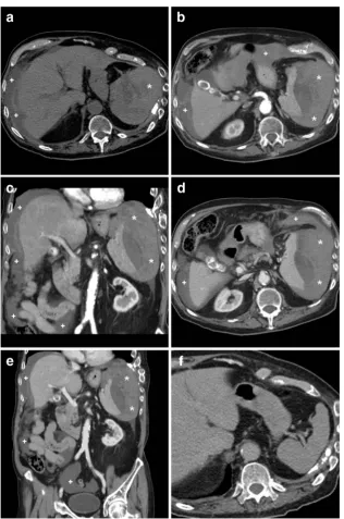
Very uncommonly (7 % of all ASRs),“idiopathic”rupture may occur in a normal spleen (Fig.2). The two hypothesised mechanisms involve: (a) intrasplenic cellular or reticuloendo-thelial hyperplasia leading to parenchymal engorgement and vascular occlusion, and (b) compression by the abdominal musculature during physiological activities such as sneezing, coughing, or defecation. Idiopathic ASR may be suggested when hemoperitoneum and high-grade splenic injury occur without CT imaging evidence of splenomegaly, focal masses, or splenic lesions. The consistent history is negative for recent trauma or surgery, known disease affecting the spleen, coag-ulopathy and anticoagulation, and signs and symptoms of sys-temic infection. The diagnosis is generally confirmed by neg-ative viral serology and normal spleen at gross inspection and histology [4,6–8].
In our opinion, general radiologists and emergency physicians should be well aware that splenic rupture with or without hemoperitoneum may occur in the ab-sence of trauma and of previously diagnosed diseases involving the spleen. ASR should be strongly suspected when acute abdominal manifestations occur in young patients with acute infections, or in the setting of haemopoietic and lympho-reticular disorders. When anticoagulation and known systemic diseases are exclud-ed, ASR is likely to be the manifesting complaint of an underlying infectious, immunologic, or neoplastic disor-der: therefore, a quick but thorough history-taking and laboratory search for infections are required [2, 3, 7].
Early imaging diagnosis of ASR is warranted to limit mortality and to provide correct triage between “ watch-ful waiting”, interventional, and surgical treatment. Ultrasound is a quick, noninvasive first-line technique to detect hemoperitoneum, which appears as complex h y p o e c h o i c e ff u s i o n w i t h r e g i o n s o f i n c r e a s e d echogenicity. However, borrowing from experience in trauma, ultrasound has moderate sensitivity (72-78 %) for the detection of splenic rupture. Furthermore, sono-graphic evaluation may be limited by large body habitus and bowel gas, has limited specificity due to the vari-able echogenicity of abscesses and hematomas, and does not provide a panoramic investigation of the entire ab-domen and pelvis [2, 3, 16, 17].
As correctly stated by Unal et al. [1], due to wide-spread availability and extreme acquisition speed, mul-tidetector CT represents the ideal imaging modality to consistently assess patients presenting to the emergency department with acute abdomen and signs of hemody-namic instability. Since physical signs are often unclear and laboratory findings do not accurately reflect the entity of bleeding, CT is warranted to investigate suspected intra-abdominal bleeding [3, 12].
Depending on the patient’s haematocrit, recent extravascu-lar blood measures at 35 to 60 Hounsfield Units (HU) of Table 1 Causes of pathologic atraumatic splenic rupture
Abnormal coagulation Therapeutic anticoagulation - Heparin
- Oral warfarin, rivaroxaban - Systemic tissue plasminogen
activator (tPA) thrombolysis Idiopathic thrombocytopenic purpura Platelet deficiencies
Uremia - haemodialysis
Infections Malaria
Mononucleosis from Epstein–Barr virus infection
Endocarditis
Human immunodeficiency virus (HIV) infection
Cytomegalovirus (CMV) infection Typhoid fever
Babesiosis Dengue fever Non-infectious inflammatory
disorders
Systemic lupus erythematosus Polyarteritis nodosa
Haematologic malignancies Acute and chronic myelogenous leukaemia
Acute lymphoblastic leukaemia Waldenstrom’s disease Solid malignancies Non-Hodgkin lymphoma
Hodgkin’s disease Splenic metastases Splenic angiosarcoma
Miscellaneous Pregnancy
Acute pancreatitis
Vascular Ehlers-Danlos Syndrome Amyloidosis
Ruptured benign splenic lesions (cyst, infarction, hamartoma, hemangioma, peliosis)
attenuation, and becomes even denser (60..80 HU) from clotting within a few hours (Figs. 1 and 2). The highest attenuation “sentinel clot” located nearest to the site of bleeding allows confident identification of the spleen as the injured organ. Furthermore, CT reliably allows detecting coexistent haemoperitoneum and ongoing bleeding, two features that are strongly associated with the risk of failed nonsurgical management. Haemoperitoneum (Figs. 1 and 2) is heralded by higher-than-water attenuation (30–45 HU) peritoneal ef-fusion, often with a mixed appearance or fluid-fluid level. Noncontained active haemorrhage (Fig. 1) appears as serpiginous or jet-like extravasation of injected con-trast medium, which follows the attenuation of blood vessels in all acquisition phases and generally pro-gresses from the arterial to the venous phase. Finally, CT allows the differentiation of ASR from other rare
non-traumatic causes of abdominal haemorrhage includ-ing ruptured liver (mostly hepatocellular adenoma or carcinoma rather than metastases) or kidney (particularly angiomyolipomas) tumours, visceral aneurysms and pseudo-aneurysms, and gynecologic conditions such as ectopic pregnancy, ruptured corpus luteum cysts, and HELLP syndrome [18–20].
Treatment guidelines for splenic injury from blunt abdominal trauma cannot be directly applied to ASR, since the latter commonly occurs in a diseased spleen and patients are generally older than those experiencing traumas. The correct therapeutic choice should consider the presence of haemodynamic instability, the amount of blood products used, the degree of haemoperitoneum, the underlying pathology, and the extent of splenic dam-age [21]. As is well known to radiologists who are familiar with polytrauma imaging, multidetector CT is Fig. 1 A 75-year-old male presented to emergency department with
generalised acute abdominal pain and physical signs of haemodynamic impairment. His past medical history included coronary stenting and endovascular treatment of aneurysmal dilatation of the thoracic aorta, respectively, 13 and 5 years earlier. He denied trauma and unusual efforts, and was on regular warfarin anticoagulation. Bedside ultrasound (not shown) revealed echogenic peritoneal effusion. Emergency multidetector CT including unenhanced (a), arterial- (b,c), and portal venous (D) post-contrast acquisitions showed a normal-sized spleen,
crucial in this setting as it detects active bleeding, and reliably measures and categorises splenic injuries
according to the American Association for the Surgery of Trauma (AAST) scale as either subcapsular and Fig. 2 An elderly 89-year-old male with chronic heart failure, previous
transurethral resection of non-muscle-invasive urinary bladder carcinoma, and lung emphysema experienced sudden hypotension and fainting. He was not on anticoagulants. Physical findings included palpation tenderness in the left hemiabdomen, tachycardia, and hypotension. Laboratory tests revealed severe blood loss: haemoglobin dropped from 9.2 to 6.4 g/dl within four hours. Platelet count, prothrombin time, and activated partial thromboplastin time were within normal range. Urgent CT including unenhanced (a), arterial- (b,c) and venous-phase (d,e) post-contrast images revealed mixed attenuation peritoneal effusion (+) consistent with haemoperitoneum. The spleen
was surrounded, medially dislocated, and compressed by massive, fresh hyperattenuating (up to 55 Hounsfield units), partly subcapsular haemorrhage (*). Pseudoaneurysms and active bleeding were not seen. The spleen showed homogeneous parenchymal enhancement without focal lesions or signs of diffuse infiltrating disease. Retrospectively, contrast-enhanced CT obtained four months earlier (F) for bladder cancer staging revealed a normal, homogeneous spleen. The patient underwent urgent splenectomy and eventually recovered. Surgical pathology revealed medium-sized spleen with reactive lymphoid hyperplasia, and excluded acute infectious or neoplastic changes. Final diagnosis was idiopathic splenic rupture
intraparenchymal hematomas, variably deep parenchy-mal and/or capsular lacerations, or devascularisation and fragmentation [22–25].
The majority (over 80 %) of reported ASR cases are treated surgically, and splenectomy remains the treat-ment of choice for patients with underlying malignan-cies. However, similarly to low-grade splenic traumas in haemodynamically stable patients, there is an increasing trend towards non-operative management for ASR as well, which achieves a high (80 %) success rate with correct patient selection. Conservative treatment includ-ing bed rest, intravenous fluids, and blood transfusions is particularly appealing in young and pediatric patients with acute infections, since preserving the spleen pre-vents long-term infectious morbidity. Strict clinical, lab-oratory, and imaging monitoring is required during non-operative management: multidetector CT consistently
allows assessment of changes in size and attenuation of haematomas over time [4, 5, 8, 16, 17]. Albeit operator-dependent and lacking panoramicity, the use of contrast-enhanced ultrasound allows younger patients to avoid irradiation from repeated CT studies [26].
Increasingly adopted to manage traumatic splenic in-juries, interventional treatment with transcatheter arterial embolisation (TAE) may also prove a valuable non-surgical option for ASR (Fig. 3), particularly in cases associated with anticoagulation, malaria, and mononu-cleosis, and when CT detects active arterial bleeding [2, 17, 27, 28]. In selected patients, TAE is useful as a temporary stabilising measure. Combined with inten-sive care, interventional radiology may allow a more rapid and safer haemostasis than surgery alone [29].
The prognosis of ASR is generally related to the underlying disease. The non-negligible fatality rate Fig. 3 A 73-year-old male on anticoagulation suffered from spontaneous
acute abdominal pain. His medical history included congestive heart failure, complete atrioventricular block treated with an implantable cardioverter-defibrillator, previous angioplasty and coronary stenting for acute myocardial infarction, and surgically treated colon carcinoma. Physical examination revealed hypotension and tender left hypocondrium. The international normalized ratio (INR) was 2.6; haemoglobin level was 6.5 g/dl. CT (a,b) revealed a large, roundish
approaches 15 % of patients. Risk factors associated with increased ASR-related mortality include spleno-megaly, advanced age, and neoplastic disorders [8].
Open AccessThis article is distributed under the terms of the Creative C o m m o n s A t t r i b u t i o n 4 . 0 I n t e r n a t i o n a l L i c e n s e ( h t t p : / / creativecommons.org/licenses/by/4.0/), which permits unrestricted use, distribution, and reproduction in any medium, provided you give appro-priate credit to the original author(s) and the source, provide a link to the Creative Commons license, and indicate if changes were made.
References
1. Unal E, Onur MR, Akpinar E et al (2016) Imaging findings of splenic emergencies: a pictorial review. Insights Imaging 7(2): 215–222
2. Alabousi A, Patlas M, Scaglione M et al (2014) Cross-Sectional Imaging of nontraumatic emergencies of the spleen. Curr Probl Diagn Radiol 42:254–267
3. Tonolini M, Bianco R (2013) Nontraumatic splenic emergencies: cross-sectional imaging findings and triage. Emerg Radiol 20:323– 332
4. Gedik E, Girgin S, Aldemir M et al (2008) Non-traumatic splenic rupture: report of seven cases and review of the literature. World J Gastroenterol 14:6711–6716
5. Kocael PC, Simsek O, Bilgin IA et al (2014) Characteristic of patients with spontaneous splenic rupture. Int Surg 99:714–718 6. Debnath D, Valerio D (2002) Atraumatic rupture of the spleen in
adults. J R Coll Surg Edinb 47:437–445
7. Aubrey-Bassler FK, Sowers N (2012) 613 cases of splenic rupture without risk factors or previously diagnosed disease: a systematic review. BME Emerg Med 12:11
8. Renzulli P, Hostettler A, Schoepfer AM et al (2009) Systematic review of atraumatic splenic rupture. Br J Surg 96:1114–1121 9. Imbert P, Rapp C, Buffet PA (2009) Pathological rupture of the
spleen in malaria: analysis of 55 cases (1958–2008). Travel Med Infect Dis 7:147–159
10. Kumar BG, Shetty MA, Chakrapani (2008) Splenic complications in malaria: a case series. Southeast Asian J Trop Med Public Health 39:791–794
11. Tonolini M, Bianco R (2012) Hemoperitoneum from splenic rup-ture in an expatriate. J Emerg Trauma Shock 5:100–102
12. Tonolini M, Ippolito S, Patella F et al (2012) Hemorrhagic compli-cations of anticoagulant therapy: role of multidetector computed tomography and spectrum of imaging findings from head to toe. Curr Probl Diagn Radiol 41:233–247
13. Pretorius ES, Fishman EK, Zinreich SJ (1997) CT of hemorrhagic complications of anticoagulant therapy. J Comput Assist Tomogr 21:44–51
14. Nazarian LN, Lev-Toaff AS, Spettell CM et al (1999) CT assess-ment of abdominal hemorrhage in coagulopathic patients: impact on clinical management. Abdom Imaging 24:246–249
15. Robertson F, Leander P, Ekberg O (2001) Radiology of the spleen. Eur Radiol 11:80–95
16. Gorg C, Colle J, Gorg K et al (2003) Spontaneous rupture of the spleen: ultrasound patterns, diagnosis and follow-up. Br J Radiol 76:704–711
17. Hadary A, Dashkovsky I, Rapaport A et al (2008) Non-traumatic rupture of spleen: can splenectomy be applied selectively? Isr Med Assoc J 10:889–891
18. Lucey BC, Varghese JC, Anderson SW et al (2007) Spontaneous hemoperitoneum: a bloody mess. Emerg Radiol 14:65–75 19. Lubner M, Menias C, Rucker C et al (2007) Blood in the belly: CT
findings of hemoperitoneum. Radiographics 27:109–125 20. Furlan A, Fakhran S, Federle MP (2009) Spontaneous abdominal
hemorrhage: causes, CT findings, and clinical implications. AJR Am J Roentgenol 193:1077–1087
21. Peitzman AB, Heil B, Rivera L et al (2000) Blunt splenic injury in adults: multi-institutional study of the Eastern Association for the surgery of trauma. J Trauma 49:177–189
22. Soto JA, Anderson SW (2012) Multidetector CT of Blunt Abdominal Trauma. Radiology 265:678–693
23. Moore EE, Cogbill TH, Jurkovich GJ et al (1995) Organ injury scaling: spleen and liver (1994 revision). J Trauma 38:323–324 24. Clark TJ, Cardoza S, Kanth N (2011) Splenic trauma: pictorial
review of contrast-enhanced CT findings. Emerg Radiol 18:227– 234
25. Marmery H, Shanmuganathan K, Alexander MT et al (2007) Optimization of selection for nonoperative management of blunt splenic injury: comparison of MDCT grading systems. AJR Am J Roentgenol 189:1421–1427
26. Miele V, Piccolo CL, Galluzzo M et al (2016) Contrast-enhanced ultrasound (CEUS) in blunt abdominal trauma. Br J Radiol 89: 20150823
27. Sekikawa Z, Takebayashi S, Kurihara H et al (2004) Factors affect-ing clinical outcome of patients who undergo transcatheter arterial embolisation in splenic injury. Br J Radiol 77:308–311
28. Popovic P, Stanislavljevic D, Jeromel M (2010) Percutaneous trans-catheter arterial embolization in haemodynamically stable patients with blunt splenic injury. Radiol Oncol 44:30–33
29. Van der Vlies CH, Hoekstra J, Ponsen KJ et al (2012) Impact of splenic artery embolization on the success rate of nonoperative management for blunt splenic injury. Cardiovasc Intervent Radiol 35:76–81