R E S E A R C H
Open Access
Role of VEGF-A in angiogenesis promoted
by umbilical cord-derived mesenchymal
stromal/stem cells: in vitro study
Irina Arutyunyan
1,2, Timur Fatkhudinov
1,3,4*, Evgeniya Kananykhina
1,2, Natalia Usman
1,3, Andrey Elchaninov
1,2,
Andrey Makarov
1,3, Galina Bolshakova
1, Dmitry Goldshtein
5and Gennady Sukhikh
1Abstract
Background:Mesenchymal stromal/stem cells derived from human umbilical cord (UC-MSCs) uniquely combine
properties of embryonic and postnatal MSCs and may be the most acceptable, safe, and effective source for allogeneic cell therapy e.g. for therapeutic angiogenesis. In this report we describe pro-angiogenic properties of UC-MSCs as manifestedin vitro.
Methods:UC-MSCs were isolated from human Wharton’s jelly by enzymatic digestion. Presence of soluble forms
of VEGF-A in UC-MSC-conditioned media was measured by ELISA. Effects of the conditioned media on human umbilical vein-derived endothelial EA.hy926 cells proliferation were measured by MTT-assay; changes in cell motility and directed migration were assessed by scratch wound healing and transwell chamber migration assays. Angiogenesis was modeledin vitroas tube formation on basement membrane matrix. Progressive differentiation of MSCs to endothelioid progeny was assessed by CD31 immunostaining.
Results:Although no detectable quantities of soluble VEGF-A were produced by UC-MSCs, the culture medium,
conditioned by the UC-MSCs, effectively stimulated proliferation, motility, and directed migration of EA.hy926 cells. In 2D culture, UC-MSCs were able to acquire CD31+endothelial cell-like phenotype when stimulated by EA.hy926-conditioned media supplemented with VEGF-A165. UC-MSCs were capable of forming unstable 2D tubular networks either by themselves or in combinations with EA.hy926 cells. Active spontaneous sprouting from cell clusters, resulting from disassembling of such networks, was observed only in the mixed cultures, not in pure UC-MSC cultures. In 3D mode of sprouting experimentation, structural support of newly formed capillary-like structures was provided by UC-MSCs that acquired the CD31+phenotype in the absence of exogenous VEGF-A.
Conclusion:These data suggest that a independent paracrine mechanism and at least partially
VEGF-A-independent differentiation mechanism are involved in the pro-angiogenic activity of UC-MSCs.
Keywords:Mesenchymal stromal cells, Multipotent, Wharton jelly, Endothelial cells, In vitro techniques, Vascular endothelial growth factor-A, Angiogenesis inducing agents, CD31 antigen, Cell migration assays, Extracellular matrix
* Correspondence:tfat@yandex.ru
1Research Center for Obstetrics, Gynecology and Perinatology of Ministry of
Healthcare of the Russian Federation, 4 Oparina Street, Moscow 117997, Russia
3Pirogov Russian National Research Medical University, Ministry of Healthcare
of the Russian Federation, 1 Ostrovitianov Street, Moscow 117997, Russia Full list of author information is available at the end of the article
Background
The concept of therapeutic angiogenesis stems from un-derstanding the importance of adequate microvascular supply for growth and regeneration of affected tissues; it refers to actions performed to facilitate revascularization of ischemic tissues. As long as the direct delivery of ex-ogenous cytokines and growth factors is ineffective, pri-marily because of their rapid elimination in vivo [1], expert opinions agree that the most promising approach for therapeutic angiogenesis is represented by stem cell therapy using multipotent mesenchymal stromal/stem cells (MSCs) because it comprises simultaneous activa-tion of multiple mechanisms (paracrine, replacement, trophic, immunomodulatory) to provide support on dif-ferent stages of formation and maturation of blood ves-sels [2–4]. Most of the research in this field is performed
on bone marrow-derived MSCs (which represent a‘gold
standard’) or adipose tissue-derived MSCs; both lineages have certain angiogenic potential and implement it in a similar manner [2, 5]. The field of therapeutic angiogen-esis is currently expanded by using MSCs from other sources, importantly from the umbilical cord and pla-centa. Perinatal stem cells share characteristics with both embryonic and adult stem cells because they may exhibit pluripotency as well as multipotent tissue maintenance, thus representing a bridge between embryonic and adult stem cells [6]. Umbilical cord-derived MSCs (UC-MSCs) have distinct biological properties: they are highly prolif-erative and enriched in transcriptionally active genes re-lated to liver or cardiovascular system development and function. Besides, UC-MSCs exhibit superior grades of plasticity and immunomodulatory activity, lack tumori-genicity, and are considered the best resource for allo-geneic transplantation [7–10].
Although therapeutic efficacy of UC-MSC transplant-ation to ischemic tissue is demonstrated in vivo [11], the understanding of how these cells implement their pro-angiogenic potential is far from complete, and there is a certain controversy among reports on this subject. The inner space of umbilical cord, net of large vessels, is oc-cupied by special connective tissue, the Wharton jelly, which is very loose and rich in gel-like ground sub-stance. Complete absence of microcirculatory vessels from Wharton jelly may indicate some anti-angiogenic properties of this microenvironment. The assumption is partially supported by recent in vitro studies. For in-stance, Kuchroo et al. [12] show that UC-MSCs do not produce detectable amounts of soluble vascular endothe-lial growth factor (VEGF)-A protein (strictly speaking, they probably do because the corresponding gene is ac-tively transcribed, but apparently this secreted VEGF-A is saturated by soluble VEGF-A-specific receptors that are also secreted by UC-MSC and act as a buffer); at the same time, the authors observe certain stimulating
influences of UC-MSCs on umbilical vein-derived endo-thelial cells in vitro and suggest the existence of some
alternative, VEGF-independent mechanism for this
stimulation. A comparative study by Amable et al. [13] describe a shifted balance of pro-angiogenic and anti-angiogenic factors in UC-MSC secretome, as compared with MSCs from other conventional sources. Ways in which UC-MSCs interact with endothelial cells and their own responses to inducers of endothelial differentiation represent an open issue. In the current study, the pro-angiogenic activity of human UC-MSCs is challenged in vitro by modeling of angiogenesis using 2D and 3D artificial matrices. Several specifically addressed prob-lems include the importance of VEGF-A, modes of UC-MSC cooperation with umbilical vein-derived endothe-lial EA.hy926 cells, and UC-MSC ability to acquire CD31+phenotypes under various stimulations.
Methods Cell cultures
The study involving human material was approved by the Ethics Committee at the Research Center for Obstet-rics, Gynecology and Perinatology. Written informed consent was obtained from all participants prior to the study.
MSCs were isolated from human umbilical cords (n= 5). The material was rinsed in phosphate-buffered saline (PBS) with 1 mg/ml cefazolin (Sintez, Kurgan, Russia) and cut into 3–4 cm pieces. After removal of blood vessels and am-nion, the Wharton jelly was chopped into smaller frag-ments with scissors. The fragfrag-ments were incubated with 200 U/ml collagenase type I (PanEco, Moscow, Russia) and 40 U/ml dispase (Invitrogen, Waltham, MA, USA) for 60 minutes at 37 °C. After the addition of fetal bovine serum (FBS; GE Healthcare, Pittsburg, PA, USA), the digested mixture was centrifuged at 1000 ×gfor 10 minutes at room temperature. Finally, the digested pieces were washed with serum-free Dulbecco’s modified Eagle’s medium (DMEM; PanEco) and cultured in growth medium (DMEM/F12 supplemented with 10 % FBS and 1 % penicillin–streptomycin (PanEco)) in a humidified in-cubator at 37 °C under a 5 % CO2atmosphere.
UC-MSCs were characterized according to the min-imal criteria to define human MSCs as proposed by the Mesenchymal and Tissue Stem Cell Committee of the International Society for Cellular Therapy [14]. For immunophenotype analysis, cells were labeled for 30
mi-nutes at room temperature using the BD Stemflow™
hMSC Analysis Kit (BD Biosciences, Pharmingen, San Diego, CA, USA). After being fixed with 4 % paraformal-dehyde (SERVA Electrophoresis, Heidelberg, Germany), the cells were analyzed on a FACScalibur using CellQuest software (BD Biosciences). The StemPro® Adipogenesis
Differentiation Kit, and the StemPro® Chondrogenesis Differentiation Kit (Gibco, Life Technologies, Carlsbad, CA, USA) were used to demonstrate the differentiation capacity of UC-MSCs in accordance with the manufac-turer’s instructions.
Human endothelial EA.hy926 cells were derived from the American Type Culture Collection (Manassas, VA, USA). Established in 1983 by fusing primary human umbilical vein endothelial cells (HUVEC) with a thioguanine-resistant clone of the human lung adenocarcinoma cell line A549/8, EA.hy926 cells represent a widely-used endothelial cell line expressing endothelin-1, Weibel-Palade bodies, prostacyc-lin, factor VIII-related antigen, and endothelial adhesion molecules ICAM-1 and VCAM-1 [15]. This line was chosen for its highly specific functions that are characteris-tic of the human vascular endothelium combined with ad-vantages of immortality, stability through passage number, and high reproducibility of the properties [16, 17].
Immunofluorescence
Cells were fixed with 4 % paraformaldehyde (SERVA Electrophoresis) for 10 minutes at room temperature. After two washes with PBS, the cells were blocked for 5 minutes with Protein Block (Abcam, Cambridge, MA, USA) at room temperature and then incubated over-night at 4 °C with antibodies against CD31 (ab24590; Abcam). After washing with PBS, the cells were incu-bated with fluorescein isothiocyanate (FITC)-conjugated antimouse IgG (ab6810; Abcam) for 1 hour in the dark. Cell nuclei were stained with 4′,6-diamidino-2-phenylin-dole (DAPI; Sigma-Aldrich, St. Louis, MO, USA). The cells were observed under the Leica DM 4000 B fluorescent microscope (Leica Microsystems, Heidelberg, Germany).
Preparation of conditioned media
At 100 % confluence, the cells (UC-MSCs or EA.hy926) were washed with serum-free DMEM, and the media were replaced with fresh growth media. After 24, 48, or 72 hours, the media were collected and centrifuged at 2800 ×g for 5 minutes, filtered through a 0.22μm filter (GE Osmonics Labstore, Minnetonka, MN, USA), and were then stored at–70 °C until VEGF-A quantification. The media conditioned by UC-MSCs or EA.hy926 cells for 72 hours were used in subsequent experiments.
VEGF-A quantification
Media conditioned by EA.hy926 cells or UC-MSCs were collected after 24, 48, or 72 hours. VEGF-А-121 and VEGF-A-165 were quantified using a commercial enzyme-linked immunosorbent assay kit (#8784; Vector-Best, Novosibirsk, Russia) in accordance with the instructions of the manufacturer. Data analysis was performed using the online application (http://elisaanalysis.com/app).
Endothelial cell proliferation assay
EA.hy926 cells were seeded in a 96-well plate (3 × 10³cells in 200μl of growth media per well). After 1, 2, or 3 days the media were replaced with UC-MSC-conditioned media, UC-MSC-conditioned media supplemented with 200 ng/ml anti-VEGF antibody (ab9570; Abcam), or fresh growth media (control wells). At day 4 the cell proliferation was measured by 3-(4,5-dimethylthiazol-2-yl)-2,5-diphenyltetrazolium bromide (MTT) assay. MTT (Sigma-Aldrich) stock solution was added to each well (to a final MTT concentration of 1.5 mg/ml). The plate was returned to a cell culture incubator for 2 hours. When the purple precipitate was clearly visible
under the microscope, 100 μl of dimethyl sulfoxide
(DMSO; Sigma-Aldrich) were added. After 15 minutes, the absorbance in each well was measured at 570 nm in a Multiskan GO microplate spectrophotometer (Thermo Fisher Scientific, Waltham, MA, USA). The reference wavelength was 650 nm.
Endothelial cell transwell migration assay
The migration of EA.hy926 cells to UC-MSC-released chemoattractants was measured by transwell chamber migration assay. UC-MSCs were seeded in a 24-well plate (105 cells in 600 μl of growth media per well). One-half of the wells with the cells contained anti-VEGF antibody (ab9570; Abcam) in 200 ng/ml final concentra-tion. The same volume of growth media without cells was added to control wells. After 24 hours, inserts with a polycarbonate membrane (pore size of 8 μm) (#35224; SPL Life Sciences, Pochun, South Korea) were installed in the plate. EA.hy926 cells were seeded in the upper chambers (105cells in 250μl of growth media). After 24, 48, or 72 hours, nonmigrating cells in the upper cham-ber were attentively removed with cotton swabs, and cells on the lower surface of the membrane were fixed with 4 % paraformaldehyde (SERVA Electrophoresis) and stained with DAPI (Sigma-Aldrich). The total num-bers of migrated cells were then counted in eight ran-domly selected fields for each insert (magnification × 100) using LAS AF v.3.1.0 build 8587 (Leica Microsystems).
Endothelial cell scratch healing assay
EA.hy926 cells were seeded in a 96-well plate (3 × 10⁴cells in 100μl of growth media per well). After 24 hours, each confluent cell monolayer was scratched with a
Wound-Maker™ tool (Essen Bioscience, Ann Arbor, MI, USA),
(Essen BioScience). Wound confluence was measured using an automated Cell Migration software module (Essen BioScience).
In vitro tube formation assay
In this experiment we used BD Matrigel™ Basement
Membrane Matrix Phenol Red Free (#356237; BD Bio-sciences). This matrix is highly enriched in laminin-1, collagen IV, heparan sulfate, proteoglycan, entactin/nido-gen, and various growth factors. Although it does not contain all of the signature components of an endothe-lial basement membrane, Matrigel promotes tube forma-tion in vitro for all endothelial cells tested to date [18].
Prior to the experiment, UC-MSCs were labeled with PKH26 (yellow–orange fluorescent dye) and EA.hy926 cells were labeled with PKH67 (green fluorescent dye) (Sigma-Aldrich) according to the manufacturer’s instruc-tions. A total of 150 μl of chilled Matrigel was added to a 48-well plate and incubated at 37 °C for 30 minutes. UC-MSCs, or EA.hy926 cells, or 1:1 mixed
UC-MSC-EA.hy926 cells (total 35 × 103 cells per well) were
suspended in 500μl of growth media and were added to the solidified Matrigel. Additionally, EA.hy926 cells
sus-pended in 500μl of MSC-conditioned media or
UC-MSC-conditioned media supplemented with 200 ng/ml of anti-VEGF antibody (ab9570; Abcam) were used. After in-cubation on Matrigel at 37 °C in a 5 % CO2chamber,
mor-phological changes were observed under an Axiovert 40 CFL inverted microscope (Carl Zeiss, Jena, Germany). Six representative fields for each well were photographed. Images were analyzed using AxioVs40 4.8.2.0 (Zeiss, Oberkochen, Germany) to determine the length of the tubes and the number of branch points (magnification × 50).
Endothelial differentiation of UC-MSCs in monolayer
After UC-MSCs formed the confluent monolayer, the growth media were replaced with the induction media. Three endothelial induction media were used for UC-MSC culture: EA.hy926-conditioned media mixed 1:1 with growth media; EA.hy926-conditioned media mixed 1:1 with growth media supplemented with VEGF-A-165 (#583702; BioLegend, San Diego, CA, USA), 50 ng/ml; and growth media supplemented with VEGF-A-165. Contents of FCS in the control and differentiation media were reduced to 5 % to avoid excessive cell growth. The media were replaced twice a week. At day 21, UC-MSCs were fixed with 4 % paraformaldehyde (SERVA Electrophoresis) and stained with CD31 anti-bodies as already described.
Endothelial differentiation of UC-MSCs in Matrigel
Whole mount immunostaining of the 3D structure, formed by the cells in Matrigel, proved impossible due to nonspecific absorption of the antibodies by the matrix.
For this reason, the analysis was performed on cryosec-tions of secondary sprouting networks. Structures formed in Matrigel were embedded in Tissue-Tek® OCT Com-pound (Sakura Finetek, Torrance, CA, USA) and cut into
5–7 μm sections using a cryostat. The sections were
stained with CD31 antibodies as already described.
Statistical analysis
Data are expressed as mean ± standard deviation (SD).
Student’s t test was used for pairwise comparisons
between groups of normally distributed values, whereas the Mann–Whitney test was applied for distributions differing from normal. Multiple comparisons were done by either one-way analysis of variance (ANOVA) or ANOVA on ranks (for the cases of unconfirmed normal-ity);p<0.05 was considered statistically significant.
Results
Characterization of cell cultures
The cells isolated from Wharton jelly of the human um-bilical cord were plastic adherent with a spindle-shaped, fibroblast-like morphology (Fig. 1b). Flow cytometry analysis showed that these cells were positive for the MSC markers CD105, CD73, and CD90 and were nega-tive for CD11b, CD19, CD45, CD34, and HLA-DR (Fig. 1a).
UC-MSCs demonstrated multipotent differentiation potential. Sudan III staining of neutral lipid vacuoles showed that UC-MSCs could differentiate into adipo-cytes. Alizarin Red S staining of calcium compound crystals showed that UC-MSCs could differentiate into osteoblasts. Positive staining of mucopolysaccharides by Alcian blue indicated that UC-MSCs could differentiate into chondrocytes (Fig. 1b).
EA.hy926 cells formed a monolayer of closely apposed small polygonal cells. EA.hy926 cells were found to be positive for CD31 as endothelial marker (Fig. 1c).
VEGF-A-121 and VEGF-A-165 quantification in the conditioned media
Within 3 days, the concentration of soluble forms of VEGF-A (VEGF-A-121 and VEGF-A-165) in EA.hy926-conditioned media gradually increased from 57.3 ± 7.7 pg/ ml to 229.0 ± 24.9 pg/ml, whereas in UC-MSC-conditioned media the contents of VEGF-A did not change, remaining at the level of the growth medium (Fig. 1d).
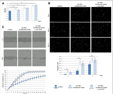
The influence of UC-MSC-conditioned media on endothelial cell proliferation
two media increased, and 72 hours of incubation resulted in almost 1.6-fold excess absorption in wells with UC-MSC-conditioned media compared with growth media. The addition of the VEGF-neutralizing antibody to the UC-MSC-conditioned media did not significantly attenu-ate the EA.hy926 cell proliferation as compared with the
UC-MSC-conditioned media treatment (p < 0.05)
(Fig. 2a).
The influence of UC-MSC-conditioned media on endothelial cell migration
Endothelial cell migration was evaluated using a trans-well chamber migration assay. EA.hy926 cells migrated from the upper chamber to the lower surface of mem-brane through 8μm pores when the lower chamber con-tained only the growth culture medium. When the lower chamber was seeded with UC-MSCs, the efficacy of EA.hy926 cell-directed migration increased significantly
at all time points (p <0.05). The addition of the VEGF-neutralizing antibody to the lower chambers seeded with UC-MSCs did not significantly attenuate the EA.hy926 cell migration as compared with UC-MSC-conditioned media treatment (p<0.05) (Fig. 2b).
with representative images from the time-lapse record-ing, is given in Fig. 2c.
Tube formation assay
The angiogenic capability of various cell types was assessed using an in vitro capillary-like structure (tube) formation assay on basement membrane matrix. As shown in Fig. 3a, both UC-MSCs and endothelial EA.hy926 cells were able
tubes, while the UC-MSCs and UC-MSC-EA.hy926 cell mix formed coarse meshes with fewer branch points and longer tubes. The addition of UC-MSC-conditioned media (regardless of the presence of the VEGF-neutralizing anti-body) to the wells with EA.hy926 cells did not significantly alter the parameters of the networks, but contributed to their formation 1 hour earlier (Fig. 3a).
Surprisingly, we found that PKH26-labeled UC-MSCs became the basis of a mixed culture network, while PKH67-labeled EA.hy926 cells were only associated with it (Fig. 3b).
In all groups, the networks were unstable and disinte-grated into tight clusters for 24 hours. These clusters were not stationary structures. In a few days, they were capable of limited movement and fusion. The movement of cell clusters stopped in about 5–7 days, and the num-ber of clusters was different between the groups: 430.0 ± 21.2 per well for EA.hy926 cells, 137.1 ± 9.2 per well for MSC-EA.hy926 cell mix, and 93.0 ± 9.2 for UC-MSCs (Fig. 3c).
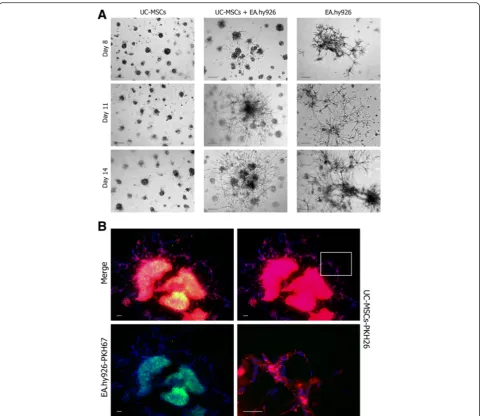
Further, we observed that the clusters formed by the EA.hy926 cells and UC-MSC-EA.hy926 cell mix, but not by UC-MSCs alone, became centers of secondary sprouting. Sprouting cells had a typical elongated shape. Gradually, the isolated sprouting centers joined into a single, very stable (follow-up of more than 30 days) 3D network with a plurality of branch points, often dichotomic (Fig. 4a). Moreover, in this mixed cul-ture network only PKH26-labeled UC-MSCs formed sprouts while PKH67-labeled EA.hy926 cells stayed in the centers of the clusters (Fig. 4b).
Endothelial differentiation of UC-MSCs in monolayer
UC-MSCs cultured in complete fresh growth medium exhibited both the shape and the wave-like arrangement of the MSCs in the monolayer; the cell growth was restricted by mutual contact inhibition, and neither of them differentiated to the CD31+phenotype.
When cultured in EA.hy926-conditioned medium, the cells retained the same characteristics but grew to higher densities. When cultured in EA.hy926-condi-tioned medium supplemented with VEGF-A-165, they formed distinct tubular structures (three to five per
35 mm dish), assembled from several dozens of narrow stretched cells with elongated nuclei, positively stained with CD31 antibody.
Finally, using VEGF-A-165 as a single growth supple-ment (except for the serum), added to complete fresh medium, led to a mosaic loss of contact inhibition. The cells started to grow in multiple layers, but formed no tubular structures and stayed CD31–(Fig. 5a).
Endothelial differentiation of UC-MSCs in Matrigel
Immunostaining of cryosections showed that, upon coculturing with EA.hy926 in Matrigel, the UC-MSCs started to express CD31 spontaneously, without add-itional VEGF-A-165 supplement (Fig. 5b).
Discussion
Weak secretion of VEGF-A by UC-MSCs derived from Wharton jelly may be related to the unusual structure of loose connective tissue forming Wharton jelly, and, in particular, the lack of blood capillaries in it. Although early events of hematopoiesis and capillary formation in this tissue are described in detail, by 7–9 weeks of devel-opment the hematopoiesis in Wharton jelly ceases, and the capillaries undergo regression [19]. It is plausible that these changes, as well as subsequent maintenance of the anti-angiogenic environment, are accompanied, or mediated, by low concentrations of soluble VEGF-A in the intercellular spaces. According to some authors, the Wharton jelly-derived UC-MSCs are able to secrete sol-uble forms of VEGF-A [20]; however, the majority of the reports (including this one) mention the almost complete absence of VEGF-A protein from the UC-MSC-conditioned culture medium as a specific feature reflecting VEGF-A deficiency of UC-MSC secretome. Typical levels of VEGF-A secretion reported for UC-MSCs are 102less than for adipose tissue-derived MSCs and 103less than for bone marrow-derived MSCs, despite detectable levels of transcription of the corresponding gene [12, 13]. Nevertheless, UC-MSCs can effectively accelerate migration and promote tube formation from endothelial cells in vitro—the effect is mediated by the UC-MSC-conditioned media; that is, by in vitro modeling of in vivo paracrine mechanisms [12].
(See figure on previous page.)
UC-MSCs implement their pro-angiogenic potential via some VEGF-A-independent mechanism. Why should this matter? The problem is that intermediate results of clinical trials using VEGF-A-121 or VEGF-A-165 (as active exogenous proteins or in the form of genetic constructs) have been qualified as rather contradictory: the effects sometimes deviate from those expected [1, 21]. This may be explained by duality that originates from the level of VEGF-A binding to its receptor, vascu-lar endothelial growth factor receptor (VEGFR). By acting via VEGFR2, VEGF-A increases survival and pro-liferation of endothelial cells, as well as recruitment of other progenitors to the site of injury, thus supporting formation and maturation of new blood vessels; in
predictable results when supported by some additional VEGF-A-independent line of intervention (e.g., using UC-MSCs).
Published evidence for the stimulating influence of MSCs on endothelial cell proliferation is rather contro-versial because of the variety of sources and methods of obtaining the cells [2]. For example, it is shown that bone marrow-derived MSCs (including cells cultured under hypoxic conditions) have no effect on EA.hy926 cell growth [25]. In our experiments, the UC-MSC-conditioned media stimulated proliferation of EA.hy926 cells; this is consistent with results reported by Choi et al. [26] for a different endothelial line (HUVEC). The absence of VEGF-A from UC-MSC-conditioned media suggests that the endothelial cells respond to a different sort of inducer (possibly VEGF-B, the positive effect of which on EA.hy926 cell proliferation was confirmed in a
recent study [27], but it is still questionable whether VEGF-B is produced by MSCs).
The transwell systems are widely used to assess chemotaxis, which plays an important role during the early stages of angiogenesis. In our setting, UC-MSCs secreted chemoattractants for EA.hy926 cells. Similar results have been reported previously for other cell
lines – HUVEC, human microvascular endothelial cell
line HMEC1, and mouse neural crest-derived cell line N2a, the effect of UC-MSCs being more pronounced compared with bone marrow-derived MSCs [26, 28, 29].
in vitro [30]; exactly these processes play a central role in angiogenesis [31].
UC-MSCs thus secrete factors that may attract endo-thelial and progenitor cells, while stimulating their mo-bility; what kind of factors in particular could be partly deduced from the literature. Interleukin (IL)-8 is shown to induce cytoskeleton rearrangement and directed mi-gration of EA.hy926 cells by activation ofр38 mitogen-activated protein kinase signaling [32]. The rate of migration of endothelial cells in vitro is also shown to depend on hepatocyte growth factor (HGF) and monocyte chemoattractant protein-1 (MCP-1) levels in UC-MSC-conditioned media [28]. These findings are consistent with other research showing that secretion of IL-8, HGF, and MCP-1 by UC-MSCs is significantly more intensive than by MSCs derived from bone marrow or adipose tissue [13, 20].
Published data on the possibilities of endothelial differ-entiation of MSCs themselves are rather contradictory. There exist several protocols that differ in the compos-ition of inducers (most widely used is VEGF-А-165 at 50 ng/ml), duration of the process of differentiation (takes from 2 to 28 days), and selection of molecular markers for the control immunostaining (CD31, von Willebrand factor (vWF), vascular endothelial cadherin (VE-cadherin), and VEGFR2 are the most common); ac-cordingly, the final products of these protocols vary greatly [33]. At the same time, designating the differenti-ated MSCs as fully mature and functional endothelial cells is considered inaccurate; it is therefore more cor-rect to define these cells as endothelial-like cells [2].
In our experiments, the UC-MSCs were capable of dif-ferentiation to the CD31+phenotype under the influence of differentiation medium containing VEGF-A-165 as an essential, although insufficient, inducer. In contrast, Choi et al. [26] observe no expression of endothelial markers by UC-MSCs after treatment with complex dif-ferentiation media containing epidermal growth factor (EGF), VEGF, basic fibroblast growth factor, insulin-like growth factor-1, hydrocortisone, and some other poten-tial inducers. In yet another study, UC-MSCs treated with media containing VEGF, EGF, and hydrocortisone started to express endothelial markers (vWF, VE-cadherin, and VEGFR2) uniformly, without any changes in cell organization or cell morphology [34]. Possibil-ities of endothelial differentiation of MSCs in vivo are even more questionable [2]. One of the reasons for this is the low level of VEGF-A in ischemic tissues: it is about 103 times lower than in standard endothelial differentiation media (50 ng/ml) [35], and roughly corresponds to the VEGF-A level in the EA.hy926-conditioned media (Fig. 1d). The networks formed in coculture of UC-MSCs with EA.hy926 cells on Matrigel were similar to the net-works formed by pure UC-MSCs (judging by their
assembly time, length of the tubes, and branching point number). The core of the mixed networks was com-posed of the PKH26-labeled MSCs, while the PKH67-labeled EA.hy926 cells were associated with the outer surface of this core (Fig 3a). Such an arrangement of cell types in mixed networks differs from that reported previously: other authors attribute only a minor role to MSCs [26, 36, 37]. The inconsistency probably relates to a different proportion of cell types taken for the net-work priming.
All tubular networks observed in our setting were un-stable. Independently of whether they were formed by UC-MSCs combined with EA.hy926 cells or either of the cell types on their own, the networks underwent spontaneous disassembling in the course of 24 hours, producing tight cell clusters; this is consistent with pre-viously published data [26, 36, 38]. These clusters, resulting from 2D network disassembly, subsequently turned into sprouting centers producing a single stable 3D network. Similar results are reported by Portalska et al. [38], who observed in vitro assembly of blood vessel-like structures from bone marrow-derived MSCs predifferentiated to an endothelial-like phenotype: the cells started to form a network with a 20-hour delay (as compared with the native undifferentiated MSCs), and this network remained stable for at least 7 days. In pure cultures of endothelial cells, as well in two of the five mixed cultures, the sprouting occurred in-variably, but all five pure UC-MSC cultures showed no signs of the sprouting. This confirms the idea that individual MSC cultures, equally complying with the standards, may show morphological and functional variation [39].
Overall, the results indicate that the ability of UC-MSCs to participate in sprouting, manifested in cocul-tures with EA.hy926 cells on Matrigel, is a consequence of their differentiation to an endothelial-like phenotype (especially given that the signals from the local environ-ment, either through cell–cell contact, soluble factors, or cell–matrix interactions, profoundly influence MSC endothelial differentiation [37]). We also assume that the low reproducibility of sprouting indexes between individual cultures of UC-MSCs is caused mainly by unequal susceptibility of these cultures to specific endothelial differentiation stimuli. Notably, acquisition of the CD31+
Conclusions
Many of the studies investigating the paracrine factors secreted from MSCs derived from various sources have reported the presence of VEGF-A and have implicated its importance in angiogenesis. In this study, we con-firmed that MSCs derived from Wharton jelly of the human umbilical cord produced no detectable quantities of soluble VEGF-A (VEGF-A-121 and VEGF-A-165); despite this, culture medium conditioned by UC-MSCs effectively stimulated the proliferation, motility, and directed migration of endothelial EA.hy926 cells. These data suggest that a VEGF-A-independent paracrine mechanism is involved in the pro-angiogenic activity of UC-MSCs.
In our experiments, the UC-MSCs were capable of dif-ferentiation to an endothelial cell-like CD31+ phenotype under the influence of differentiation medium contain-ing VEGF-A-165 as an essential, although insufficient, inducer. However, we found that the ability of UC-MSCs to participate in secondary sprouting, as manifested in long-term cocultures with EA.hy926 cells on Matrigel, is a consequence of their differentiation to an
endothelial-like CD31+ phenotype in the absence of exogenous
VEGF-A. We can assume that signals from the local environment, either through UC-MSC–EA.hy926 cell contact or UC-MSC–basement membrane matrix inter-actions, profoundly influenced UC-MSC differentiation. Endothelial differentiation as one of the proposed mech-anisms of action for UC-MSC transplantation can thus also be partially VEGF-A independent.
The conclusions of this study have practical applications in the field of pro-angiogenic therapy: VEGF-A-based therapy supported by an additional VEGF-A-independent line of intervention (e.g., using UC-MSC transplantation) may have higher efficacy.
Abbreviations
ANOVA:Analysis of variance; DAPI: 4′,6-Diamidino-2-phenylindole; DMEM: Dulbecco’s modified Eagle’s medium; DMSO: Dimethyl sulfoxide; EGF: Epidermal growth factor; FBS: Fetal bovine serum; FITC: Fluorescein isothiocyanate; HGF: Hepatocyte growth factor; HUVEC: Human umbilical vein endothelial cells; IL: Interleukin; MCP-1: Monocyte chemoattractant protein-1; MSC: Mesenchymal stromal/stem cell; MTT: 3-(4,5-Dimethylthiazol-2-yl)-2,5-diphenyltetrazolium bromide; PBS: Phosphate-buffered saline; SD: Standard deviation; UC-MSC: Umbilical cord-derived mesenchymal stromal/stem cell; VE-cadherin: Vascular endothelial cadherin; VEGF: Vascular endothelial growth factor; VEGFR: Vascular endothelial growth factor receptor; vWF: Von Willebrand factor.
Competing interests
The authors declare that they have no competing interests.
Authors’contributions
IA and TF designed the study, coordinated the research, wrote most of the text, and composed the figures. IA, EK, AE, NU, and AM performed the experiments, and collected and analyzed the data. AE performed the statistical analysis. NU, GB, and DG participated in the manuscript writing. GS conceived of the study. All authors have been involved in drafting and consequent critical revision of all versions of the manuscript and the figures for
important intellectual content and way of presentation. All authors read and approved the manuscript.
Acknowledgements
The authors thank Irina Teveleva for careful proofreading of the text. This work was supported by Ministry of Education and Science of the Russian Federation (unique identification code: RFMEFI61314X0008).
Author details
1
Research Center for Obstetrics, Gynecology and Perinatology of Ministry of Healthcare of the Russian Federation, 4 Oparina Street, Moscow 117997, Russia.2Scientific Research Institute of Human Morphology, 3 Tsurupa Street, Moscow 117418, Russia.3Pirogov Russian National Research Medical
University, Ministry of Healthcare of the Russian Federation, 1 Ostrovitianov Street, Moscow 117997, Russia.4Laboratory of Regenerative Medicine,
Research Center for Obstetrics, Gynecology and Perinatology, 4 Oparin Street, Moscow 117997, Russia.5Research Center of Medical Genetics, 1
Moskvorechie Street, Moscow 115478, Russia.
Received: 10 November 2015 Revised: 25 February 2016 Accepted: 3 March 2016
References
1. Ylä-Herttuala S, Rissanen TT, Vajanto I, Hartikainen J. Vascular endothelial growth factors: biology and current status of clinical applications in cardiovascular medicine. J Am Coll Cardiol. 2007;49(10):1015–26. 2. Bronckaers A, Hilkens P, Martens W, Gervois P, Ratajczak J, Struys T, et al.
Mesenchymal stem/stromal cells as a pharmacological and therapeutic approach to accelerate angiogenesis. Pharmacol Ther. 2014;143(2):181–96. 3. Liew A, O’Brien T. Therapeutic potential for mesenchymal stem cell
transplantation in critical limb ischemia. Stem Cell Res Ther. 2012;3(4):28. 4. Yan J, Tie G, Xu TY, Cecchini K, Messina LM. Mesenchymal stem cells as a
treatment for peripheral arterial disease: current status and potential impact of type II diabetes on their therapeutic efficacy. Stem Cell Rev. 2013;9(3):360–72. 5. Lin RZ, Moreno-Luna R, Zhou B, Pu WT, Melero-Martin JM. Equal modulation
of endothelial cell function by four distinct tissue-specific mesenchymal stem cells. Angiogenesis. 2012;15(3):443–55.
6. Taghizadeh RR, Cetrulo KJ, Cetrulo CL. Wharton’s Jelly stem cells: future clinical applications. Placenta. 2011;32 Suppl 4:S311–5.
7. De Kock J, Najar M, Bolleyn J, Al Battah F, Rodrigues RM, Buyl K, et al. Mesoderm-derived stem cells: the link between the transcriptome and their differentiation potential. Stem Cells Dev. 2012;21(18):3309–23.
8. Gauthaman K, Fong CY, Suganya CA, et al. Extra-embryonic human Wharton’s jelly stem cells do not induce tumorigenesis, unlike human embryonic stem cells. Reprod Biomed Online. 2012;24(2):235–46. 9. Li X, Bai J, Ji X, Li R, Xuan Y, Wang Y. Comprehensive characterization of
four different populations of human mesenchymal stem cells as regards their immune properties, proliferation and differentiation. Int J Mol Med. 2014;34(3):695–704.
10. Lv F, Lu M, Cheung KM, Leung VY, Zhou G. Intrinsic properties of mesemchymal stem cells from human bone marrow, umbilical cord and umbilical cord blood comparing the different sources of MSC. Curr Stem Cell Res Ther. 2012;7(6):389–99.
11. Santos Nascimento D, Mosqueira D, Sousa LM, et al. Human umbilical cord tissue-derived mesenchymal stromal cells attenuate remodeling after myocardial infarction by proangiogenic, antiapoptotic, and endogenous cell-activation mechanisms. Stem Cell Res Ther. 2014;5(1):5.
12. Kuchroo P, Dave V, Vijayan A, Viswanathan C, Ghosh D. Paracrine factors secreted by umbilical cord-derived mesenchymal stem cells induce angiogenesis in vitro by a VEGF-independent pathway. Stem Cells Dev. 2015;24(4):437–50. 13. Amable PR, Teixeira MV, Carias RB, Granjeiro JM, Borojevic R. Protein
synthesis and secretion in human mesenchymal cells derived from bone marrow, adipose tissue and Wharton’s jelly. Stem Cell Res Ther. 2014;5(2):53. 14. Dominici M, Le Blanc K, Mueller I, Slaper-Cortenbach I, Marini F, Krause D,
et al. Minimal criteria for defining multipotent mesenchymal stromal cells. The International Society for Cellular Therapy position statement. Cytotherapy. 2006;8(4):315.
16. Aranda E, Owen GI. A semi-quantitative assay to screen for angiogenic compounds and compounds with angiogenic potential using the EA.hy926 endothelial cell line. Biol Res. 2009;42(3):377–89.
17. Bouïs D, Hospers GA, Meijer C, Molema G, Mulder NH. Endothelium in vitro: a review of human vascular endothelial cell lines for blood vessel-related research. Angiogenesis. 2001;4(2):91–102.
18. Arnaoutova I, George J, Kleinman HK, Benton G. The endothelial cell tube formation assay on basement membrane turns 20: state of the science and the art. Angiogenesis. 2009;12(3):267–74.
19. Robaczyński J. Development of capillaries in Wharton’s jelly of the human umbilical cord in the course of ontogenesis. Folia Morphol (Warsz). 1967; 26(4):371–82.
20. Edwards SS, Zavala G, Prieto CP, Elliott M, Martínez S, Egaña JT, et al. Functional analysis reveals angiogenic potential of human mesenchymal stem cells from Wharton’s jelly in dermal regeneration. Angiogenesis. 2014;17(4):851–66. 21. Shimamura M, Nakagami H, Taniyama Y, Morishita R. Gene therapy for
peripheral arterial disease. Expert Opin Biol Ther. 2014;14(8):1175–84. 22. Imoukhuede PI, Popel AS. Quantification and cell-to-cell variation of vascular
endothelial growth factor receptors. Exp Cell Res. 2011;317(7):955–65. 23. Imoukhuede PI, Dokun AO, Annex BH, et al. Endothelial cell-by-cell profiling
reveals temporal dynamics of VEGFR1 and VEGFR2 membrane-localization following murine hindlimb ischemia. Am J Physiol Heart Circ Physiol. 2013; 304(8):H1085–93.
24. Saito T, Takeda N, Amiya E, Nakao T, Abe H, Semba H, et al. VEGF-A induces its negative regulator, soluble form of VEGFR-1, by modulating its alternative splicing. FEBS Lett. 2013;587(14):2179–85.
25. Burlacu A, Grigorescu G, Rosca AM, Preda MB, Simionescu M. Factors secreted by mesenchymal stem cells and endothelial progenitor cells have complementary effects on angiogenesis in vitro. Stem Cells Dev. 2013;22(4):643–53.
26. Choi M, Lee HS, Naidansaren P, Kim HK, O E, Cha JH, et al. Proangiogenic features of Wharton's jelly-derived mesenchymal stromal/stem cells and their ability to form functional vessels. Int J Biochem Cell Biol. 2013;45(3):560–70. 27. Zhang GH, Qin R, Zhang SH, Zhu H. Effects of vascular endothelial growth
factor B on proliferation and migration in EA.Hy926 cells. Mol Biol Rep. 2014; 41(2):779–85.
28. Shen C, Lie P, Miao T, Yu M, Lu Q, Feng T, et al. Conditioned medium from umbilical cord mesenchymal stem cells induces migration and
angiogenesis. Mol Med Rep. 2015;12(1):20–30.
29. Hsieh JY, Wang HW, Chang SJ, Liao KH, Lee IH, Lin WS, et al. Mesenchymal stem cells from human umbilical cord express preferentially secreted factors related to neuroprotection, neurogenesis, and angiogenesis. PLoS One. 2013;8(8):e72604. 30. Rodriguez LG, Wu X, Guan JL. Wound-healing assay. Methods Mol Biol.
2005;294:23–9.
31. Isner JM, Vale P, Symes J, Losordo DW, Asahara T. Angiogenesis and cardiovascular disease. Dialog Cardiovasc Med. 2001;6(3):145–72. 32. Lai Y, Liu XH, Zeng Y, Zhang Y, Shen Y, Liu Y. Interleukin-8 induces the
endothelial cell migration through the Rac 1/RhoA-p38MAPK pathway. Eur Rev Med Pharmacol Sci. 2012;16(5):630–8.
33. Vater C, Kasten P, Stiehler M. Culture media for the differentiation of mesenchymal stromal cells. Acta Biomater. 2011;7(2):463–77.
34. Chen MY, Lie PC, Li ZL, Wei X. Endothelial differentiation of Wharton’s jelly-derived mesenchymal stem cells in comparison with bone marrow-jelly-derived mesenchymal stem cells. Exp Hematol. 2009;37(5):629–40.
35. Jiang Q, Ding S, Wu J, Liu X, Wu Z. Norepinephrine stimulates mobilization of endothelial progenitor cells after limb ischemia. PLoS One. 2014;9(7):e101774. 36. Blocki A, Wang Y, Koch M, Peh P, Beyer S, Law P, et al. Not all MSCs can act as
pericytes: functional in vitro assays to distinguish pericytes from other mesenchymal stem cells in angiogenesis. Stem Cells Dev. 2013;22(17):2347–55. 37. Lozito TP, Kuo CK, Taboas JM, Tuan RS. Human mesenchymal stem cells
express vascular cell phenotypes upon interaction with endothelial cell matrix. J Cell Biochem. 2009;107(4):714–22.
38. Portalska K, Leferink A, Groen N, Fernandes H, Moroni L, van Blitterswijk C, et al. Endothelial differentiation of mesenchymal stromal cells. PLoS One. 2012;7(10):e46842.
39. Pacini S, Petrini I. Are MSCs angiogenic cells? New insights on human nestin-positive bone marrow-derived multipotent cells Front. Cell Dev Biol. 2014;2:20.
40. Ball SG, Shuttleworth CA, Kielty CM. Vascular endothelial growth factor can signal through platelet-derived growth factor receptors. J Cell Biol. 2007; 177(3):489–500.
• We accept pre-submission inquiries
• Our selector tool helps you to find the most relevant journal
• We provide round the clock customer support
• Convenient online submission
• Thorough peer review
• Inclusion in PubMed and all major indexing services
• Maximum visibility for your research
Submit your manuscript at www.biomedcentral.com/submit