C R I T I C A L R E V I E W
Open Access
Imaging of non-neuronopathic Gaucher
disease: recent advances in quantitative
imaging and comprehensive assessment of
disease involvement
Andrew J. Degnan
1,2*, Victor M. Ho-Fung
1,2, Rebecca C. Ahrens-Nicklas
3,4, Christian A. Barrera
1, Suraj D. Serai
1,
Dah-Jyuu Wang
1and Can Ficicioglu
3,4Abstract
Gaucher disease is an inherited metabolic disorder resulting in deficiency of lysosomal enzyme β -glucocerebrosidase causing the accumulation of abnormal macrophages (“Gaucher cells”) within multiple organs, most conspicuously affecting the liver, spleen, and bone marrow. As the most common glycolipid metabolism disorder, it is important for radiologists encountering these patients to be familiar with advances in imaging of organ and bone marrow involvement and understand the role of imaging in clinical decision-making. The recent advent of commercially available, reliable, and reproducible quantitative MRI acquisitions to measure fat fractions prompts revisiting the role of quantitative assessment of bone marrow involvement. This manuscript reviews the diverse imaging manifestations of Gaucher disease and discusses more optimal quantitative approaches to ascertain solid organ and bone marrow involvement with an emphasis on future applications of other quantitative methods including elastography.
Keywords:Gaucher disease, Lysosomal storage disorder, Bone marrow infiltration, Quantitative MRI, Treatment monitoring
Key points
Non-neuronopathic Gaucher disease most conspicuously involves bone marrow, liver, and spleen.
Organ volumes are commonly used for disease severity and treatment response.
Anatomic imaging is relatively insensitive to marrow infiltration in Gaucher disease.
Quantitative marrow fat-fractions indicate musculo-skeletal severity and show early treatment response.
Elastography may aid in assessing fibrosis in Gaucher disease.
Background
Classification of Gaucher disease
As the most common lysosomal storage disorder, Gaucher disease manifests with abnormal accumulation of glucosylceramide within macrophage lysosomes in multiple organs resulting from an absence or deficiency of β-glucocerebrosidase (also termed β-glucosidase, a glycosphingolipid) enzyme activity [1,2]. This disease is inherited in an autosomal recessive fashion with over 350 mutations in the glucosylcerebrosidase (GBA) gene identified to-date [3].
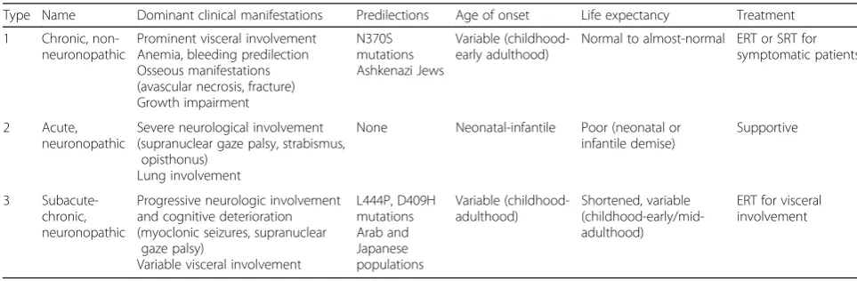
Although this condition is attributed to a single gene, there is substantial variability in the phenotypic presen-tation of Gaucher disease with three classically described types [3]. This article focuses on the chronic, non-neuronopathic form (type 1), manifesting with prom-inent involvement of the reticuloendothelial system with liver, spleen, and marrow involvement, whereas the other types (acute neuronopathic type 2 and subacute-chronic
© The Author(s). 2019Open AccessThis article is distributed under the terms of the Creative Commons Attribution 4.0 International License (http://creativecommons.org/licenses/by/4.0/), which permits unrestricted use, distribution, and reproduction in any medium, provided you give appropriate credit to the original author(s) and the source, provide a link to the Creative Commons license, and indicate if changes were made.
* Correspondence:[email protected]
1
Department of Radiology, Children’s Hospital of Philadelphia, 3401 Civic Center Blvd., Philadelphia, PA 19104, USA
2Department of Radiology, Perelman School of Medicine at the University of
neuronopathic type 3, summarized in Table1) are far less common [4]. It is worth noting that some experts propose conceptualizing these disease types as a phenotypic spectrum rather than as distinct entities. Some work has suggested subclinical neurological involvement in the
“non-neuronopathic” type with abnormal brain magnetic resonance spectroscopy (MRS) findings reported in type 1 Gaucher disease patients, although subsequent investiga-tions have failed to replicate these observainvestiga-tions and further work is needed [5,6]. Others have suggested an in-creased risk of Parkinsonism in individuals withGBA mu-tations that reflects a complex relationship between glucocerebrosidase and Lewy body disorders [7].
While type 1 Gaucher disease is most often diagnosed in childhood or early adulthood with a little under one-third of cases diagnosed by 10 years of age [8], this condition can come to clinical attention at any age due to gradations of residual enzyme activity and phenotypic variability [9]. Pediatric phenotypic expression, however, tends to be particularly severe [10]. The diversity of clin-ical manifestation and spectrum of disease severity underscore the importance of accurate assessment of multisystemic involvement at diagnosis and across the lifespan [9]; as this article will further discuss, diagnostic imaging plays an instrumental role in clinical evaluation and management of Gaucher disease.
Epidemiology, clinical evaluation, and treatment
While Gaucher disease is relatively uncommon overall with an estimated prevalence of approximately 1/40,000, certain groups have much higher carrier and disease prevalence with a carrier frequency of as high as 6–10% in Ashkenazi Jewish individuals [11,12]. The majority of type 1 Gaucher disease patients are diagnosed during childhood, and type 1 Gaucher disease is most com-monly due to biallelic p.N370Smutations in the Western world [4].
Detailed description of the diagnostic workup and clinical management of Gaucher disease is beyond the
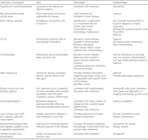
scope of this article, but it is helpful for radiologists evaluating patients with Gaucher disease to be familiar with commonly utilized clinical tests and available ther-apies. Laboratory tests and biomarkers are summarized in Table2; these clinical tests are part of comprehensive patient assessment that also involves diagnostic imaging. Clinical severity scoring is often performed using the Gaucher Disease Type 1 Severity Scoring System (GD-DS3) [13], which incorporates bone involvement, hematologic parameters, and organ enlargement. The Pediatric Gaucher Severity Scoring System (PGS3) [14] additionally accounts for growth disturbance as a key consideration in the younger population.
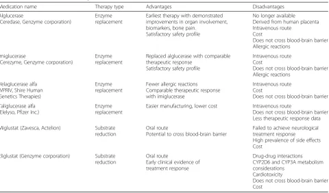
Traditionally, Gaucher disease has been conceptualized as a single gene-related disease due to one enzyme with one treatment consisting of enzyme replacement therapy (ERT) [3]. Prior to the advent of ERT, treatment was largely based on addressing symptoms with splenectomy performed for extensive splenic infiltration in severe pa-tients with poor prognosis. The earliest effective ERT, alglucerase, was introduced in the 1990s and has dramat-ically changed the prognosis of this condition. Subsequent newer forms of ERT with improved manufacturing tech-niques and safety profiles have been approved. Newer oral substrate reduction therapy (SRT) functions earlier in the biochemical pathway by mitigating the accumulation of glucosylceramide [15]. Both types of therapies are expen-sive, and each have disadvantages to consider with ERT requiring intravenous infusion every few weeks and SRT possessing a narrower safety profile with CYP2D6 and CYP3A metabolism considerations as well as being only approved for adult use [3, 4, 16]. Historic and currently available treatment options are summarized in Table 3, but we refer interested readers elsewhere for detailed de-scription of clinical trials and advances in Gaucher disease treatment [17].
Many asymptomatic patients with mild disease may not receive treatment at first; most children with milder genotypes perhaps may not require treatment initially.
Table 1Clinical classifications of Gaucher disease
Type Name Dominant clinical manifestations Predilections Age of onset Life expectancy Treatment
1 Chronic, non-neuronopathic
Prominent visceral involvement Anemia, bleeding predilection Osseous manifestations (avascular necrosis, fracture) Growth impairment
N370S mutations Ashkenazi Jews
Variable (childhood-early adulthood)
Normal to almost-normal ERT or SRT for symptomatic patients
2 Acute, neuronopathic
Severe neurological involvement (supranuclear gaze palsy, strabismus,
opisthonus) Lung involvement
None Neonatal-infantile Poor (neonatal or infantile demise)
Supportive
3 Subacute-chronic, neuronopathic
Progressive neurologic involvement and cognitive deterioration (myoclonic seizures, supranuclear
gaze palsy)
Variable visceral involvement
L444P, D409H mutations Arab and Japanese populations
Variable (childhood-adulthood)
Shortened, variable (childhood-early/mid-adulthood)
However, active bone disease, even if asymptomatic, and significant hepatosplenomegaly are considered indica-tions for initiating ERT in asymptomatic children [18]. Therefore, monitoring of patients not currently on therapy with imaging is clearly important in guiding cli-nicians as to early disease involvement [18].
As some patients may respond poorly to particular ther-apies, imaging can be important to inform clinicians regarding changes in an individual patient’s response to specific therapies to provide personalized therapy deci-sions. The management of treated individuals who are asymptomatic despite biomarker and imaging findings of Gaucher disease poses a unique challenge, leading some clinicians to propose a concept of minimal disease activity
wherein some treated patients may have mild persistent abnormalities on imaging and laboratory investigations without clinically significant disease [19]. Moreover, the substantial financial burden incurred by these therapies with annual treatment costs ranging between 70,000 and 380,000 USD [8,20] may also motivate the use of imaging assessment of treatment response in well-controlled patients to assist in verifying efficacy and safety of reduced dosing schedules or alternative strategies to reduce the overall cost of therapy.
Imaging of Gaucher disease involvement
Due to the reliance on ERT and SRT for the long-term management of patients with Gaucher disease, Table 2Laboratory investigations in Gaucher disease
Laboratory investigation Basis Advantages Disadvantages
Angiotensin-converting enzyme Increased in the plasma of affected patients
Decreases with treatment Nonspecific
Beta-glucocerebrosidase activity assay
Direct assessment of enzyme responsible for disease
Gold standard test Elevated in active disease
Expense
Bone marrow aspirate Visualization of Gaucher cells in marrow
Identification of alternative or concomitant disease entities with similar presentations (e.g., hematologic malignancy)
Not routinely recommended if Gaucher diagnosis is highly suspected
Nonspecific (pseudo-Gaucher cells) Discomfort
Expense
CCL18 Produced by Gaucher cells as macrophage chemokine
Elevated in active disease Suitable in chitotriosidase deficient individuals More closely reflects organ volumes than chitotriosidase
Expense
No head-to-head comparison with chitotriosidase
Chitotriosidase Released by glucocerebrosidase-laden Gaucher cells
Elevated in active disease Reduction from baseline values indicates treatment response
Increasing values are consistent with active disease
Normal individuals occasionally may not produce chitotriosidase Can vary widely between patients Expense
DNA sequencing Testing for genetic mutations (known and de novo) in the
GBAgene
Provides detailed information regarding genotype, which may be associated with specific forms of the disease
Identifies carriers
Expense
Variable phenotypic expression
Ferritin, serum iron, iron binding capacity
Iron overload occurs in patients. Uncertain etiology with possible association withHFEgene mutations, chronic inflammation
Correlates with hepatomegaly Decreases with treatment
Nonspecific with poor correlation with organ iron deposition on imaging and disease severity scoring
Glucosylsphingosine Byproduct related to glucosylceramide, reflecting beta-glucocerebrosidase function
Correlates with other markers of disease activity, organomegaly, platelet levels
Decreases with treatment
Expense, availability
Liver function tests (AST, ALT, bilirubin, albumin, total protein)
Hepatic dysfunction related to liver infiltration is common
Provides assessment of active hepatic involvement
May be insensitive to early hepatic involvement
Routine hematological tests (hemoglobin, platelet count, coagulation parameters)
Anemia and thrombocytopenia hallmark features of this disease
Provides information regarding hematologic involvement that may prompt other treatment
Nonspecific for overall disease severity
Tartrate-resistant acid phosphatase
Marker of osteoclasts and Gaucher cells
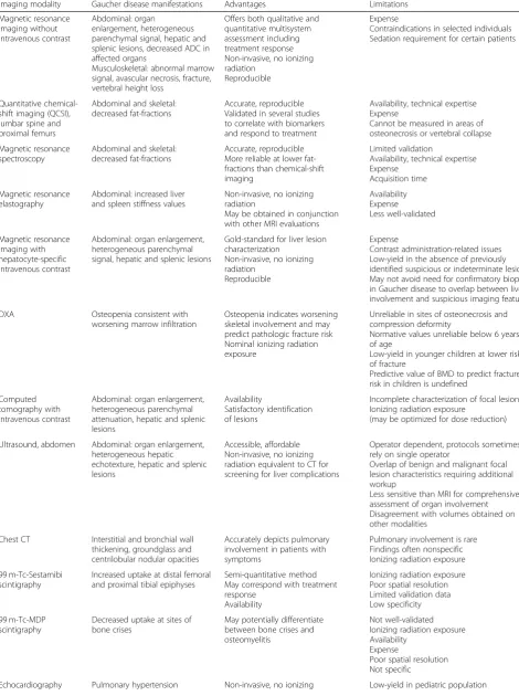
non-invasive imaging methods are essential in ascer-tainment of response to therapy and detection of disease-related complications. Therefore, understand-ing the role of different imagunderstand-ing modalities in the con-text of disease processes is important to provide clinicians and radiologists with guidance in assessing disease severity and treatment response. General strengths and limitations of each imaging modality are summarized in Table4[18,21–24].
Hepatic and splenic involvement Organomegaly and infiltration
While the inciting pathophysiological mechanism of Gaucher disease, enzymatic deficiency with accumula-tion of glucocerebroside, has been elucidated, the extent to which substrate accumulation accounts for liver en-largement has been called into question [25]. Originally, visceromegaly was attributed to simple accumulation glucocerebroside in the reticuloendothelial system with buildup of this material within Gaucher cells in the spleen as well as Kupffer cells in the liver [25]. However, newer evidence suggests that pro-inflammatory and pro-infiltrative chemotaxis factors may also account for organ enlargement with Gaucher cells contributing only partially to enlarged liver and spleen volumes [25, 26]. Regardless, severe hepatic involvement generally corre-sponds with additional organ system manifestations. Hepatosplenomegaly is a hallmark of Gaucher disease and uniformly present beginning in childhood, and liver
disease is a major contributor to Gaucher disease-related mortality [27]. As such, traditional imaging of disease severity has been based on hepatic and splenic visceral organ enlargement [24,25,28].
Organ sizes are interpreted from a clinical standpoint in the context of volume relative to the expected volume based on body weight, expressed as multiples of normal (MN) [18]. While there are a variety of available calcula-tions for normal liver and spleen volumes in the imaging literature incorporating age, sex, and body mass index, most Gaucher disease studies utilize a simple weight-based formula for these normative values (normal liver volume (mL) = 25 mL/kg × weight (kg); normal spleen volume (mL) = 2 mL/kg × weight (kg)) that should be consistently employed to avoid confusion across studies [29]. Treatment objectives often include specific targets such as liver volumes of less than 1–1.5 MN and spleen volumes of < 2–8 MN [8,30].
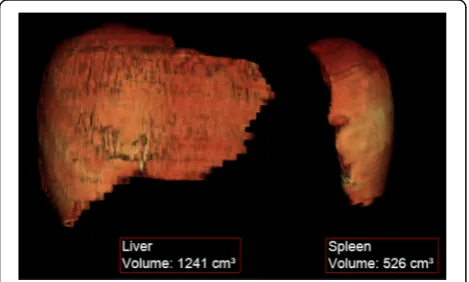
Organ volumes may be calculated using a variety of methodologies with the simplest being a volumetric esti-mate from orthogonal dimensions on conventional ultra-sound. Conventional ultrasound has the benefit of being non-invasive and relatively inexpensive; most notably, ultrasound may avoid the use of sedation in younger children [31, 32]. While the longitudinal axis measure-ments on US modestly correlate with CT-derived vol-umes, others note that sonographic-derived values differ significantly from true volumetric measurements (Fig.1) obtained on cross-sectional imaging, namely CT and Table 3Historic and available therapies for Gaucher disease
Medication name Therapy type Advantages Disadvantages
Alglucerase
(Ceredase, Genzyme corporation)
Enzyme replacement
Earliest therapy with demonstrated improvements in organ involvement, biomarkers, bone pain.
Satisfactory safety profile
No longer available
Derived from human placenta Intravenous route
Cost
Does not cross blood-brain barrier Allergic reactions
Imiglucerase
(Cerezyme, Genzyme corporation)
Enzyme replacement
Replaced alglucerase with comparable therapeutic response
Satisfactory safety profile
Intravenous route Cost
Does not cross blood-brain barrier Allergic reactions
Velaglucerase alfa (VPRIV, Shire Human Genetics Therapies)
Enzyme replacement
Fewer allergic reactions Comparable therapeutic response with imiglucerase
Intravenous route Cost
Does not cross blood-brain barrier
Taliglucerase alfa (Elelyso, Pfizer Inc.)
Enzyme replacement
Easier manufacturing, lower cost Intravenous route
Does not cross blood-brain barrier Less therapeutic response data
Miglustat (Zavesca, Actelion) Substrate reduction
Oral route
Potential to cross blood-brain barrier
Failed to achieve neurological treatment response
High prevalence of side effects Cost
Eliglustat (Genzyme corporation) Substrate reduction
Oral route
Early clinical evidence of treatment response
Drug-drug interactions CYP2D6 and CYP3A metabolism considerations
Cardiotoxicity
Table 4Imaging modalities relevant to Gaucher disease
Imaging modality Gaucher disease manifestations Advantages Limitations
Magnetic resonance imaging without intravenous contrast
Abdominal: organ
enlargement, heterogeneous parenchymal signal, hepatic and splenic lesions, decreased ADC in affected organs
Musculoskeletal: abnormal marrow signal, avascular necrosis, fracture, vertebral height loss
Offers both qualitative and quantitative multisystem assessment including treatment response Non-invasive, no ionizing radiation
Reproducible
Expense
Contraindications in selected individuals Sedation requirement for certain patients
Quantitative chemical-shift imaging (QCSI), lumbar spine and proximal femurs
Abdominal and skeletal: decreased fat-fractions
Accurate, reproducible Validated in several studies to correlate with biomarkers and respond to treatment
Availability, technical expertise Expense
Cannot be measured in areas of osteonecrosis or vertebral collapse
Magnetic resonance spectroscopy
Abdominal and skeletal: decreased fat-fractions
Accurate, reproducible More reliable at lower fat-fractions than chemical-shift imaging
Limited validation
Availability, technical expertise Expense
Acquisition time
Magnetic resonance elastography
Abdominal: increased liver and spleen stiffness values
Non-invasive, no ionizing radiation
May be obtained in conjunction with other MRI evaluations
Availability Expense Less well-validated Magnetic resonance imaging with hepatocyte-specific intravenous contrast
Abdominal: organ enlargement, heterogeneous parenchymal signal, hepatic and splenic lesions
Gold-standard for liver lesion characterization
Non-invasive, no ionizing radiation
Reproducible
Expense
Contrast administration-related issues Low-yield in the absence of previously identified suspicious or indeterminate lesion May not avoid need for confirmatory biopsy in Gaucher disease to overlap between liver involvement and suspicious imaging features
DXA Osteopenia consistent with worsening marrow infiltration
Osteopenia indicates worsening skeletal involvement and may predict pathologic fracture risk Nominal ionizing radiation exposure
Unreliable in sites of osteonecrosis and compression deformity
Normative values unreliable below 6 years of age
Low-yield in younger children at lower risk of fracture
Predictive value of BMD to predict fracture risk in children is undefined
Computed tomography with intravenous contrast
Abdominal: organ enlargement, heterogeneous parenchymal attenuation, hepatic and splenic lesions
Availability
Satisfactory identification of lesions
Incomplete characterization of focal lesions Ionizing radiation exposure
(may be optimized for dose reduction)
Ultrasound, abdomen Abdominal: organ enlargement, heterogeneous hepatic echotexture, hepatic and splenic lesions
Accessible, affordable Non-invasive, no ionizing radiation equivalent to CT for screening for liver complications
Operator dependent, protocols sometimes rely on single operator
Overlap of benign and malignant focal lesion characteristics requiring additional workup
Less sensitive than MRI for comprehensive assessment of organ involvement Disagreement with volumes obtained on other modalities
Chest CT Interstitial and bronchial wall thickening, groundglass and centrilobular nodular opacities
Accurately depicts pulmonary involvement in patients with symptoms
Pulmonary involvement is rare Findings often nonspecific Ionizing radiation exposure
99 m-Tc-Sestamibi scintigraphy
Increased uptake at distal femoral and proximal tibial epiphyses
Semi-quantitative method May correspond with treatment response
Availability
Ionizing radiation exposure Poor spatial resolution Limited validation data Low specificity
99 m-Tc-MDP scintigraphy
Decreased uptake at sites of bone crises
May potentially differentiate between bone crises and osteomyelitis
Not well-validated Ionizing radiation exposure Availability
Expense
Poor spatial resolution Not specific
MRI, and ultrasound may also depend on a single reader over serial evaluations for consistency [31, 33, 34]. Therefore, many consensus recommendations caution against the use of ultrasound [18]. In more recent years, there is widespread agreement upholding the use volu-metric MRI over CT, given concerns regarding ionizing radiation exposure from repeated examinations, and additional information obtainable with MRI [24]. Semi-automated techniques of measuring organ volumes on MRI also afford more reliable measurements and avoid dependence on specialized readers [35].
Organ volumes decrease substantially with therapy ini-tiation, although some have noted that well-controlled disease may result in plateauing of organ volumes after the first 3 years of successful treatment [34].
Diffusion-weighted imaging (DWI) offers another quantitative assessment of organ infiltration as a surro-gate of tissue cellularity [36]. Apparent diffusion coeffi-cient (ADC) values in the liver and spleen appear to
correlate well with chitotriosidase as a marker of disease severity in one pediatric study with decreased ADC con-noting greater infiltration and worse involvement [37].
Liver fibrosis
While cirrhosis and portal hypertension have been trad-itionally regarded as rare in Gaucher disease [38], there is increased risk for liver fibrosis, cirrhosis, and hepato-cellular carcinoma (HCC) in this condition, especially in previously splenectomized individuals [39–41]. Gaucher cell deposition may establish a fibrogenic microenviron-ment due to chronic low-grade inflammation [25,27]. In Gaucher disease, liver fibrosis is thought to increase the increased risk of hepatic cirrhosis, HCC risk, and liver disease-related mortality.
Advances in the understanding of Gaucher disease and observations of elevated ferritin levels and increased risk of hepatic fibrosis suggest greater importance of imaging assessment for complications of liver involvement beyond simple organ enlargement. Ultrasound has been traditionally used to monitor development of cirrhotic morphology (Fig. 2) but is relatively insensitive in detecting earlier fibrotic changes. Some means of non-invasively assessing hepatic fibrosis include non-imagi-ng-based transient elastography (TE), ultrasound shear wave elastography (SWE), and magnetic resonance elas-tography (MRE) [42]. These elastography modalities pro-vide reliable measurements that can detect significant liver fibrosis each with unique strengths and limitations summarized in Table4[42].
Liver stiffness may be only mildly elevated in Gaucher disease patients without cirrhosis, likely reflecting treatment-related reductions in the extent of liver fibro-sis [43]. However, Gaucher disease patients with cirrho-sis exhibit markedly elevated liver stiffness values [43], and other studies of patients who were previously sple-nectomized (with more severe disease) demonstrated greater liver stiffness compared with patients with milder disease [40, 44]. In a recent study, MRE-measured liver Table 4Imaging modalities relevant to Gaucher disease(Continued)
Imaging modality Gaucher disease manifestations Advantages Limitations
and cardiac MRI (mostly adults on treatment) Valvular calcifications (D409H homozygous mutation)
radiation
Definitive investigations for cardiac involvement
Expense
Limited data to support widespread use, particularly for cardiac MRI
Acoustic radiation force impulse/shear wave elastography (US)
Increased liver and spleen stiffness values
Similar or higher performance compared with transient elastography Non-invasive, no ionizing radiation May be combined with conventional US evaluation of organ involvement No sedation
Availability
Measurement variability
Transient elastography Increased liver and spleen stiffness values
Non-invasive, no ionizing radiation Expense
No imaging guidance to assess most affected regions of organs
No imaging component for further characterization of organ parenchyma quality or lesions
stiffness values correlated with Gaucher clinical severity (GD-DS3) scores, highlighting the potential clinical util-ity of evaluating liver stiffness in this disease [44]. These findings suggest a role for elastography-based modalities in identifying early hepatic fibrosis in Gaucher disease patients, although further study is needed to better understand the extent of fibrosis in Gaucher disease and the clinical utility of earlier identification.
Iron overload
Other important clinical manifestations of Gaucher dis-ease include hematologic abnormalities such as anemia and hyperferritinemia. The precise etiology of hyperferri-tinemia in Gaucher disease is poorly understood, al-though it is a frequent finding that may correspond with other indicators of disease severity and respond to ERT [45, 46]. Ferritin levels are also noted to be higher in
asplenic patients, a finding that may be confounded by greater disease severity in these patients also often re-quiring blood transfusions [45]. From an imaging stand-point, hyperferritinemia with iron deposition such as within the liver may appear on with increased attenu-ation on CT, signal drop-out on out-of-phase gradient T1-weighted MRI and low signal on T2*-weighted MRI [47]. A group of investigators highlighted substantial iron deposition (as indicated by R2* values) in treated Gaucher disease patients compared with controls [48]. However, the precise relationship between hyperferriti-nemia and visceral iron deposition is poorly understood in Gaucher disease and the subject of continued investigation.
Liver lesions
The majority of liver lesions encountered in Gaucher disease are thought to be related to focal deposition of glucocerebroside within the Kupffer cells [39]; however, Gaucher disease confers substantial increased risk of malignancy and HCC [49,50].
In one large ultrasound study of Gaucher disease patients, focal liver lesions were identified in just under 5% of individuals; the majority of which were multiple hypere-choic lesions [39]. Authors have suggested that such small, hyperechoic lesions do not merit biopsy if slowly growing, as they are thought to reflect focal accumulation of Gaucher cells [39]. On the other hand, early studies of MRI in Gaucher disease identified focal signal abnormal-ities in about one-fifth of patients [51]. Such focal accumu-lations of Gaucher cells are thought to be hypoattenuating on CT, hypointense on T1-weighted imaging, and hetero-geneous on T2-weighted imaging (Fig.3).
In addition, reports of biopsy-proven hepatic amyloidosis have been made in rare numbers of patients [39, 52, 53]. More worrisome, HCC can develop at younger ages in Gaucher disease; the previously mentioned large series Fig. 2Liver cirrhosis in pediatric Gaucher disease. Sagittal grayscale
ultrasound demonstrates an enlarged liver with nodular cirrhotic morphology and perihepatic ascites in a 12-year-old male with Gaucher disease
identified only one case of HCC, which was found in the setting of rapidly progressive cirrhotic morphology on serial US evaluations [39].
Therefore, screening evaluation for development of suspicious liver lesions is warranted in Gaucher disease. Identification of more suspicious (e.g., hypoechoic, large, irregular, hypervascular) lesions may warrant further in-vestigation with dedicated liver protocol CT, hepatocyte-specific contrast-enhanced MRI, or contrast-enhanced US, which may offer a viable alternative especially for children [54].
Spleen fibrosis
Gaucher patients also demonstrate significantly ele-vated spleen stiffness values using a variety of elastogra-phy techniques [43]. Elevated stiffness values observed in Gaucher disease (Fig.4) may reflect progressive infil-tration and inflammatory changes culminating in fibro-sis. Elevated spleen stiffness has been deemed a harbinger of portal hypertension in non-Gaucher dis-ease conditions [42]; however, prospective assessment of the clinical significance of splenic fibrosis in Gaucher disease is needed.
Splenic lesions
Similar to the previously described hepatic lesions thought to represent focal Gaucher cell buildup, patients with Gaucher disease are often noted to have hypere-choic splenic lesions on ultrasound in one-fifth to one-third of cases [43]. These lesions tend to be low in attenuation with occasional peripheral calcification on CT [55]. On MRI, Gaucher-related splenic nodules usu-ally demonstrate low T1-weighted and high T2-weighted signal intensity, although focal areas of necrosis are also
encountered which can be hypointense on T1-weighted imaging [32, 55]. Early MRI studies demonstrated that numbers of splenic nodules correlated with spleen size; these investigators posited that these lesions often repre-sent areas of infarction in the setting of splenic infiltra-tion [56]. These splenic nodules appear to be more common in adults, likely reflecting longstanding infil-tration needed to result in ischemia [51]. Similarly, sub-capsular splenic infarcts were commonly noted as well prior to introduction of effective ERT [51]. In especially severe cases, splenic necrosis may occur with replace-ment with fluid attenuation material and peripheral calcification (Fig.5).
Skeletal involvement and complications
In patients with type 1 Gaucher disease, bone marrow involvement represents a spectrum of disease (Fig. 6) with some level of involvement in nearly every patient [4, 57, 58]. Recognition that musculoskeletal manifesta-tions may be the presenting symptom of Gaucher dis-ease is important to avoid confusion with other conditions with overlapping symptoms and signs such as hematopoietic malignancies and rheumatologic condi-tions [59]. Infiltration of the bone marrow with Gaucher cells with concomitant osteopenia progressively compro-mises the bone density of the axial then appendicular skeleton with prominent lower extremity involvement of the proximal femurs and tibias [22, 60, 61]. Upper extremity involvement, while less commonly recognized, is also a substantial contributor to musculoskeletal mor-bidity with many children and adolescents reporting upper extremity pain.
Greater severity of musculoskeletal involvement often corresponds with severe organ involvement. Early inves-tigations of bone involvement in Gaucher disease believed bone changes were irreversible [56]. However, newer studies have shown that treatment results in sub-stantial improvement of early bone marrow changes in many patients. The prognosis of skeletal involvement once complications have occurred remains poor and bone marrow responses take longer than organ responses [55, 62]. Bone turnover biomarkers have largely failed to risk stratify patients, making imaging evaluation of musculoskeletal involvement indispensable to clinical management [57].
Marrow infiltration
Abnormal bone marrow involvement in Gaucher disease is a prominent finding, almost uniformly noted in type 1 patients and most conspicuously involves the spine, femurs, tibias, and humeri. Anemia commonly encoun-tered in this condition results in greater hematopoietic marrow than expected for age, and direct accumulation of Gaucher cells also contributes to abnormal bone Fig. 4Spleen fibrosis in pediatric Gaucher disease. Axial MR
marrow. Marrow involvement follows a pattern of cen-trifugal spread with the epiphyses and apophyses mostly spared except in the most severe of marrow infiltration [61]. Bone marrow involvement has long been regarded as more difficult to treat than visceral involvement. A previous study showed through histological examina-tions a decreased uptake of administered alglucerase within the bone marrow [63]. Marrow infiltration of the lumbar spine is thought to be more responsive to ther-apy, whereas some authors regard femoral marrow reflecting more static disease [64].
Subjective imaging assessment Radiographs are
in-sensitive for detection of marrow infiltration; substantial medullary cavity expansion is needed to result in the classically reported Erlenmeyer flask deformity of the distal femur (Fig.7) [22,65]. In addition, the Erlenmeyer
flask finding is nonspecific for Gaucher disease, being encountered in a variety of other conditions reviewed at-length elsewhere [66]. Initial marrow infiltration can be patchy, also making radiographic detection impos-sible [67].
Marrow infiltration manifests with low signal on T1-weighted imaging (Fig. 8). Early qualitative MRI assessments of marrow involvement in patients on ERT-based noted T1-weighted signal increases corre-sponded with reduced organ volumes with changes observed as early as 1 year [46, 62]. This increase in T1-weighted signal is thought to reflect greater accumu-lation of yellow marrow with decreased concentration of Gaucher cells and hematopoietic marrow. Interpretation of qualitative marrow changes in children should be interpreted within the context of developmental changes in hematopoietic marrow [10,68].
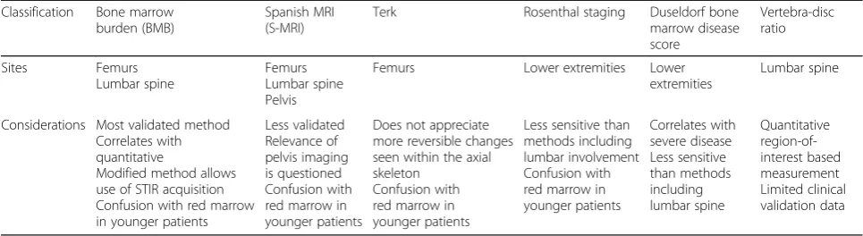
Semi-quantitative imaging assessment Numerous
semi-quantitative scoring methods have been developed to report overall extent of bone marrow involvement based on signal intensity alterations on MRI, each with specific objectives and limitations summarized in Table 5 [61, 69]. Semi-quantitative region-of-interest (ROI)-based techniques have been offered up as slightly more objective alternatives to signal intensity based methods; the most commonly performed of these methods is the vertebra disc ratio (VDR) in which ROIs are placed on T1-weighted images of the lumbar spine [70, 71]. Out of these methods, the bone marrow bur-den (BMB) score (Table6) based on conventional lum-bar spine and femur MRI is the most validated and correlates well with disease severity, splenectomy status (indicative of more severe disease), treatment response, and quantitative fat fractions [20, 23, 64, 72–75]. An-other method, the Spanish-MRI (S-MRI) score, also in-corporates imaging of the pelvis [76]. More recent investigations have applied some of these semi-quantitative Fig. 5Splenic necrosis in pediatric Gaucher disease. Transverse grayscale abdominal ultrasound (a) of a 2-year-old male Gaucher disease patient with marked splenomegaly shows replacement of normal splenic parenchyma with fluid and hyperechoic regions corresponding to dystrophic calcification. Contrast-enhanced abdominal CT (b) of this patient demonstrates enlarged spleen replaced with liquefying necrosis and peripheral dystrophic calcifications
scoring techniques to whole-body MRI to assess treat-ment response [77–79]. While the reliability of these measures is promoted by several small studies, more recent work has called reliability of BMB scoring into question with poor interobserver agreement and others have pointed out issues related to later fat marrow conversion in children [77, 80].
Quantitative imaging assessmentMultiple quantitative
factors have been proposed to objectively assess marrow involvement in Gaucher disease and are supported by histopathologic evidence of an inverse relationship be-tween marrow glucocerebroside and triglyceride concen-trations [72]. Some of earliest quantitative assessments of bone marrow composition were performed using dual-energy CT and noted low amounts of marrow fat in patients [81, 82]. These initial investigations envisioned fat fractions as a marker of bone marrow disease severity with lower values representing greater infiltration [81]. Early MRI investigations noted longer T1 relaxation times in the bone marrow as well as decreases in these values with treatment [83, 84]. The emergence of MRI tech-niques shortly following these CT investigations led to the abandonment of CT for this indication, and fat fractions calculated from MRI have become the dominant quantita-tive variable to report marrow involvement [61,84].
Currently, quantitative MRI-based fat fraction mea-surements have been applied to objectively assess marrow involvement in Gaucher disease. Quantitative chemical-shift imaging (QCSI) using Dixon-based tech-niques and MRS are both capable of reliably measuring bone marrow fat fraction [85–90]. Work by Maas and colleagues has suggested a cut-off of 0.23 for a fat frac-tion ratio—values lower than this suggest a markedly elevated risk of bone-related complications [91]. Early studies also identified a correlation between marrow fat Fig. 7Erlenmeyer flask deformity. Frontal radiograph of the right
knee in a 12-year-old-male Gaucher disease patient with widening of the distal femoral diaphysis and metaphysis resulting in Erlenmeyer flask deformity
fractions and spleen size, suggesting an association between bone marrow and visceral disease severity [84]. One small MRS study noted significantly lower fat frac-tions in the proximal femurs but not the lumbar spine of mostly asymptomatic children with Gaucher children compared with age-matched controls [92]. Treatment-related responses in bone marrow fat fractions have been demonstrated in response to ERT (Fig.9) [93–95]. A fat fraction of 0.37 (derived from normal controls) or more is proposed as a reference goal for treatment response [72]. Marrow fat quantification may serve as a means of prognostication and identifying nonresponders with responses noted as early as 1 year [91,93,95].
The importance of understanding developmental in-creases in marrow fat over the lifespan beginning first in the peripheral skeleton as well as normal variation in bone marrow fat fractions cannot be understated.
Patient age, sex, and body mass index should be consid-ered in the interpretation of these values, ideally in the context of normative data relevant to the measurement method [88,96–98]. Importantly, untreated Gaucher pa-tients do not seem to exhibit normal expected age-related increases in fat fractions [93]. Adoption of QCSI for fat fraction measurements has been stymied for decades outside a few selected academic medical centers due to technical requirements, but the recent release of commercially available sequences may lead to a revival of fat fraction quantification [88,99].
DWI may also hold promise with some evidence of decreased vertebral marrow ADC reflecting increased cellularity corresponding with disease status [100]. Last, iron filtration in marrow has been quantified in Gaucher disease with elevated R2* values in femoral and vertebral marrow compared to matched healthy controls [48]. Table 5Semi-quantitative Gaucher disease bone marrow involvement scoring systems
Classification Bone marrow burden (BMB)
Spanish MRI (S-MRI)
Terk Rosenthal staging Duseldorf bone marrow disease score
Vertebra-disc ratio
Sites Femurs Lumbar spine
Femurs Lumbar spine Pelvis
Femurs Lower extremities Lower extremities
Lumbar spine
Considerations Most validated method Correlates with quantitative
Modified method allows use of STIR acquisition Confusion with red marrow in younger patients
Less validated Relevance of pelvis imaging is questioned Confusion with red marrow in younger patients
Does not appreciate more reversible changes seen within the axial skeleton
Confusion with red marrow in younger patients
Less sensitive than methods including lumbar involvement Confusion with red marrow in younger patients
Correlates with severe disease Less sensitive than methods including lumbar spine
Quantitative region-of-interest based measurement Limited clinical validation data
Table 6Bone marrow burden (BMB) classification
Femurs Lumbar spine
Involvement Site Score Site Score
Diaphysis 1 Patchy 1
Proximal epiphysis/apophysis 2 Diffuse 2
Distal epiphysis 3 Absence of fat in basiverteral region 1
Sequence Signal intensitya Score Signal intensityb Score
T2-weighted, (or, STIR)c Hyperintense 2 (2) Hyperintense 2 (2)
Slightly hyperintense 1 (1) Slightly hyperintense 1 (1)
Isointense 0 (0) Isointense 0 (0)
Slightly hypointense 1 (N/A) Slightly hypointense 1 (N/A)
Hypointense 2 (N/A) Hypointense 2 (N/A)
Mixed type 3 (3)
T1-weighted Slightly hyperintense or isointense 0 Slightly hyperintense 0
Slightly hypointense 1 Isointense 1
Hypointense 2 Slightly hypointense 2
Hypointense 3
Sum Sum
a
Relative to subcutaneous fat
b
Relative to normal intervertebral disc
c
Metabolic imaging assessment Nuclear medicine stud-ies are not routinely used for routine follow-up assess-ment of Gaucher disease at most institutions, although 99 m-Tc-Sestamibi scintigraphy has been studied as an alternative means of quantifying bone marrow involvement with good agreement with MRI-based semi-quantitative scoring [101, 102]. Treatment-related changes have been successfully observed using 99 m-Tc-Sestamibi scintigraphy [103]. Other scintigraphy methods are largely ineffective at quantifying marrow in-volvement as typical bone radiotracers are only taken up by normal marrow, but 99 m-Tc-MDP could potentially be helpful in discerning between bone crises and osteo-myelitis, although this observation is not well validated
[22]. Nuclear medicine-based techniques are hampered by limited spatial resolution and inherent ionizing radi-ation exposure. While sestamibi patterns do not appear to be affected by age-related marrow conversion, ioniz-ing radiation exposure incumbent in this technique pre-cludes adoption for pediatric imaging [32]. In the future, targeted assessment of disease involvement may be pos-sible with promising experimental positron emission tomography data using a 18F-labeled glucocerebrosidase [104]. In current practice, superior assessment of both marrow, osseous complications, and soft tissues has made MRI the modality of choice for the evaluation of musculoskeletal manifestations of Gaucher disease.
Osteopenia
One of the earliest skeletal manifestations of Gaucher disease type 1 in children is osteopenia [31]. The devel-opment of osteopenia in Gaucher disease is multifactor-ial with abnormal osteoclast function and decrease in bone formation as possible etiologies reviewed in detail elsewhere [105]. Abnormal trabecular bone density has been observed in the absence of focal abnormalities or remodeling and cortical thinning is noted with marrow expansion on CT [81, 82]. Decreased bone density is appreciated qualitatively on radiographic examinations and quantitatively assessed using dual-energy X-ray absorptiometry (DXA) assessment of bone mineral dens-ity (BMD). Z-scores from multiple sites are lower for Gaucher disease patients compared with healthy individ-uals [106]. Bone density has also been observed to increase in response to treatment [28, 107, 108]. Tech-nical difficulties may be encountered in interpreting BMD in younger children, patients with compression fractures, and bone infarction [61]. Nonetheless, BMD retains an important role in predicting the risk of osteo-necrosis and pathologic fracture in adult patients [109].
Osseous complications
Progressive osseous involvement in type 1 Gaucher dis-ease may begin with painful bone crises that may even-tually result in osteonecrosis (also termed avascular necrosis and bone infarction) in about half of patients that may be attributed to packing of the marrow with Gaucher cells leading to thrombosis in situ within the marrow [57, 110]. Osteonecrosis most commonly involves the femoral head and neck, proximal humeri, tibias, and vertebrae [22]. Upper extremity involvement seems to occur in patients with greater global bone mar-row involvement [78]. Incumbent pathologic fractures occur in about one-tenth to one-quarter of patients and often require specialized orthopedic management including arthroplasty detailed elsewhere [110–113]. The frequency of osseous complications is thought to mirror that of other organ involvement severity; one study Fig. 9Magnetic resonance spectroscopy assessment of marrow
showed a significant difference in liver volumes between patients with and without osteonecrosis [56]. Prior to the advent of ERT, thoracic kyphosis and vertebral body collapse following severe osteopenia were commonly noted complications in affected children with resultant morbidity and deformity [114]. Bone crises are now infrequently encountered in pediatric patients in the era of ERT [10]. Osteomyelitis is a relatively infrequent com-plication noted in 6% of patients in one cohort [110].
Radiographic detection of early osseous complications such as osteonecrosis and bone infarct may be difficult, often being occult on initial imaging with subsequent de-velopment periosteal reaction that may be confused with osteomyelitis (“pseudo-osteomyelitis”) [22, 67]. Focal ac-cumulation can result in lytic lesions visible radiographic-ally [32]. Later stages of osteonecrosis demonstrate serpentine sclerosis radiographically. Radiographs are helpful, however, in the initial workup of acute pain to as-sess for pathologic fracture [115]. Late sequelae including collapse of the femoral heads, proximal humeri, and verte-bral bodies can also be appreciated on radiographs along with early onset osteoarthritis [10].
Given these limitations, assessment of acute osseous complications is best performed with initial radiographs followed by prompt MRI assessment [72]. Bone crises may be seen with focal marrow edema on fluid-sensitive sequences [10]. With progressive ischemic insults, osteo-necrosis can develop and is evident as T1-weighted hypointense signal with heterogeneous T2-weighted hyperintense signal often with a“double line”of low and high signal reflecting bone infarction [67]. This focal bone involvement can progress further with collapse of the femoral heads and vertebral bodies encountered in
severe marrow disease [10]. Other associated complica-tions can be seen including cortical disruption and sub-periosteal hemorrhage (Fig. 10) in the setting of massive medullary expansion and cortical thinning, which may also mimic osteomyelitis [116,117].
Bone-related malignancies may be slightly more com-mon in Gaucher disease, perhaps related to osteonecro-sis as a predisposing factor. MRI may often be warranted for evaluation of discrete lytic lesions given the incidence of malignant bone lesions such as multiple myeloma, osseous lymphoma, sarcomas, and malignant epithelioid hemangioendothelioma [118–120]. Particular attention should be paid to enlarging lesions and those with morph-ology dissimilar to more common Gaucher disease-related osteopenia, marrow infiltration, and osteonecrosis.
Cardiopulmonary involvement Pulmonary infiltration
As with the liver, spleen, and bone marrow, Gaucher cells can infiltrate in the lungs, typically depositing within interstitial spaces. Such lung involvement is quite rare, more common in the neuronopathic type, nd not likely to occur as an initial manifestation of this disease [10]. Reticulonodular opacities may be seen on chest radiography, but chest CT evaluation is more useful in depicting centrilobular nodules, ground glass opacities, interstitial, and bronchial wall thickening [22].
Pulmonary hypertension
There are multiple proposed etiologies for pulmonary hypertension in Gaucher patients. Pulmonary hyperten-sion is attributable to perivascular infiltration of Gaucher cells or secondary to hepatic disease [22]. An early
report of echocardiography in 134 adult Gaucher disease patients identified pulmonary hypertension in approxi-mately one in ten treated patients and none in untreated patients, raising concern for possible treatment-related pulmonary hypertension [121]; however, a subsequent follow-up of children screened with echocardiography did not find pulmonary hypertension in this younger patient population [122]. Therefore, routine screening of pediatric Gaucher disease patients for pulmonary hyper-tension is not indicated.
Cardiac involvement
The extent of cardiac involvement in Gaucher disease is uncertain with small numbers of mitral valve prolapse and valvular calcifications reported in a few patients with homozygous D409H mutations [123]. Given the infre-quent occurrence of cardiac involvement, most authors only recommend baseline echocardiographic screening in children with Gaucher disease to exclude abnormal-ities with others also recommending follow-up echocar-diography in children with homozygous D409H mutations [122,123]. A more recent investigation of car-diac MRI in a small number of Gaucher disease patients noted left atrial enlargement in three of nine patients [124], and others have implied an association between Gaucher disease and left ventricular diastolic dysfunc-tion on echocardiography, suggesting myocardial infiltra-tion as a possible etiology [125]. Further systematic investigation is needed to assess the frequency of cardiac involvement in Gaucher disease, and the possible rele-vance of imaging for screening for subclinical disease.
Recommended imaging protocols
There is a clear need for an objective and reproducible scoring system capable of assessing disease severity to standardize patient monitoring. While clinical scoring methods (PGS3 and GD-DS3) are useful tools for estimating disease severity, these scoring methods do not assess diffuse hepatic disease such as fibrosis, iron accumulation, fat infiltration, and portal hypertension, nor do they assess the full extent of asymptomatic mar-row involvement with inclusion of only subjective im-aging assessment of marrow involvement.
While ultrasound is affordable and may be sufficient for the assessment of organ volumes and liver lesion screening, there is increasing use of MRI in Gaucher dis-ease with most authorities considering MRI the standard of care [24, 45]. As highlighted throughout this article, there are additional findings on newer imaging methods worth assessing in Gaucher disease patients that cannot be imaged using conventional MRI, ultrasound, and/or radiography. For this reason, we advocate the routine use of an MRI protocol that provides both qualitative and quantitative information of multiple organ systems
to provide a comprehensive assessment of disease in-volvement in Gaucher disease. In younger children under the age of 6 years, sedation may be needed to per-form MRI; abbreviated protocols, child life, and distrac-tion techniques may reduce the need for sedadistrac-tion in children [23]. In institutions where particular resources are constrained or unavailable, suitable alternative imaging techniques may be employed acknowledging potential limitations summarized in Table 4. Some ex-amples of alternatives to MRI-based modalities include carefully acquired ultrasound measurements of organ volumes using standardized methods by selected readers and ultrasound elastography methods. However, reliable determination of marrow infiltration requires MRI or nuclear medicine studies.
Liver and spleen MRI
Diagnostic imaging of hepatic and splenic involvement should consist of sequences tailored for volumetric organ measurement and screening for focal lesions. In conjunction with these conventional acquisitions, MR elastography should be considered, when available, to provide non-invasive assessment of liver and splenic stiffness. While the role of elastography in the clinical management of hepatic fibrosis in Gaucher disease is not yet established, acquisition of organ stiffness mea-surements may afford insights into disease and treat-ment effects in the future with wider clinical adoption. It is important to note that elastography results should be interpreted in the context of known limitations includ-ing inaccuracy of gradient recalled echo(GRE)-based elastography in patients with iron deposition and massive ascites [42]; as such, performance of both GRE and spin-echo echo-planar imaging elastography sequences is recommended. In addition, assessment of fat fractions and iron quantification may be helpful in providing add-itional quantitative information for disease severity and treatment response assessment, although these methods also require additional validation in larger populations.
Newer proton density fat fraction (PDFF) sequences allow for single breath-hold acquisition of the entire liver to provide triglyceride fat fractions, R2* mapping, and some also provide automated liver volumes (mDIXON Quant, Philips Healthcare; LiverLab, Siemens Healthi-neers; IDEAL-IQ, GE Healthcare) [126–128]. These mea-surements are reliable and highly reproducible across vendors, making them ideal for multi-center investigations that are essential in studying rare conditions such as Gaucher disease [127,128]. However, these measurement packages often require significant capital and are not yet widely available outside of major research institutions.
PDFF sequences are not available. DWI is included in our protocol as it may also be provide indirect assess-ment of organ cellularity with ADC as a possible quanti-tative marker of disease severity [37].
If suspicious focal lesions are noted on this surveil-lance evaluation, dedicated lesion work-up with dynamic hepatocyte-specific contrast-enhanced MRI, multiphase contrast-enhanced CT, or contrast-enhanced ultrasound should be recommended, depending on institutional resources [54,129,130].
Bone marrow MRI
Qualitative imaging of both the axial (lumbar spine) and appendicular skeleton (proximal femurs) is important for assessment of global marrow involvement, as these sites may respond differently to treatment [64]. While a few recent studies have explored the use of whole-body MRI to screen the entire skeleton, none of the cases of humeral involvement were asymptomatic and therefore screening of the upper extremities may be less salient [78]. As emphasized throughout this article, quantitative measurement of bone marrow fat fractions is of increas-ing importance in estimatincreas-ing disease severity and track-ing treatment response. Similar PDFF acquisitions (mDIXON Quant, Philips Healthcare; LiverLab, Siemens Healthineers; IDEAL-IQ, GE Healthcare) employed to measure fat fractions in the liver may be employed to measure fat fractions in the proximal femur (neck) and lumbar spine (L5), although marrow measurements using these sequences have not been validated in Gaucher disease [88]. Single-voxel MRS is a reliable al-ternative quantitative method that has been used in Gaucher disease and shows good concordance with Dixon-based methods [92, 131], although acquisition times may be greater for this method. MRS additionally requires expertise in analysis for fat fraction determin-ation, but may be more reliable for lower fat fractions. Both PDFF and MRS generally require the contributions of a medical physicist dedicated to the successful imple-mentation of these protocols. Therefore, institutions without such expertise in quantitative imaging may rely on conventional acquisitions and subjective assessment of marrow involvement by radiologists familiar with semi-quantitative scoring methods such as BMB.
Frequency of comprehensive MRI evaluation
Discrete parameters for the frequency of follow-up of Gaucher disease patients with imaging are informed by consensus recommendations and institution-specific practices [18, 24, 132, 133]. Comprehensive imaging evaluation of the liver, spleen, and bone marrow of the femurs and lumbar spine should be performed at initial diagnosis, annually for asymptomatic, untreated pediatric patients and biannually for asymptomatic,
untreated adults. While some recommend less frequent skeletal imaging evaluation of asymptomatic patients, subclinical marrow involvement appears to be relatively common and asymptomatic individuals may benefit from regular monitoring [24, 58]. Abdominal imaging is generally recommended on a more frequent basis, every 6 months, in younger pediatric patients [18]. Given the convenience of the previously described comprehensive MRI evaluation (when appropriate sequences and equip-ment are available) and the observation that musculo-skeletal complications indicative of treatment failure sometimes only brought to clinical attention by serial imaging [110], we recommend annual MRI of the bone marrow and abdominal organs. Follow-up evaluation in actively treated patients is typically performed annually as well, although marrow responses may plateau after 4 to 5 years of successful therapy [24, 93]. Ultimately, the frequency and nature of imaging follow-up may need to be adjusted on an individual basis dependent on the availability of imaging techniques, severity of involve-ment, and trends on prior imaging assessments.
Bone density assessment
As part of clinical severity scoring and recommended care for patients, bone mineral density measurement is recommended beginning at 6 years of age with follow-up obtained every 1 to 2 years [14,18, 31]. DXA in children should be performed at institutions with technical and interpretive expertise specific to pediatric populations [18, 23]. DXA may be performed less frequently in adults depending on extent of marrow in-filtration and risk of osteoporosis-related complications.
Assessment of bone complications
Regional radiographic assessment of acute bone pain should be the first-line examination to assess for the presence of pathologic fracture [24]. However, we advo-cate a low threshold for the evaluation of affected osse-ous structures with MRI, given the markedly improved sensitivity for osteonecrosis and collapse [72].
Other imaging assessments
syndrome and may be helpful in symptomatic patients, but routine use of chest CT is not recommended due to the rarity of these abnormalities in asymptomatic type 1 patients [10].
Conclusions
Conventional imaging of non-neuronopathic Gaucher disease underestimates multisystem organ involvement in Gaucher disease, and recent clinical and experimental evidence shows a significant role of multifaceted pro-cesses of inflammation, chemotaxis, iron deposition, and fibrosis, even in treated patients. Quantitative bone mar-row fat fraction measurements have been touted as a reliable marker of skeletal involvement for treatment re-sponse assessment but have been relegated to selected academic medical centers previously. Newer commer-cially available quantitative MRI methods enable efficient quantification of liver, spleen, and bone marrow fat frac-tion values and a comprehensive imaging protocol is introduced in this article to take advantage of this op-portunity to better characterize this complex disease and guide personalized therapy.
Abbreviations
ADC:Apparent diffusion coefficient; BMB: Bone marrow burden; BMD: Bone mineral density; CCL18: Chemokine ligand 18; DWI: Diffusion-weighted imaging; ERT: Enzyme replacement therapy; GBA: Glucocerebrosidase; GD-DS3: Gaucher Disease Type 1 Severity Scoring System; HCC: Hepatocellular carcinoma; MRE: Magnetic resonance elastography; MRS: Magnetic resonance spectroscopy; PDFF: Proton density fat-fraction; PGS3: Pediatric Gaucher Severity Scoring System; QCSI: Quantitative chemical shift imaging; S-MRI: Spanish MRI; SRT: Substrate reduction therapy; SWE: Shear wave elastography; TE: Transient elastography
Acknowledgements
The authors thank Cauleen Nöel, PhD for her writing assistance in editing the manuscript.
Funding
Some of the included imaging studies were made possible through the support of a Society of Pediatric Radiology Research and Education Foundation Seed Grant (2018).
Availability of data and materials
Not applicable.
Authors’contributions
AJD conceptualized the manuscript and was the main contributor in writing the manuscript. VMHF and CAB contributed to the imaging portions of this review. SDS and DJW provided assistance with protocol development and contributed technical expertise. RCAN and CF contributed clinical expertise. All authors provided edits, read, and approved the final manuscript.
Ethics approval and consent to participate
The Institutional Review Board (local ethics committee) of Children’s Hospital of Philadelphia approved the review and publication of clinical studies, IRB 18-015509.
Consent for publication
De-identified images included in this manuscript are permitted through local ethics committee approval.
Competing interests
CF has received consulting fees and/or honoraria from Genzyme corporation, Pfizer Inc., and Shire Pharmaceutical, makers of therapies for Gaucher disease. The remaining authors declare that they have no competing interests.
Publisher’s Note
Springer Nature remains neutral with regard to jurisdictional claims in published maps and institutional affiliations.
Author details
1Department of Radiology, Children’s Hospital of Philadelphia, 3401 Civic
Center Blvd., Philadelphia, PA 19104, USA.2Department of Radiology, Perelman School of Medicine at the University of Pennsylvania, 3400 Civic Center Blvd., Philadelphia, PA 19104, USA.3Division of Human Genetics, The
Children’s Hospital of Philadelphia, Colket Translational Research Building, 3501 Civic Center Blvd, Floor 9, Philadelphia, PA 19104, USA.4Department of Pediatrics, Perelman School of Medicine at the University of Pennsylvania, 3400 Civic Center Blvd., Philadelphia, PA 19104, USA.
Received: 20 March 2019 Accepted: 29 April 2019
References
1. Thomas AS, Mehta A, Hughes DA (2014) Gaucher disease: haematological presentations and complications. Br J Haematol 165(4):427–440
2. Meikle PJ, Hopwood JJ, Clague AE, Carey WF (1999) Prevalence of lysosomal storage disorders. JAMA. 281(3):249–254
3. Zimran A (2011) How I treat Gaucher disease. Blood. 118(6):1463–1471 4. Dandana A, Ben Khelifa S, Chahed H, Miled A, Ferchichi S (2016)
Gaucher disease: clinical, biological and therapeutic aspects. Pathobiology. 83(1):13–23
5. Mercimek-Mahmutoglu S, Gruber S, Rolfs A et al (2007) Neurological and brain MRS findings in patients with Gaucher disease type 1. Mol Genet Metab 91(4):390–395
6. Gruber S, Bogner W, Stadlbauer A, Krssak M, Bodamer O (2011) Magnetic resonance spectroscopy in patients with Fabry and Gaucher disease. Eur J Radiol 79(2):295–298
7. Aflaki E, Westbroek W, Sidransky E (2017) The complicated relationship between Gaucher disease and parkinsonism: insights from a rare disease. Neuron. 93(4):737–746
8. Grabowski GA (2008) Phenotype, diagnosis, and treatment of Gaucher's disease. Lancet. 372(9645):1263–1271
9. Cassinerio E, Graziadei G, Poggiali E (2014) Gaucher disease: a diagnostic challenge for internists. Eur J Intern Med. 25(2):117–124
10. McHugh K, Olsen E ØE, Vellodi A (2004) Gaucher disease in children: radiology of non-central nervous system manifestations. Clin Radiol 59(2): 117–123
11. Horowitz M, Pasmanik-Chor M, Borochowitz Z et al (1998) Prevalence of glucocerebrosidase mutations in the Israeli Ashkenazi Jewish population. Hum Mutat 12(4):240–244
12. Beutler E, Nguyen NJ, Henneberger MW et al (1993) Gaucher disease: gene frequencies in the Ashkenazi Jewish population. Am J Hum Genet 52(1):85–88 13. Weinreb NJ, Cappellini MD, Cox TM et al (2010) A validated disease severity
scoring system for adults with type 1 Gaucher disease. Genet Med. 12(1):44–51 14. Kallish S, Kaplan P (2013) A disease severity scoring system for children with
type 1 Gaucher disease. Eur J Pediatr 172(1):39–43
15. Weinreb NJ, Finegold DN, Feingold E et al (2015) Evaluation of disease burden and response to treatment in adults with type 1 Gaucher disease using a validated disease severity scoring system (DS3). Orphanet J Rare Dis 10:64 16. Belmatoug N, Di Rocco M, Fraga C et al (2017) Management and
monitoring recommendations for the use of eliglustat in adults with type 1 Gaucher disease in Europe. Eur J Intern Med 37:25–32
17. Shemesh E, Deroma L, Bembi B et al (2015) Enzyme replacement and substrate reduction therapy for Gaucher disease. Cochrane Database Syst Rev (3):CD010324
18. Kaplan P, Baris H, De Meirleir L et al (2013) Revised recommendations for the management of Gaucher disease in children. Eur J Pediatr 172(4):447–458 19. Di Rocco M, Andria G, Bembi B et al (2012) Minimal disease activity in
Gaucher disease: criteria for definition. Mol Genet Metab 107(3):521–525 20. de Fost M, Hollak CE, Groener JE et al (2006) Superior effects of high-dose
involvement and chitotriosidase levels: a 2-center retrospective analysis. Blood. 108(3):830–835
21. Vom Dahl S, Poll L, Di Rocco M et al (2006) Evidence-based recommendations for monitoring bone disease and the response to enzyme replacement therapy in Gaucher patients. Curr Med Res Opin 22(6): 1045–1064
22. Katz R, Booth T, Hargunani R, Wylie P, Holloway B (2011) Radiological aspects of Gaucher disease. Skeletal Radiol 40(12):1505–1513 23. Maas M, Hangartner T, Mariani G et al (2008) Recommendations for the
assessment and monitoring of skeletal manifestations in children with Gaucher disease. Skeletal Radiol 37(3):185–188
24. Charrow J, Esplin JA, Gribble TJ et al (1998) Gaucher disease:
recommendations on diagnosis, evaluation, and monitoring. Arch Intern Med 158(16):1754–1760
25. Adar T, Ilan Y, Elstein D, Zimran A (2018) Liver involvement in Gaucher disease—review and clinical approach. Blood Cells Mol Dis 68:66–73 26. Pandey MK, Jabre NA, Xu YH, Zhang W, Setchell KD, Grabowski GA (2014)
Gaucher disease: chemotactic factors and immunological cell invasion in a mouse model. Mol Genet Metab 111(2):163–171
27. Weinreb NJ, Barbouth DS, Lee RE (2018) Causes of death in 184 patients with type 1 Gaucher disease from the United States who were never treated with enzyme replacement therapy. Blood Cells Mol Dis 68:211–217 28. Lukina E, Watman N, Arreguin EA et al (2010) Improvement in
hematological, visceral, and skeletal manifestations of Gaucher disease type 1 with oral eliglustat tartrate (Genz-112638) treatment: 2-year results of a phase 2 study. Blood. 116(20):4095–4098
29. Zimran A, Gonzalez-Rodriguez DE, Abrahamov A et al (2015) Safety and efficacy of two dose levels of taliglucerase alfa in pediatric patients with Gaucher disease. Blood Cells Mol Dis 54(1):9–16
30. Pastores GM, Weinreb NJ, Aerts H et al (2004) Therapeutic goals in the treatment of Gaucher disease. Semin Hematol 41(4 Suppl 5):4–14 31. Yang AC, Bier L, Overbey JR et al (2017) Early manifestations of type 1
Gaucher disease in presymptomatic children diagnosed after parental carrier screening. Genet Med 19(6):652–658
32. Green BA, Alexander AA, Hill PR, Lowe LH (2011) Imaging findings in pediatric type 1 Gaucher disease: what the clinician needs to know. J Pediatr Hematol Oncol 33(4):301–305
33. Elstein D, Tiomkin M, Hadas-Halpern I, Zimran A (2011) Organ volume by computed tomography correlates with longitudinal axis on ultrasound in patients with Gaucher disease. Ultrasound Q 27(4):225–228
34. Patlas M, Hadas-Halpern I, Abrahamov A, Zimran A, Elstein D (2002) Repeat abdominal ultrasound evaluation of 100 patients with type I Gaucher disease treated with enzyme replacement therapy for up to 7 years. Hematol J 3(1):17–20
35. Bracoud L, Ahmad H, Brill-Almon E, Chertkoff R (2011) Improving the accuracy of MRI spleen and liver volume measurements: a phase III Gaucher disease clinical trial setting as a model. Blood Cells Mol Dis 46(1):47–52 36. Lewis S, Dyvorne H, Cui Y, Taouli B (2014) Diffusion-weighted imaging of
the liver: techniques and applications. Magn Reson Imaging Clin N Am 22(3):373–395
37. Razek AAKA, Abdalla A, Barakat T, El-Taher H, Ali K (2018) Assessment of the liver and spleen in children with Gaucher disease type I with diffusion-weighted MR imaging. Blood Cells Mol Dis 68:139–142
38. Lachmann RH, Wight DG, Lomas DJ et al (2000) Massive hepatic fibrosis in Gaucher's disease: clinico-pathological and radiological features. QJM. 93(4): 237–244
39. Hadas-Halpern I, Deeb M, Abrahamov A, Zimran A, Elstein D (2010) Gaucher disease: spectrum of sonographic findings in the liver. J Ultrasound Med 29(5):727–733
40. Bohte AE, van Dussen L, Akkerman EM et al (2013) Liver fibrosis in type I Gaucher disease: magnetic resonance imaging, transient elastography and parameters of iron storage. PLoS One 8(3):e57507
41. Rosenbloom BE, Weinreb NJ (2013) Gaucher disease: a comprehensive review. Crit Rev Oncog 18(3):163–175
42. Kennedy P, Wagner M, Castéra L et al (2018) Quantitative Elastography methods in liver disease: current evidence and future directions. Radiology. 286(3):738–763
43. Webb M, Zimran A, Dinur T et al (2018) Are transient and shear wave elastography useful tools in Gaucher disease? Blood Cells Mol Dis 68:143–147 44. Serai SD, Naidu AP, Andrew Burrow T, Prada CE, Xanthakos S, Towbin AJ
(2018) Correlating liver stiffness with disease severity scoring system (DS3)
values in Gaucher disease type 1 (GD1) patients. Mol Genet Metab 123(3): 357–363
45. Stein P, Yu H, Jain D, Mistry PK (2010) Hyperferritinemia and iron overload in type 1 Gaucher disease. Am J Hematol 85(7):472–476
46. Poll LW, Koch JA, Willers R et al (2002) Correlation of bone marrow response with hematological, biochemical, and visceral responses to enzyme replacement therapy of nonneuronopathic (type 1) Gaucher disease in 30 adult patients. Blood Cells Mol Dis 28(2):209–220
47. Queiroz-Andrade M, Blasbalg R, Ortega CD et al (2009) MR imaging findings of iron overload. Radiographics. 29(6):1575–1589
48. Regenboog M, Bohte AE, Akkerman EM, Stoker J, Hollak CEM (2017) Iron storage in liver, bone marrow and splenic Gaucheroma reflects residual disease in type 1 Gaucher disease patients on treatment. Br J Haematol 179(4):635–647 49. de Fost M, Vom Dahl S, Weverling GJ et al (2006) Increased incidence of
cancer in adult Gaucher disease in Western Europe. Blood Cells Mol Dis 36(1):53–58
50. Mistry PK, Taddei T, vom Dahl S, Rosenbloom BE (2013) Gaucher disease and malignancy: a model for cancer pathogenesis in an inborn error of metabolism. Crit Rev Oncog 18(3):235–246
51. Hill SC, Damaska BM, Ling A et al (1992) Gaucher disease: abdominal MR imaging findings in 46 patients. Radiology. 184(2):561–566
52. Elstein D, Rosenmann E, Reinus C, Paz J, Altarescu G, Zimran A (2003) Amyloidosis and gastric bleeding in a patient with Gaucher disease. J Clin Gastroenterol 37(3):234–237
53. Kaloterakis A, Filiotou A, Koskinas J et al (1999) Systemic AL amyloidosis in Gaucher disease. A case report and review of the literature. J Intern Med 246(6):587–590
54. Anupindi SA, Biko DM, Ntoulia A et al (2017) Contrast-enhanced US assessment of focal liver lesions in children. Radiographics. 37(6):1632–1647 55. Simpson WL, Hermann G, Balwani M (2014) Imaging of Gaucher disease.
World J Radiol 6(9):657–668
56. Terk MR, Esplin J, Lee K, Magre G, Colletti PM (1995) MR imaging of patients with type 1 Gaucher's disease: relationship between bone and visceral changes. AJR Am J Roentgenol 165(3):599–604
57. Pastores GM, Meere PA (2005) Musculoskeletal complications associated with lysosomal storage disorders: Gaucher disease and Hurler-Scheie syndrome (mucopolysaccharidosis type I). Curr Opin Rheumatol 17(1):70–78 58. Balwani M, Fuerstman L, Kornreich R, Edelmann L, Desnick RJ (2010) Type 1
Gaucher disease: significant disease manifestations in“asymptomatic” homozygotes. Arch Intern Med 170(16):1463–1469
59. Aldenhoven M, Sakkers RJ, Boelens J, de Koning TJ, Wulffraat NM (2009) Musculoskeletal manifestations of lysosomal storage disorders. Ann Rheum Dis 68(11):1659–1665
60. Rosenthal DI, Scott JA, Barranger J et al (1986) Evaluation of Gaucher disease using magnetic resonance imaging. J Bone Joint Surg Am 68(6):802–808 61. Maas M, Poll LW, Terk MR (2002) Imaging and quantifying skeletal
involvement in Gaucher disease. Br J Radiol 75(Suppl 1):A13–A24 62. Terk MR, Dardashti S, Liebman HA (2000) Bone marrow response in treated
patients with Gaucher disease: evaluation by T1-weighted magnetic resonance images and correlation with reduction in liver and spleen volume. Skeletal Radiol 29(10):563–571
63. Beutler E, Kuhl W, Vaughan LM (1995) Failure of alglucerase infused into Gaucher disease patients to localize in marrow macrophages. Mol Med 1(3): 320–324
64. DeMayo RF, Haims AH, McRae MC, Yang R, Mistry PK (2008) Correlation of MRI-based bone marrow burden score with genotype and spleen status in Gaucher’s disease. AJR Am J Roentgenol 191(1):115–123
65. Carter A, Rajan PS, Deegan P, Cox TM, Bearcroft P (2012) Quantifying the Erlenmeyer flask deformity. Br J Radiol 85(1015):905–909
66. Faden MA, Krakow D, Ezgu F, Rimoin DL, Lachman RS (2009) The Erlenmeyer flask bone deformity in the skeletal dysplasias. Am J Med Genet A 149A(6):1334–1345
67. States LJ (2001) Imaging of metabolic bone disease and marrow disorders in children. Radiol Clin North Am 39(4):749–772
68. Chan BY, Gill KG, Rebsamen SL, Nguyen JC (2016) MR imaging of pediatric bone marrow. Radiographics. 36(6):1911–1930
69. Poll LW, Willers R, Häussinger D, Mödder U, vom Dahl S (2010) MRI bone marrow findings in 63 patients with type I Gaucher disease. Rofo. 182(11):979–985 70. Komninaka V, Kolomodi D, Christoulas D et al (2015) Evaluation of bone