Effect of Bioglass 45S5 air‑abrasion
on dentin bonding: evaluation of microtensile
bond strength and confocal microscopy
Mário Alexandre Coelho Sinhoreti
1, Rafael Pino Vitti
2*, Gabriel Abuna
1and Victor Pinheiro Feitosa
3Background
Biomaterials are natural or synthetic materials used in biological systems to repair or replace tissues, organs or bodily functions, improving the quality of life of patients [1]. These materials are often used in dentistry due to their ability to interact with dental tis-sues, inducing the remineralization process [2–4].
It is very important to create a favorable environment to repair the demineralized den-tal tissues, aiming the minimally invasive dentistry through restorations that can solve the caries process and remineralize the dental tissues [5].
Of all dental tissues, the dentin is the most critical, mainly because the hybrid layer. The dentin is a permeable and tubular structure composed of organic matrix of collagen
Abstract
The aim of this study was to evaluate the influence of air-abrasion executed with Bio-glass 45S5 on the bonding performance of total and self-etching techniques. Human molars were divided into four groups (n = 5) according to the surface treatment: G1: self-etching without air-abrasion; G2: total-etching without air-abrasion; G3: self-etch-ing with air-abrasion Bioglass 45S5 for 1 min; and G4: total-etchself-etch-ing with air-abrasion Bioglass 45S5 for 1 min. The Single Bond Universal (3 M ESPE, St. Paul, Minnesota, USA) was used in both etching techniques. The adhesive was photo-activated by a LED with 800 mW/cm2 irradiance (Ultralume 5, Ultradent Products, South Jordan, UT, USA). Composite resin blocks were made on the dentin using Filtek Z100 (3M ESPE, St. Paul, MN, USA). Then, the samples were cut to obtain sticks attached to a jig in a universal testing machine (EZ Test, Shimadzu, Kyoto, Kansai, Japan) for testing microtensile bond strength (μTBS). The values μTBS of microtensile bond strength were submitted to two-way ANOVA and means were compared by Tukey test (p < 0.05). The results showed significant differences in μTBS between G1 and G2. No differences were found between G3 and G4. For the self-etching technique no differences were found for G1 and G3. In total-etching, G2 showed greater μTBS than G4. The application of Bioglass 45S5 on dentin did not increase the microtensile bond strength in self-etching groups and decreased the bond strength values for total-etching groups, but improved the quality of the hybrid layer.
Keywords: Dental materials, Microscopy confocal, Microtensile strength, Dentin, Calcium phosphates
Open Access
© 2015 Sinhoreti et al. This article is distributed under the terms of the Creative Commons Attribution 4.0 International License (http://creativecommons.org/licenses/by/4.0/), which permits unrestricted use, distribution, and reproduction in any medium, provided you give appropriate credit to the original author(s) and the source, provide a link to the Creative Commons license, and indicate if changes were made.
RESEARCH
*Correspondence: rafapvitti@gmail.com
2 Department
of Prosthodontics, School of Dentistry, University of Taubaté, 09 Rua Dos Operários, Taubaté, SP 12020-270, Brazil
and other proteins, in which crystalline apatite mineral is embedded [6]. The increase of organic acids in the oral cavity from biofilm or cariogenic diet, jeopardize the physiolog-ical balance between remineralization and demineralization processes, which may cause the mineral loss from the dental tissues jeopardizing the tooth integrity [7, 8].
Studies show the addition of mineral in demineralized dentin to promote the remin-eralization process [4, 6, 9, 10]. Calcium phosphate-based materials have the potential to stimulate dentin remineralization [11–14]. The bioactive glasses are biomaterials that have calcium phosphate in their composition. These materials are widely studied and used in several areas due their remineralization capacity and the formation of crystalline apatite [3, 8, 15].
Currently the bioactive glasses are used in dentistry to treatment and prevention of dentin hypersensitivity [16] and in abrasive systems used as an alternative to the high-speed and slow-high-speed burs in removing the dental hard tissues (i.e. enamel, dentine, and cementum), promoting the ultraconservative cavities preparation with rounded internal line angles, thus decreasing the shrinkage stress of dental composites [17]. Moreover, bioactive glasses can be used on the restoration procedure together with the application of adhesive system on dentin to remineralize the hybrid layer, improving the quality of this layer and consequently the adhesion of the restoration to the tooth [15, 17]. How-ever, there is little information in the literature on the remineralization process of the hybrid layer.
The aim of this study was to evaluate the effect of Bioglass 45S5 air-abrasion on the dentin surface in the tooth-restoration bonding interface through the microtensile bond strength test (μTBS) and confocal microscopy analysis. The hypothesis was that the Bio-glass 45S5 air-abrasion would improve microtensile bond strength.
Methods
Intact human third molars were used in this study, the procedures of which were approved by the Research Ethics Committee of Piracicaba Dental School, University of Campinas, Brazil (Protocol No. 096/2014). The teeth were cleaned and used within a 4-month period, during which they were kept in 0.5 chloramine T at 4 °C. They were decoronated with a water-cooled diamond saw (Isomet, Buehler Ltd., Lake Bluff, IL, USA) and the crowns were then sectioned in two perpendicular cuts. One of them 1.5 mm below the cementoenamel junction and other 1.5 mm above this junction to expose flat surface of deep dentin. The exposed coronal flat dentin surface was wet-polished with 600-grit silicon carbide paper to create a standardized smear layer before adhesive system application.
Ultradent Products, South Jordan, UT, USA). A composite resin (Filtek Z100, 3M ESPE, St. Paul, MN, USA) was used to build up a crown of approximately 5.0 mm in height.
The bonded surfaces were then stored in water at 37 °C for 24 h and then serially sec-tioned in a longitudinal direction into 1-mm-thick slabs using a diamond saw. Each slab was further sectioned to produce sticks, with a cross-sectional area of approximately 1 mm2.
Microtensile bond strength
The sticks were attached to a jig in a universal testing machine (EZ Test, Shimadzu, Kyoto, Kansai, Japan). The microtensile bond strength (μTBS) was determined as the samples were submitted to a tensile force at a crosshead speed of 1 mm/min and 500 N until failure. After μTBS testing, samples were removed from the fixtures with a scalpel and the cross-sectional area at the site of fracture was measured to the nearest 0.01 mm with a digital caliper. μTBS value was divided by sample cross-sectional area and expressed in MPa.
Confocal microscopy
The teeth were bonded as previously described with the Single Bond Universal (3M ESPE) adhesive system doped with 0.3 wt % rhodamine-B and assessed by confocal microscopy. The samples were stored in deionized water for 24 h and then immersed in aqueous fluorescein solution (0.2 wt %) for 3 h. The microscopy examination was per-formed using a confocal laser scanning microscope (Leica TCS-SP5, Leica Microsystems Inc., Buffalo Grove, ILL, USA).
Statistical analysis
All data were analyzed by two-way ANOVA (etching technique x air-abrasion) and means were compared by Tukey test (p < 0.05).
Results
Two-way ANOVA (Table 1) showed statistically significant differences for both factors (etching technique and air-abrasion) concerning μTBS. For the groups without air-abra-sion (G1 and G2), G2 showed the highest μTBS mean values (p < 0.05). In other hand, the groups with air-abrasion (G3 and G4) were statistically similar (p > 0.05). The self-etching groups (G1 e G3) also presented no diferences in μTBS mean values (p > 0.05). For total-etching techniques (G2 and G4), G2 showed the highest μTBS mean values (p < 0.05).
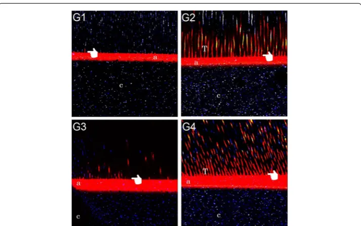
The confocal microscopy (Fig. 1) shows a regular hybrid layer with small resin tags (G1); greater infiltration of adhesive resins into dentinal tubules (G2); decrease in the resin tags length (G3); and the formation of small, but many resin tags (G4).
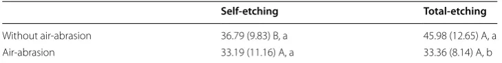
Table 1 Mean values for μTBS (±SD) of surface treatments
Data with different lowercase letters in column and capital letters in row are statistically different (p < 0.05)
Self-etching Total-etching
Without air-abrasion 36.79 (9.83) B, a 45.98 (12.65) A, a
Discussion
The bioactive glasses are used in minimally invasive techniques for caries removal because hardness of these materials very similar to those of dental tissue [18]. Moreover, the bioactive glasses also have ability to stimulate the remineralization of demineralized tissue, promoting crystal growth similar to apatite presents in dental tissue [8]. The Bio-glass 45S5 is composed by silicon dioxide, calcium oxide, sodium oxide and phosphorus pentoxide, with fillers size ranging from 30 to 90 µm [19].
The hypothesis was rejected, since after the Bioglass 45S5 air-abrasion the microten-sile bond strength value decreased in the total-etching groups (G2 and G4). According to etching technique, the results showed that for the groups without air-abrasion (G1 e G2) the total-etching technique (G2) had highest μTBS mean values. The self-etch-ing adhesive system has higher pH (~2.7) than total-etchself-etch-ing (pH = 0.6). This higher pH may not remove the same amount of mineral that phosphoric acid used in total-etching may remove. Thus the demineralization is restricted a surface layer, not reaching deep dentin [20]. The confocal microscopy evaluation (Fig. 1) shows this situation, where it can be observed a large region of dye penetration in G2 and G4 (total-etching) show-ing the complete demineralization caused by phosphoric acid etchshow-ing. Futhermore, the hydrophilic nature of the self-etching adhesive system can act as membrane permeable, attracting water and degrading faster than more hydrophobic adhesives [21]. The self-etching adhesive system is hydrolytically unstable, so the presence of porosities within the hybrid layer confirms that the nanoleakage which promote water movement in the tooth-restoration interface. This movement of water within the bonding interface may result in plasticization of the resin matrix causing a decrease in bond strength [22].
However, in the air-abrasion groups (G3 and G4) both dentin etching technique showed similar bond strength values and the air-abrasion caused a decrease on
microtensile bond strength ìn the total-etching groups (G2 and G4). The great dem-ineralization caused by acid etching exposes a large amount of collagen fibrils and the air-abrasion with Bioglass 45S5 for 1 min may cause the collapse of the demineralized collagen fibers, jeopardizing the adhesion between adhesive and dentin decreasing the bond strength. Further studies should be performed to confirm this hypothesis. The self-etching with air-abrasion Bioglass 45S5 (G3) not improve the microtensile bond strength. The presence of the functional monomer 10-MDP may have compete with Bio-glass 45S5, since this monomer shows a phosphate group attached to carbon chain [23]. In water this monomer ionizes creating covalent bonds with Ca+2 and PO4 ions pre-sent in dentin [24]. Moreover, the alcohol (adhesive solvent) may also interfere on bond strength [25].
Although the Bioglass 45S5 air-abrasion has not improved the bond strength, the bio-activity of this material may induce hydroxyapatite formation within the bonding inter-face, protecting the demineralized dentin collagen against endogenous dentin proteases [18] improving the quality of the hybrid layer. It can be observed also in Fig. 1 that Bio-glass 45S5 air-abrasion promoted a hybrid layer with a good inter–diffusion zone (G3). In this study the teeth were stored only for 24 h after the Bioglass 45S5 air-abrasion. This time is impracticable for that this material induces the dentin remineralization pro-cess by calcium precipitation [26]. Higher storage times may improve the bond strength results, inducing the remineralization process. Futhermore, the mineralization con-sumes the dentin water [27] and may reduce the hydrolytic degradation that occurs in this layer [28].
Conclusions
The Bioglass 45S5 air-abrasion did not promote improvements on μTBS values in self-etching groups and decreased the μTBS values for total-self-etching groups, however caused the formation of a hybrid layer with a continuous inter–diffusion zone, greater infiltra-tion of adhesive resins into dentinal tubules and many resin tags.
Authors’ contributions
MA: analysis and interpretation of data and performed statistical evaluation; RP: substantial contributions to introduc-tion and discussion topics, drafting the work and wrote the manuscript; GA: performed the tests and acquisiintroduc-tion of data, interpretation of data for the research; VP: contributed to idea of this study, designed research. All authors read and approved the final manuscript.
Author details
1 Department of Operative Dentistry, Piracicaba Dental School, University of Campinas, Av. Limeira 901, Piracicaba, SP
13414-903, Brazil. 2 Department of Prosthodontics, School of Dentistry, University of Taubaté, 09 Rua Dos Operários,
Taubaté, SP 12020-270, Brazil. 3 Department of Operative, School of Dentistry, Federal University of Ceará, Fortaleza, Rua
Alexandre Baraúna 949, Fortaleza, CE 60430-160, Brazil. Competing interests
The author(s) declare that they have no competing interests.
Received: 19 November 2015 Accepted: 3 December 2015
References
1. LeGeros RZ. Properties of osteoconductive biomaterials: calcium phosphates. Clin Orthop Relat Res. 2002;395:81–98. 2. Ryou H, Niu LN, Dai L, Pucci CR, Arola DD, Pashley DH, Tay FR. Effect of biomimetic remineralization on the dynamic
nanomechanical properties of dentin hybrid layers. J Dent Res. 2011;90:1122–8.
4. Kim YK, Gu LS, Bryan TE, Kim JR, Chen L, Liu Y, Yoon JC, Breschi L, Pashley DH, Tay FR. Mineralisation of reconstituted collagen using polyvinylphosphonic acid/polyacrylic acid templating matrix protein analogues in the presence of calcium, phosphate and hydroxyl ions. Biomaterials. 2010;31:6618–27.
5. Peters MC, Bresciani E, Barata TJ, Fagundes TC, Navarro RL, Navarro MF, Dickens SH. In vivo dentin remineralization by calcium-phosphate cement. J Dent Res. 2010;89:286–91.
6. Liu Y, Kim YK, Dai L, Li N, Khan SO, Pashley DH, Tay FR. Hierarchical and non-hierarchical mineralisation of collagen. Biomaterials. 2011;32:1291–300.
7. Narongdej T, Sakoolnamarka R, Boonroung T. The effectiveness of a calcium sodium phosphosilicate desensitizer in reducing cervical dentin hypersensitivity: a pilot study. J Am Dent Assoc. 2010;141:995–9.
8. Vollenweider M, Brunner TJ, Knecht S, Grass RN, Zehnder M, Imfeld T, Stark WJ. Remineralization of human dentin using ultrafine bioactive glass particles. Acta Biomater. 2007;3:936–43.
9. Kinney JH, Habelitz S, Marshall SJ, Marshall GW. The importance of intrafibrillar mineralization of collagen on the mechanical properties of dentin. J Dent Res. 2003;82:957–61.
10. Bertassoni LE, Habelitz S, Marshall SJ, Marshall GW. Mechanical recovery of dentin following remineralization in vitro—an indentation study. J Biomech. 2011;44:176–81.
11. Dickens SH, Flaim GM. Effect of a bonding agent on in vitro biochemical activities of remineralizing resin-based calcium phosphate cements. Dent Mater. 2008;24:1273–80.
12. Ngo HC, Mount G, Mc Intyre J, Tuisuva J, Von Doussa RJ. Chemical exchange between glass-ionomer restorations and residual carious dentine in permanent molars: an in vivo study. J Dent. 2006;34:608–13.
13. Arends J, Ruben JL, Inaba D. Major topics in quantitative microradiography of enamel and dentin: R parameter, mineral distribution visualization, and hyper-remineralization. Adv Dent Res. 1997;11:403–14.
14. Dickens SH, Flaim GM, Takagi S. Mechanical properties and biochemical activity of remineralizing resin-based Ca-PO4 cements. Dent Mater. 2003;19:558–66.
15. Ana ID, Matsuya S, Ohta M, Ishikawa K. Effects of added bioactive glass on the setting and mechanical properties of resin-modified glass ionomer cement. Biomaterials. 2003;24:3061–7.
16. Banerjee A, M Hajatdoost-Sani, S Farrell, Thompson I. A clinical evaluation and comparison of bioactive glass and sodium bicarbonate air-polishing powders. J Dent. 2010;38:475–9.
17. Banerjee A, Paolinelis G, Socker M, McDonald F, Watson TF. An in vitro investigation of the effectiveness of bioactive glass air-abrasion in the ‘selective’ removal of orthodontic resin adhesive. Eur J Oral Sci. 2008;116:488–92. 18. Sauro S, Watson TF, Thompson I, Toledano M, Nucci C, Banerjee A. Influence of air-abrasion executed with
poly-acrylic acid-Bioglass 45S5 on the bonding performance of a resin-modified glass ionomer cement. Eur J Oral Sci. 2012;120:168–77.
19. Wang Z, Jiang T, Sauro S, Wang Y, Thompson I, Watson TF, Sa Y, Xing W, Shen Y, Haapasalo M. Dentine remineraliza-tion induced by two bioactive glasses developed for air abrasion purposes. J Dent. 2011;39:746–56.
20. Van Meerbeek B, Kanumilli P, Munck JD, Landuyt KV, Lambrechts P, Peumans M. A randomized controlled study evaluating the effectiveness of a two-step self-etch adhesive with and without selective phosphoric-acid etching of enamel. Dent Mater. 2005;21:375–83.
21. Tanaka J, Ishikawa K, Yatani H, Yamashita A, Suzuki K. Correlation of dentin bond durability with water absorption of bonding layer. Dent Mater J. 1999;18:11–8.
22. Hashimoto M, Tay FR, Ito S, Sano H, Kaga M, Pashley DH. Permeability of adhesive resin films. J Biomed Mater Res B Appl Biomater. 2005;74:699–705.
23. Van Landuyt KL, Snauwaert J, De Munck J, Peumans M, Yoshida Y, Poitevin A, Coutinho E, Suzuki K, Lambrechts P, Van Meerbeek B. Systematic review of the chemical composition of contemporary dental adhesives. Biomaterials. 2007;28:3585–757.
24. Fukegawa D, Hayakawa S, Yoshida Y, Suzuki K, Osaka A, Van Meerbeek B. Chemical interaction of phosphoric acid ester with hydroxyapatite. J Dent Res. 2006;85:941–4.
25. Sauro S, Watson T, Thompson I, Banerjee A. One-bottle self-etching adhesives applied to dentine air- abraded using bioactive glasses containing polyacrylic acid: an in vitro microtensile bond strength and confocal microscopy study. J Dent. 2012;40:896–905.
26. Gandolfi MG, Taddei P, Siboni F, Modena E, De Stefano ED, Prati C. Biomimetic remineralization of human dentin using promising innovative calcium-silicate hybrid “smart” materials. Dent Mater. 2011;2011(27):1055–69.
27. Wang Y, Von Euw S, Fernandes MF, Cassanignon S, Selmane M, Laurent G, Pehau-Arnaudet G, Coelho C, Bonhomme-Coury L, Giraud-Guille MM, Babonneau F, Azaïs T, Nassif N. Water-mediated structuring of bone apatite. Nat Mater. 2013;12:1144–53.