R E S E A R C H
Open Access
Separating mouse malignant cell line (EL4)
from neonate spermatogonial stem cells
utilizing microfluidic device in vitro
Behnaz Ashtari
1,2,3, Azar Shams
4, Narges Esmaeilzadeh
3, Sara Tanbakooei
5, Morteza Koruji
4,5,
Mojtaba Johari Moghadam
6, Javad Mohajer Ansari
4, Adel Johari Moghadam
7and Ronak Shabani
4,5*Abstract
Background:Some children who have survived cancer will be azoospermic in the future. Performing isolation and purification procedures for spermatogonial stem cells (SSC) is very critical. In this regard, performing the process of decontamination of cancerous cells is the initial step. The major objective of the present study is to separate the malignant EL4 cell line in mice and spermatogonial stem cells in vitro.
Methods:The spermatogonial stem cells of sixty neonatal mice were isolated, and the procedure of co-culturing was carried out by EL4 which were classified into 2 major groups: (1) the control group (co-culture in a growth medium) and (2) the group of co-cultured cells which were separated using the microfluidic device. The percentage of cells was assessed using flow cytometry technique and common laboratory technique of immunocytochemistry and finally was confirmed through the laboratory technique of reverse transcription-polymerase chain reaction (RT-PCR).
Results: The actual percentage of EL4 and SSC after isolation was collected at two outlets: the outputs for the smaller outlet were 0.12% for SSC and 42.14% for EL4, while in the larger outlet, the outputs were 80.38% for SSC and 0.32% for EL4; in the control group, the percentages of cells were 21.44% for SSC and 23.28% for EL4 (based on t test (p≤0.05)).
Conclusions: The present study demonstrates that the use of the microfluidic device is effective in separating cancer cells from spermatogonial stem cells.
Keywords: Microfluidic device, Spermatogonial stem cells, EL4 cancer cell, Cell sorting, Purification
© The Author(s). 2020Open AccessThis article is licensed under a Creative Commons Attribution 4.0 International License, which permits use, sharing, adaptation, distribution and reproduction in any medium or format, as long as you give appropriate credit to the original author(s) and the source, provide a link to the Creative Commons licence, and indicate if changes were made. The images or other third party material in this article are included in the article's Creative Commons licence, unless indicated otherwise in a credit line to the material. If material is not included in the article's Creative Commons licence and your intended use is not permitted by statutory regulation or exceeds the permitted use, you will need to obtain permission directly from the copyright holder. To view a copy of this licence, visithttp://creativecommons.org/licenses/by/4.0/. The Creative Commons Public Domain Dedication waiver (http://creativecommons.org/publicdomain/zero/1.0/) applies to the data made available in this article, unless otherwise stated in a credit line to the data.
* Correspondence:[email protected]
4
Cellular and Molecular Research Center, Iran University of Medical Sciences, Tehran, Iran
5School of Mechanical Engineering, Iran University of Science & Technology,
Tehran, Iran
Background
The spermatogonium that are present in the testicles from birth are the precursors to the production of male sex cells. The presence of these kinds of cells is very essential for performing the process of spermatogenesis. But unfortunately, these cells are not capable of making the mature sperm before the puberty period due to the fact that they are dependent on hormonal stimuli [1]. Sometimes, this system is in difficulty, and it is possible to maintain the reproductive capacity and fertility system in men that can ejaculate. In recent years, the success rate of chemotherapy or radiotherapy for the treatment of childhood cancers has been high, whereas more than 70% of children with cancer reach adulthood and repro-ductive years. Infertility is one of the most common complications of childhood cancer in treated boys in the long run. Unfortunately, about one third of children dur-ing their puberty period would experience a considerable decrease in the number of sperms or may face with the medical condition of azoospermia [2, 3]. This can jeopardize their quality of life [4].
In recent years, many attempts have been made to cryopreserve testicular tissue and then to transplant it after chemotherapy and cancer relief, and the willingness of physicians has increased in this way [5, 6]. Cryopre-served testicular tissues have been obtained from boys with cancer before chemotherapy begins to be used to produce sperm germ cells using different culture methods. Before starting chemotherapy, the germ cell (spermatogonial stem cell) is isolated and maintained, and after the patient’s treatment period and after pu-berty, the cells can be transplanted to the patient and, as a result, fertility can be maintained in these individuals. However, there is a risk of contaminated germ cells taken by tumor cells [7]. The risk of interstitial and intravascular infiltration of testicular tissue among chil-dren would increase due to the hematological metastatic spread of childhood solid tumors. Additionally, in chil-dren with acute lymphoblastic leukemia (ALL) cancer, the risk of interstitial and intravascular infiltration of tes-ticular tissue is very substantial too. Anyway, among one fifth of patients diagnosed with this kind of tumor, microscopic infiltration of leukemic cells would be seen in their blood tests [8].
Jahnukainen reported that germ cell transplantation from leukemic mice induced tumor formation. Germ cells must, therefore, be completely separated from the tumor cells [7]. Fujita et al. isolated the germ cell from 5 different human cancer cell lines using the FACS tech-nique. In this study, human germ cells from 5 cell lines of leukemia by using anti-MHC-I and CD45+ antibodies (specific tumor cell markers) were isolated. In this study, it is shown that, with this method, spermatogonial trans-plantation is not safe enough [9]. Geens et al.
investigated the separation of germ cells from tumor cells in mice and in human testis cell suspensions using MACS and FACS. Initially, they created a cancerous model in the mouse by infusion of the EL4 tumor cell line; after co-culture isolation, separation of cell suspen-sion was performed using MACS and FACS, and after that, unfortunately one of the 20 transplanted mice got malignant after transplantation. Moreover, Hou et al. [10] have tested the MACS method and showed that the separation method of magnetic-activated cell sorting was not able to remove malignant contamination effectively [10]. As a result, MACS and FACS were not sufficiently effective for complete tumor cell removal from the tes-ticular tissue [11]. Furthermore, these methods are com-plex and costly, and the survival of cells is poorly reported. Differential plating was also suggested by some articles to enrich spermatogonia in cell suspension derived from the testis sample after enzymatic digestion [12,13]. Moreover, during this stage of studies perform-ing the identification process of specific markers, isola-tion and enrichment of undifferentiated spermatogonia from differentiated germ cells and vegetal cells were per-formed precisely by researchers. However, conducting the process of isolation of blood-related malignancies from germ cells requires a more precise assessment be-fore performing any related clinical procedures. In a study conducted by Dirami and colleagues [12], by utiliz-ing differential adhesion and sedimentation velocity (separating based on shape and size), an isolate of cells was created that contains 95 to 98% porcine type A spermatogonia. In their study, Shinohara et al. [14] dem-onstrated that by using the technique of laminin adhe-sion, the process of isolation of spermatogonial stem cells (SSCs) was improved to 3-fold [14]. In another similar study, Morena et al. [15] demonstrated that attaining an 85% isolate of type A, c-kit-positive sperm-atogonia utilized the technique of sedimentation velocity (SV-AUC) in conjunction with differential adhesion hy-pothesis (DAH) [15].
Due to the lack of specific SSC cell markers, it should focus on other methods. But it turns out that the focus on destroying cancer cells instead of focusing on healthy cells is also a new method used in recent years. Tumor cells should be targeted as well, and studies have been carried out in this regard.
with a dose of 15 mg cisplatin decreased significantly compared with the control group (p≤0.05). Besides, at different times, there was a significant difference be-tween the half maximal inhibitory concentration (IC50) in doses 10 and 15 mg/ml [16]. Chemotherapy drug re-lease method requires an intelligent tool to select and target cancer cells which today uses nanomaterials to achieve this goal.
In a study conducted by Shabani et al., the anticancer effect of cisplatin encapsulated in spermatogonial stem cells (SSCs) from in vitro and folic acid-conjugated poly(lactic-co-glycolic acid) (PLGA) nanoparticles (NPs) on malignant EL4 cell line of mice was assessed. As their main study outcomes, the rate of caspase 3 and BAX genes in EL4 cells increased, and an increase was ob-served in the TUNEL-positive cells. Cells treated with carrier nanoparticles were then grafted to the mouse, and no tumor symptoms were observed [17]. Eslahi et al. devised a new method for removing cancer cells through gold nanoparticles (AuNPs) by Folate-Silica-Gold Nanorods (F-Si-GNRs); based on their outcomes, in comparison with SSCs, an increment in the signs of F-Si-GNR toxicity was observed in EL4 cells [18]. On the other hand, as shown by Beebe et al. [19], conduct-ing the sortconduct-ing process of microfluidic cell may be one of the most critical techniques for isolating the imma-ture cells of spermatogonia based on their density and size [19].
The advancement of microfluidic technology has had a tremendous impact on the progress of cell biology sci-ence [20]. The benefits of this new method to non-traditional and non-traditional methods include controlling 3-D culture conditions, having micro-scale physical and fluid properties, and creating multiplexed nanoliter ar-rays and paths to improve biological research [21–23].
Very low sample size, very fast processing, multi-functionality, and a very large volume/volume ratio of microfluidic system are features [24, 25] that offer new opportunities for cytology and cytopathology, especially
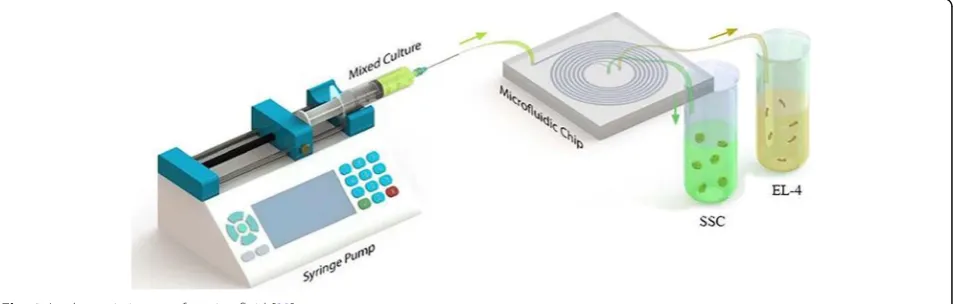
for cell sorting and detection [26–32]. Leveraging these advantages, various microfluidic platforms have been de-veloped (Fig. 1). The next important step is to expand microfluidic systems for greater and more efficient use, commercialization and ease of use, industrial improve-ments, and more effective cost reductions for a long-term continuous perfusion cell culture like a bioreactor [33].
In this current study, we presented a new technique of cell sorting of spermatogonial stem cells from cancerous cells. Based on the performed studies, the present study is the first scientific trial that has focused on research in-vestigating the process of delivering a microfluidic-based cell culture system for achieving this objective. For spreading the advantages of applying microfluidic tech-nology to a broader practical scope, the aforementioned methodology must be developed and integrated into re-search and screening laboratories.
Methods
Device designed and fabricated
With respect to the advent of the novel technique of separation, a new spiral microfluidic device was devel-oped by Warkiani et al. [34]. The pattern of the designed spiral chip device (in the CAD software environment) with an eight-loop spiral microchannel has a unique in-let and two separate outin-lets with a variable radius that varies from 8 to 24 mm. The cross section (channel width) is 600μm, and the heights of the inner and outer sections were fitted at 80 and 130μm respectively as the best values for the trapezoid cross section. Then, to cre-ate a master, the pattern was printed by a high-resolution printer and was used as a mask in photolith-ography on a SU-8 (MicroChem) spin-coated on a on a thin slice of semiconductor (silicon wafer). The device was prepared by mixing of PDMS prepolymer and the curing agent (10:1 ratio) (sylgard 184 Dow Corning) and by degassing onto the master and backing for 2 h at 70 °C. After the baking procedure, the replica of PDMS was peeled off from the master. The surface of the two
PDMS replica was oxidized by plasma machine and was bonded irreversibly together. To enhance the bonding, the device was placed inside an oven (30 min at 70 °C). Both of fluidic inlets and outlets were punched (with 1.5 mm diameter) and connected respectively to the syringe pump and the output container (two sterile 15 mL fal-con) by Tygon® tubing.
Sample processing and cell loading
Before sample processing, the spiral chip was washed by ethanol 70% and sterile medium. Using a syringe driver, all samples were split through the inlet into the spiral microfluidic device. For performing the cell separation process appropriately, the optimized flow rate of the sample in microchannels was 1.7 ml/min; typically, into the spiral microchannel inner wall flows the cells larger than 12μm and cells smaller than that flow into the outer wall. In the end, these cells will separate by inner and outer outlets. The collected cells can then be ana-lyzed by suitable downstream techniques such as immunostaining.
Isolation of mouse spermatogonial stem cells
For conducting the present examination, sixty neonatal mice models in the age range of 3 and 6 days old were chosen. All of these mice were taken from the National Medical Research Institute (Tehran, Iran). They were kept in cages made of plastic in a room at a temperature range of 22–25 °C, with a 12-h light/dark cycle. The mice could freely reach drinking water and standard la-boratory pellets. All animal experiments were approved by the Animal Ethics Committee at Iran University of Medical Sciences (code: IR-IUMS.95-04-117-29910). Nearly all germ cells of testicles were isolated by means of the aforementioned techniques along with some mod-ifications [16, 35]. Mouse testes were collected in phosphate-buffered saline (PBS, Invitrogen, USA) and penicillin/streptomycin. After decapsulation, we used a 2-step enzymatic digestion protocol to obtain a single cell suspension. The testes were mechanically dissociated by two-step enzymatic digestion. The testicles were mechanically and enzymatically digested and isolated. In the first stage, the testicles were divided into smaller pieces and incubated in enzymatic solutions. The first stage enzyme solution contains the following: Dulbecco’s modified Eagle’s medium (DMEM/f12) with 0.05 mg/ml DNase, 1 mg/ml trypsin, and 1 mg/ml collagenase for about half hour with pipetting and shaking at 37 °C for a period of 15–30 min. The digestion process of tissue was carried out by enzyme washing and then centrifugation, and finally, through draining the supernatant solution, the interstitial cells were removed from the seminiferous tubules. The remnants of non-digested seminal tubes en-tered the second stage of enzymatic digestion so that all
cells were extracted from the tubes at this stage. Finally, the isolated spermatogonium and Sertoli cells were cul-tured in special culture media in DMEM/f12 medium (DMEM/f12; Gibco, Paisley, UK), non-essential amino acids, 2% Bovine serum albumine (BSA) (Sigma, MO, USA), 100μg/ml streptomycin, and 100 IU/ml penicillin (from Gibco, Paisley, UK). It was a conventional cell condition, and it was continued for 2 weeks to increase the cell number. Finally, in the present study, three main groups were designed, and by accessing the culture col-lection of Pasteur Institute, Tehran, Iran, the mouse acute lymphoblastic leukemia cell line EL4 was prepared.
Identity confirmation of the spermatogonial cell by RT-PCR
The confirmation of the nature of spermatogonial cells in the culture medium was investigated by expressing specific genes of these cells according to previous studies.
Testicular cells before cultivation and total RNA mole-cules as a positive control obtained from the testis sam-ples were extracted by the standard extraction RNX-plus kit in accordance with guidelines presented by its manu-facturer (Cinnagen, Iran). Then, the integrity and purity RNA was examined using a ratio measurement of 26/28 nm. Aimed to eliminate residual genomic DNA (gDNA) contamination, total RNA was treated by means of de-oxyribonuclease I (DNase I). Initially, by means of SuperScript II Reverse Transcriptase (RT) system and Oligo (dT)18 Primers, strand complementary DNA (cDNA) synthesis was performed.
Cell confirmation by flow cytometry
For confirmation of the cell value, spermatogonial stem cells (SSCs) were fixed in a paraformaldehyde (PFA) product of 4% in the buffer solution of phosphate-buffered saline (PBS) with a pH of 7.4. After that, the process of washing of cells was conducted three times with paraformaldehyde (PFA) product, incubating in a 1% nonionic surfactant of Triton X-100 in phosphate-buffered saline for a period of half hour and blocking in 0.5% liquid of Bovine serum albumine (BSA) and finally in PBS buffer solution for about half hour. Then, the cells were incubated in a special antibody solution that contains a primary antibody protein of PLZF (Abcam) at 4 °C for a period of time less than 45 min and were then examined. The malignant cell line of EL4 was fixed in a paraformaldehyde (PFA) product of 4% in a PBS buffer solution with a pH of 7.4 and then for a period of half hour washed with the buffer solution of PBS and was in-cubated with a special H-2Kb antibody (Abcam) and fi-nally assessed precisely (Fig.5).
Tumorigenic evaluation of cells after microfluidic isolation
For this purpose, we first transformed healthy rats into azoospermia mice by intraperitoneal injection of busul-fan 4 weeks before transplantation. Then, for tumor evaluation, cells were transplanted into azoospermia mice after microfluidic gates, and after 8 weeks, the tumor status was checked (Fig.7).
Statistical analysis
The procedure of analyzing data was performed using SPSS Statistics V22, and the statistical significance threshold was determined to be p≤0.05. In this study, the Tukey test and an independent ttest were used for comparing the cell percentages.
Results
EL4 tumor cell culture was performed in DMEM/F12 medium with FBS 2%, and the percentage of viable cells
was about 80 ± 2.4%. The nature of this cell line during the culture was suspended cells and did not stick to the culture dish. As shown in Fig. 1, their appearance was not spherical and did not form a colony. After 24 h, there was a significant amount of cell clinging to the flask. By invert microscope examination, spermatogon-ium was circular or oval, with a large nucleus and a small cytoplasm. The isolated SSCs tend to form col-onies and form a small cell cluster. The proliferation rate of these cells was very high, with almost every 48 h of cell passage. Also, El4 cells, which were suspended in culture medium, were simultaneously cultured and stored (Fig.2).
In order to proliferate spermatogonial stem cells, these cells were cultured in DMEM/F12 medium including 2% FBS with GDNF 20 ng/ml and 10 ng/ml BFGF for 2 weeks. At the end of the first week, the process of for-mation of cluster stem cell assemblages started after about 4 h since in the first passage and a large number of stem cells were colonized in a colony (Fig. 2) culture medium.
Expression of specific genes of SSCs and EL4 cells using RT-PCR
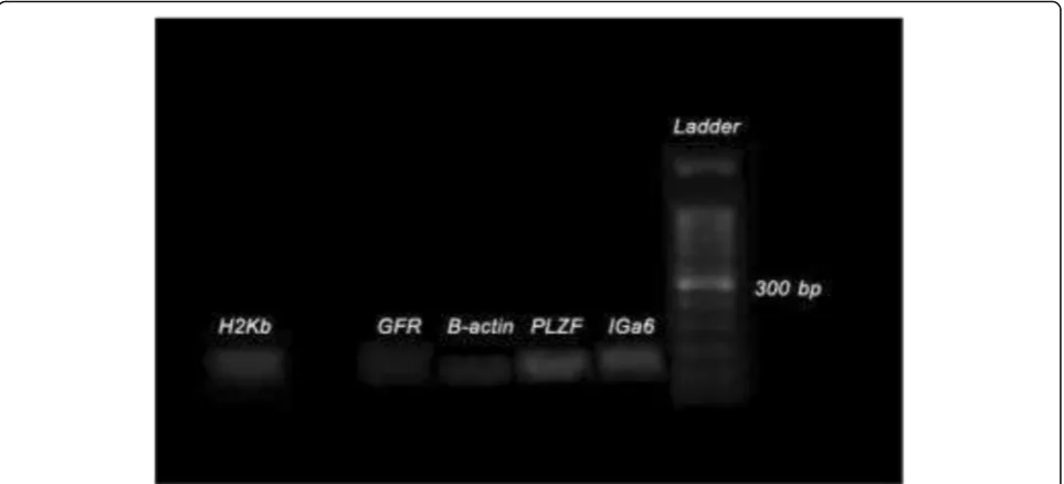
As could be seen from Fig.3, specific markers of sperm-atogonial stem cells (SSCs) (Integα-6, GFRα-1, PLZF) in cells after 2 weeks of culture (SC2) and the EL4 marker of H2K-b EL4 cells from product excretion RT-PCR have been proven. Also,β-actin was also observed as the house keeping gene in both samples.
Determination of the percentage of EL4s and SSCs after microfluidic separation by flow cytometry
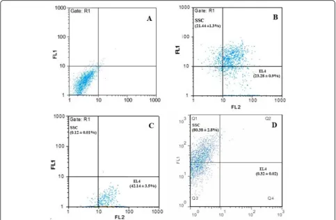
In order to evaluate the percentage of spermatogonial stem cells and tumor cells, flow cytometry was used to de-termine the percentage of the cells. As shown in Fig. 3, the percentage of tumor cells and spermatogonial stem cells after microfluidic isolation was collected at two out-lets, and the outer outlet were approximately 0.12 ± 0.01%
(SSC) and 42.14 ± 3.5% (EL4). While the outputs col-lected from the device for inner outlet were 80.38 ± 2.8% (SSC) and 0.32 ± 0.02 (EL4) in the control group, the percentages of SSC and EL4 cells were 21.44 ± 1.3% and 23.28 ± 0.9%, respectively, which did not enter the microfluidic apparatus and were individually mixed in a cell dish (based on t test (p≤0.05) (Figs.4
and 5).
Immunocytochemistry
After isolating the SSC and EL4 cellular composition using the microfluidic device, the cells were cultured in
separate plates for 1 week, and after the end of the first week, the immunocytochemistry test was performed to confirm the microfluidic cells, so for cell EL4, the conju-gated CD45 marker was PE, and for the SSCs, the PLZF conjugate marker was used with FITC, which was initially fixed at 4 °C in PBS with PH7.4 for 20 min. After three times washing with PBS, the cells were exposed to Triton X-100 for 10 min in order to penetrate the cells, and then, after three times, the PBS was incubated in 10% goat serum (Sigma, Missouri, USA) for 1 h. It was then incu-bated with 10μg/ml antibodies for CD45 and PLZF for 2 h at room temperature. Then, it was washed with 1% goat serum in PBS three times and incubated with FITC-conjugated secondary antibody for 2 h at RT away from Fig. 3Results of RT-PCR production of spermatogonial stem cells and EL4 cells. The expression of PLZF, GFRα-1, and Integα-6 in spermatogonial stem cells and H2Kb for EL4 cells.β-actin was included as a housekeeping gene
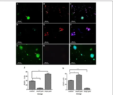
light, PE-conjugated, and stained with PBS for coloration of the cells with DAPI for 3 min. The coverslips were mounted and observed under a Nikon Eclipse TE300 Inverted microscope (country). Sample images were cap-tured by the CCD camera directly connected to the micro-scope mentioned above (Fig. 6). The ICC data were quantified on the basis of ImageJ software and were ana-lyzed by one-way ANOVA, and the results showed that for the smaller gate output, the number of CD45+ was more than the number of PLZF+ cells and was statistically significant (55 ± 2% CD45+ vs 6 ± 1% PLZF+, control 35 ± 1%,p≤0.005), and for the larger gate output, the number of PLZF+ cells was statistically significant than CD45+ (70 ± 4% PLZF+ vs 6 ± 1% CD45+, control 38 ± 2%, p≤ 0.005).
Histological assessment
The tissue sections of testis after transplantation were checked to confirm the tumorigenicity of the cells and pathological changes from the tumor. As shown in Fig. 7c, compared with the control group (a), the
arrangement of epithelial cells of the seminal tubes in the tumor model was degraded and uncertain so that the structure of tissue and tubes was severely disrupted. Other typical pathological changes associated with tes-ticular cancer, including tumor invasion and hyperplasia of the testicular tissue, high numbers of lymphocytes, and the loss of the order of the spermatozoa tubes, were seen. In histopathologic sections of the EL4 cell line and SSCs, no pathologic evidence of tumor was detected after microfluidic isolation, and seminal tubes with nor-mal appearance in histological sections were observed. In addition, spermatogonial stem cell transplantation has improved the relative position and structure of seminal tubes and the onset of spermatogenesis in many seminal tubes (b).
Discussion
Fig. 6Part A is for the PLZF marker for SSC cells and part B is for tumor cell CD45 markers and the MERGE-shaped image of the above images and DAPI staining. Based on flow cytometry, in the first row, the control group, which contains the SSC and EL4 cell composition, is seen, and both types of cells are seen. In the 2nd row is the output from the outer inlet, which indicates a higher number of EL4 cell. The 3rd row refers to the output of the inner outlet which indicates a higher number of PLZF markers, the SSC cell
preservation of male fertility is highlighted which is the destructive effect of chemotherapy on reproductive func-tion among men, especially in the process of spermato-genesis. At the present time, one of the most critical ways for preserving the natural capability of fertility among boys before their puberty period is performing the process of testicular tissue cryopreservation. This procedure is performed mainly for future isolation and transplantation of spermatogonial stem cells for restor-ing the process of spermatogenesis [36].
The main concern in the cryopreserved testicular tis-sue is the potential of cancer cells in the biopsy. Patients with non-tumor cancers are more likely to be at risk, the resumption of cancer [36]. In addition to the available risks of reintroducing lingering tumor cells, the culture system of SSCs could provide the opportunity of select-ing the best way for the isolation of cancer cells from healthy cells.
The high precision and significant amount of cell sep-aration should be the main feature of cell sepsep-aration methods. As long as traditional methods can have high efficiency in the process of cell sorting in a short period of time, the achieved progress in microfluidics has rein-forced the realization of miniaturized devices to be able to offer similar capabilities that could extract a broad range of physical principles.
Cell separation methods are rapidly expanding to allow them to target and isolate small numbers of cells such as circulating fetal cells (CFCs), hematopoietic stem cells (HSCs), and circulating tumor cells (CTCs) from the blood [37–39]. Several methods for cell sorting are cur-rently underway, with a large and clear limitations that include the following: low sampling rate and small plers that do not have the ability to work on larger sam-ples at a wider scale and in a shorter period of time (more than 500 million cells), high operating pressures that can reduce the viability and/or function of equip-ment and devices that occupy a lot of space, and enough experience to work with the above equipment and in-creased risk of safety concerns and sample contamin-ation because of performing an aerosolized sample sorting procedure [40]. Older methods for isolating cells have disadvantages, for example, in FACS, the disadvan-tage for sorting cells is electrolysis of water based on the current of electricity between the cathode and anode poles, which could be one of the main causes of generat-ing harmful compounds like hydrogen peroxide (H2O2) and bubbles, which would affect cell viability and sur-vival and also the pH of a solution seriously if not be regulated and monitored appropriately, or the use of nanoparticles to remove cancer cells and isolate healthy cells, which are considered as newer methods [41, 42]. Studies have shown that nanoparticles and exposure to them can have devastating effects on prokaryotic cells
and complex eukaryotes such as humans. Many studies have shown that nanoparticles can cause degradation of DNA, inflammatory response, oxidative stress, lipid per-oxidation, apoptosis, carcinogenicity, non-genotoxic (NGTX), immunotoxicity, alterations in gene expression, reproductive toxicity, cytotoxicity, and genotoxicity [43– 50]. The epigenetic changes are among the main avail-able mechanisms for genotoxicity and toxicity of nano-particles that happen in the specific DNA methylation patterns that may cause alternations in the process of gene expression [51]. On the other hand, although proteomic and genomic data suggest changes in the pro-file of protein and gene of cells exposed to NP, epigen-etic changes are underestimated [49,52]. Due to the fact that microchips could precisely control temporal and spatial conditions in an appropriate miniaturized droplet microarray platform, they could easily monitor and con-trol cells [20,53].
In this study, the percentages of tumor cells and spermatogonial stem cells after microfluidic isolation were collected at two outlets, and the outputs for smaller tumor cells were approximately 0.12% (SSC) and 42.14% (EL4). While the outputs collected from the device for larger cells were 80.38% (SSC) and 0.32% (EL4) in the control group, the percentages of cells were 21.44% (SSC) and 23.28% (EL4). Bleilevens et al. also showed in their study that this method is the best method for con-tinuous separation of cells without labeling of red blood cells and platelets [54]. The cell separation function was slightly comparable to that of MACS and FACS devices for whole blood, for instance, purities are ranging, but as high as 99%, and throughputs are up to 48,000 cells [32,
55, 56]. Additionally, based on the data presented by Son et al. [57], at two outer wall outlets, all isolated sperm cells were obtained from the red blood cells (RBCs) and also nearly 81% of non-progressive motility sperms were successfully recovered. On the other hand, at two inner wall outlets at a flow rate of 0.52 ml min−1 with the system, nearly 99% of the red blood cells are successfully recovered [57]. The aforementioned devices could be made by standardized superior microfabrica-tion systems that reduce the cost and complexity of commercialization efforts [58–62]. The device provides a new approach for cancer cell sorting with high through-put and purity.
Conclusion
malignant cells in an individual that has just been treated and cured of cancer.
Abbreviations
SSC:Spermatogonial stem cells; DMEM: Dulbecco’s modified Eagle’s medium; CTCs: Circulating tumor cells; HSCs: Hematopoietic stem cells;
CFCs: Circulating fetal cells; FACS: Fluorescence-activated cell sorting; MACS: Magnetic-activated cell sorting
Acknowledgements
The authors would like to kindly acknowledge all the supports from the Iran University of Medical Sciences (IUMS) grant (95-04-117-29910). All experiments were performed at the cellular and molecular research center and radiation biology research center, Tehran, Iran (IUMS).
Authors’contributions
KHA, ASH, NE, ST, MK, MJM, JMA, AJM, and RSH conceived the study and contributed to the data analysis and draft and design. All authors
contributed to the interpretation of the results and wrote the manuscript. All authors contributed to writing subsequent drafts of the manuscript. All authors read and approved the final manuscript.
Funding
This study was funded by the Iran University of Medical Sciences (IUMS) grant 95-04-117-29910.
Availability of data and materials
All data generated or analyzed during this study are included in this article.
Ethics approval and consent to participate
All procedures performed in studies involving animals were in accordance with the ethical standards of IUMS (code: IR-IUMS. 95-04-117-29910).
Consent for publication Not applicable
Competing interests
The authors declare that they have no competing interests.
Author details
1Shahdad Ronak Commercialization Company, Pasdaran Street, Tehran, Iran. 2Radiation Biology Research Center, Iran University of Medical Sciences,
Tehran, Iran.3Department of Medical Nanotechnology, Faculty of Advanced
Technologies in Medicine, Iran University of Medical Sciences, Tehran, Iran.
4Cellular and Molecular Research Center, Iran University of Medical Sciences,
Tehran, Iran.5School of Mechanical Engineering, Iran University of Science &
Technology, Tehran, Iran.6Department of Cardiology, Aja University of
Medical Sciences, Tehran, Iran.7School of Mechanical and Manufacturing
Engineering, University of New South Wales, Sydney, New South Wales 2052, Australia.
Received: 12 December 2019 Revised: 25 February 2020 Accepted: 7 April 2020
References
1. Shams A, Eslahi N, Movahedin M, Izadyar F, Asgari H, Koruji M. Future of spermatogonial stem cell culture: application of nanofiber scaffolds. Curr Stem Cell Res Ther. 2017;12(7):544–53.
2. Jemal A, Clegg LX, Ward E, et al. Annual report to the nation on the status of cancer, 1975-2001, with a special feature regarding survival. Cancer. 2004; 101(1):3–27.
3. Humpl T, Schramm P, Gutjahr P. Male fertility in long-term survivors of childhood ALL. Arch Androl. 1999;43(2):123–9.
4. Bauld C, Anderson V, Arnold J. Psychosocial aspects of adolescent cancer survival. J Paediatr Child Health. 1998;34(2):120–6.
5. Struijk RB, Mulder CL, Van Der Veen F, Van Pelt AM, Repping SJBRI. Restoring fertility in sterile childhood cancer survivors by autotransplanting spermatogonial stem cells: are we there yet? 2013 (2013).
6. Gatta G, Botta L, Rossi S, et al. Childhood cancer survival in Europe 1999– 2007: results of EUROCARE-5—a population-based study. Lancet Oncol. 2014;15(1):35–47.
7. Jahnukainen K, Hou M, Petersen C, Setchell B, Söder O. Intratesticular transplantation of testicular cells from leukemic rats causes transmission of leukemia. Cancer Res. 2001;61(2):706.
8. Kim T, Brynes R, Lui V-S, et al. Pretreatment testicular biopsy in childhood acute lymphocytic leukaemia. Lancet. 1981;318(8248):657–8.
9. Fujita K, Tsujimura A, Miyagawa Y, et al. Isolation of germ cells from leukemia and lymphoma cells in a human in vitro model: potential clinical application for restoring human fertility after anticancer therapy. Cancer Res. 2006;66(23):11166–71.
10. Hou M, Andersson M, Zheng C, Sundblad A, Söder O, Jahnukainen K. Immunomagnetic separation of normal rat testicular cells from Roser’s T-cell leukaemia cells is ineffective. Int J Androl. 2009;32(1):66–73.
11. Geens M, Van De Velde H, De Block G, Goossens E, Van Steirteghem A, Tournaye H. The efficiency of magnetic-activated cell sorting and fluorescence-activated cell sorting in the decontamination of testicular cell suspensions in cancer patients. Hum Reprod. 2007;22(3):733–42. 12. Dirami G, Ravindranath N, Pursel V, Dym M. Effects of stem cell factor and
granulocyte macrophage-colony stimulating factor on survival of porcine type A spermatogonia cultured in KSOM. Biol Reprod. 1999;61(1):225–30. 13. Sadri-Ardekani H, Mizrak SC, Van Daalen SK, et al. Propagation of human
spermatogonial stem cells in vitro. Jama. 2009;302(19):2127–34. 14. Shinohara T, Avarbock MR, Brinster RL. Functional analysis of
spermatogonial stem cells in steel and cryptorchid infertile mouse models. Dev Biol. 2000;220(2):401–11.
15. Morena AR, Boitani C, Pesce M, De Felici M, Stefanini M. Isolation of highly purified type A spermatogonia from prepubertal rat testis. J Androl. 1996; 17(6):708–17.
16. Shabani R, Ashtari K, Behnam B, et al. In vitro toxicity assay of cisplatin on mouse acute lymphoblastic leukaemia and spermatogonial stemcells. Andrologia. 2016;48(5):584–94.
17. Shabani R, Ashjari M, Ashtari K, et al. Elimination of mouse tumor cells from neonate spermatogonial cells utilizing cisplatin-entrapped folic acid-conjugated poly (lactic-co-glycolic acid) nanoparticles in vitro. Int J Nanomed. 2018;13:2943.
18. Eslahi N, Shakeri-Zadeh A, Ashtari K, et al. In vitro cytotoxicity of folate-silica-gold nanorods on mouse acute lymphoblastic leukemia and
spermatogonial cells. Cell (Yakhteh). 2019;21(1):14.
19. Beebe DJ, Mensing GA, Walker GM. Physics and applications of microfluidics in biology. Annu Rev Biomed Eng. 2002;4(1):261–86.
20. El-Ali J, Sorger PK, Jensen KF. Cells on chips. Nature. 2006;442(7101):403. 21. Griffith LG, Swartz MA. Capturing complex 3D tissue physiology in vitro. Nat
Rev Mol Cell Biol. 2006;7(3):211.
22. Walker GM, Zeringue HC, Beebe DJ. Microenvironment design considerations for cellular scale studies. Lab Chip. 2004;4(2):91–7. 23. Lee PJ, Hung PJ, Rao VM, Lee LP. Nanoliter scale microbioreactor array for
quantitative cell biology. Biotechnol Bioeng. 2006;94(1):5–14.
24. Squires TM, Quake SR. Microfluidics: fluid physics at the nanoliter scale. Rev Mod Phys. 2005;77(3):977.
25. Whitesides GM. The origins and the future of microfluidics. Nature. 2006; 442(7101):368.
26. Yi C, Li C-W, Ji S, Yang M. Microfluidics technology for manipulation and analysis of biological cells. Anal Chim Acta. 2006;560(1–2):1–23. 27. Gascoyne PR, Vykoukal J. Particle separation by dielectrophoresis.
Electrophoresis. 2002;23(13):1973–83.
28. Gonzalez CF, Remcho VT. Harnessing dielectric forces for separations of cells, fine particles and macromolecules. J Chromatogr A. 2005;1079(1–2):59–68. 29. Liu C, Stakenborg T, Peeters S, Lagae L. Cell manipulation with magnetic
particles toward microfluidic cytometry. J Appl Phys. 2009;105(10):102014. 30. Bhagat AS, Bow H, Hou HW, Tan SJ, Han J, Lim CT. Microfluidics for cell
separation. Med Biol Eng Comput. 2010;48(10):999–1014.
31. Didar TF, Tabrizian M. Adhesion based detection, sorting and enrichment of cells in microfluidic Lab-on-Chip devices. Lab Chip. 2010;10(22):3043–53. 32. Gossett DR, Weaver WM, Mach AJ, et al. Label-free cell separation and
sorting in microfluidic systems. Anal Bioanal Chem. 2010;397(8):3249–67. 33. Lee PJ, Ghorashian N, Gaige TA, Hung PJ. Microfluidic system for automated
cell-based assays. JALA. 2007;12(6):363–7.
34. Warkiani ME, Guan G, Luan KB, et al. Slanted spiral microfluidics for the ultra-fast, label-free isolation of circulating tumor cells. Lab Chip. 2014;14(1): 128–37.
testicular torsion-detorsion mice. J Assist Reprod Genet. 2016.https://doi. org/10.1007/s10815-016-0708-2.
36. Struijk RB, Mulder CL, Van Der Veen F, Van Pelt AM, Repping S. Restoring fertility in sterile childhood cancer survivors by autotransplanting spermatogonial stem cells: are we there yet? BioMed Res Int. 2013; 2013(2314–6133).
37. Armstrong AJ, Marengo MS, Oltean S et al. Circulating tumor cells from patients with advanced prostate and breast cancer display both epithelial and mesenchymal markers. Mol Cancer Res Molcanres. 0490.2010 (2011). 38. Wognum AW, Eaves AC, Thomas TE. Identification and isolation of
hematopoietic stem cells. Arch Med Res. 2003;34(6):461–75. 39. Chen Y, Li P, Huang P-H, et al. Rare cell isolation and analysis in
microfluidics. Lab Chip. 2014;14(4):626–45.
40. Shields Iv CW, Reyes CD, López GP. Microfluidic cell sorting: a review of the advances in the separation of cells from debulking to rare cell isolation. Lab Chip. 2015;15(5):1230–49.
41. Shabani R, Ashjari M, Ashtari K et al. Elimination of mouse tumor cells from neonate spermatogonial cells utilizing cisplatin-entrapped folic acid-conjugated poly (lactic-co-glycolic acid) nanoparticles in vitro. 13 2943 (2018). 42. Eslahi N, Hadjighassem MR, Joghataei MT, et al. The effects of poly L-lactic
acid nanofiber scaffold on mouse spermatogonial stem cell culture. Int J Nanomedicine. 2013;8(4563).
43. Lewinski N, Colvin V, Drezek RJS. Cytotoxicity nanoparticles 4(1), 26–49 (2008). 44. Huang Y-W, Wu C-H, RSJM A. Toxicity of transition metal oxide
nanoparticles: recent insights from in vitro studies. 2010;3(10):4842–59. 45. DjurišićAB, Leung YH, Ng AM et al. Toxicity of metal oxide nanoparticles:
mechanisms, characterization, and avoiding experimental artefacts. 11(1), 26–44 (2015).
46. Luque-Garcia JL, Sanchez-Díaz R, Lopez-Heras I, Camara C, Martin PJTTIaC. Bioanalytical strategies for in-vitro and in-vivo evaluation of the toxicity induced by metallic nanoparticles 43 254–268 (2013).
47. Khanna P, Ong C, Bay BH, Baeg GHJN. Nanotoxicity: an interplay of oxidative stress, inflammation and cell death. 5(3), 1163–1180 (2015).
48. Sarkar A, Ghosh M, Sil PCJJON, Nanotechnology. Nanotoxicity: oxidative stress mediated toxicity of metal and metal oxide nanoparticles. 14(1), 730–743 (2014). 49. Singh N, Manshian B, Jenkins GJ et al. NanoGenotoxicology: the DNA
damaging potential of engineered nanomaterials. 30(23–24), 3891–3914 (2009). 50. Zolnik BS, Gonzalez-Fernandez A, Sadrieh N, Dobrovolskaia MaJE.
Minireview: nanoparticles and the immune system. 151(2), 458–465 (2010). 51. Herceg Z, Lambert M-P, Van Veldhoven K et al. Towards incorporating
epigenetic mechanisms into carcinogen identification and evaluation. 34(9), 1955–1967 (2013).
52. Matysiak M, Kapka-Skrzypczak L, Brzóska K, Gutleb AC, Kruszewski MJJOP. Proteomic approach to nanotoxicity 137 35–44 (2016).
53. Piyasena ME, Graves SW. The intersection of flow cytometry with microfluidics and microfabrication. Lab on a Chip. 2014;14(6):1044–59. 54. Bleilevens C, Lölsberg J, Cinar A et al. Microfluidic cell sorting: towards improved
biocompatibility of extracorporeal lung assist devices 8(1), 8031 (2018). 55. Hulspas R, Villa-Komaroff L, Koksal E et al. Purification of regulatory T cells
with the use of a fully enclosed high-speed microfluidic system. 16(10), 1384–1389 (2014).
56. Karabacak NM, Spuhler PS, Fachin F et al. Microfluidic, marker-free isolation of circulating tumor cells from blood samples. 9(3), 694 (2014).
57. Son J, Murphy K, Samuel R, Gale BK, Carrell DT, Hotaling JMJaM. Non-motile sperm cell separation using a spiral channel. 7(19), 8041–8047 (2015). 58. Yang S, Ündar A, Zahn JD. A microfluidic device for continuous, real time
blood plasma separation. Lab on a Chip. 2006;6(7):871–80.
59. Mernier G, Piacentini N, Braschler T, Demierre N, Renaud P. Continuous-flow electrical lysis device with integrated control by dielectrophoretic cell sorting. Lab Chip. 2010;10(16):2077–82.
60. Sheng W, Ogunwobi OO, Chen T, et al. Capture, release and culture of circulating tumor cells from pancreatic cancer patients using an enhanced mixing chip. Lab Chip. 2014;14(1):89–98.
61. Nan L, Jiang Z, Wei X. Emerging microfluidic devices for cell lysis: a review. Lab Chip. 2014;14(6):1060–73.
62. Nilsson J, Evander M, Hammarström B, Laurell T. Review of cell and particle trapping in microfluidic systems. Anal Chim Acta. 2009;649(2):141–57.
Publisher’s Note