R E S E A R C H
Open Access
Human Wharton
’
s jelly mesenchymal stem cells
promote skin wound healing through paracrine
signaling
Anna I Arno
1,2, Saeid Amini-Nik
2, Patrick H Blit
2, Mohammed Al-Shehab
2, Cassandra Belo
2, Elaine Herer
3,
Col Homer Tien
4and Marc G Jeschke
2*Abstract
Introduction:The prevalence of nonhealing wounds is predicted to increase due to the growing aging population. Despite the use of novel skin substitutes and wound dressings, poorly vascularized wound niches impair wound repair. Mesenchymal stem cells (MSCs) have been reported to provide paracrine signals to promote wound healing, but the effect of human Wharton’s jelly-derived MSCs (WJ-MSCs) has not yet been described in human normal skin. The aim of this study is to examine the effects of human WJ-MSC paracrine signaling on normal skin fibroblasts in vitro, and in aninvivopreclinical model.
Methods:Human WJ-MSCs and normal skin fibroblasts were isolated from donated umbilical cords and normal adult human skin. Fibroblasts were treated with WJ-MSC-conditioned medium (WJ-MSC-CM) or nonconditioned medium.
Results:Expression of genes involved in re-epithelialization (transforming growth factor-β2), neovascularization (hypoxia-inducible factor-1α) and fibroproliferation (plasminogen activator inhibitor-1) was upregulated in WJ-MSC-CM-treated fibroblasts (P≤0.05). WJ-MSC-CM enhanced normal skin fibroblast proliferation (P≤0.001) and migration (P≤0.05), and promoted wound healing in an excisional full-thickness skin murine model.
Conclusions:Under our experimental conditions, WJ-MSCs enhanced skin wound healing in anin vivomouse model.
Introduction
Nonhealing or chronic wounds represent an increasingly prevalent and costly public health issue. As a consequence of longer lifespans due to improvements in acute care, the number of patients who suffer diabetes and other chronic aging-related diseases is growing considerably. There is a link with aging-associated diseases and wound healing im-pairment [1]. On the other hand, other wound healing challenges also arise at any age because of accidents, burns and other traumatic injuries.
Mesenchymal stem cells (MSCs) appear to emerge as a promising wound healing therapy. Paracrine signaling, such as the release of factors that promote angiogenesis, immunomodulation and recruitment of endogenous
tissue stem/progenitor cells, as well as differentiation, have been described as possible mechanisms underlying the positive wound healing effects of MSCs [2,3]. From the different available sources of MSCs, the umbilical cord represents a cost-effective, productive, feasible, ac-cepted, and universal source to isolate MSCs, and is considered advantageous compared with bone marrow-derived mesenchymal stem cells (BM-MSCs) and adipose-derived MSCs for some researchers [4]. Previous studies with umbilical cord Wharton’s jelly-derived mes-enchymal stem cells (WJ-MSCs) have demonstrated that they represent a high-yield source of young, nontu-morigenic and immunomodulatory cells that may be allo-transplanted to regenerate liver, heart, bone, cartilage, fat, pancreas, neural, vascular/endothelial and skin compo-nents [5]. WJ-MSCs isolated from goats have been demonstrated to accelerate wound closure in animals from the same species, while minimizing granulation tis-sue and inflammation [6]. Human WJ-MSCs decrease * Correspondence:marc.jeschke@sunnybrook.ca
2Ross Tilley Burn Centre and Sunnybrook Research Institute, Sunnybrook
Health Sciences Centre, University of Toronto, 2075 Bayview Avenue, Toronto, ON M4N 3M5, Canada
Full list of author information is available at the end of the article
lung [7], kidney [8] and liver [9] fibrosis, and have been shown to be able to differentiate into sweat gland-like cells and may therefore promote skin regeneration [10]. WJ-MSCs secrete proangiogenic and wound healing promot-ing factors, such as transformpromot-ing growth factor beta (TGF-β), vascular endothelial growth factor (VEGF), platelet-derived growth factor, insulin-like growth factor-I, interleukin (IL)-6 and IL-8, among others [11]. Paracrine effects appear to be responsible for the wound healing promoting effects of WJ-MSCs, at least in mice [12]. However, to date, there are no re-ports regarding the use of human WJ-MSCs in hu-man skin wounds.
The novelty of this study lies in the use of a promising stem cell type, the WJ-MSC, which has yet not been studied in the context of human skin, and investigating thein vitro effect on human skin fibroblasts, as a means to develop a new therapeutic strategy to aid in wound healing. Specifically, the aim of this study is to analyze the wound healing effects of human WJ-MSC paracrine sig-naling on human normal skin fibroblastsin vitro, and to examine the application of WJ-MSC conditioned medium (CM) into an in vivo mouse wound healing model.
Materials and methods
Tissue sources
Normal skin was obtained from healthy donors undergoing plastic surgery procedures (mostly dermolipectomies), both men and women, excluding pregnant females. Umbilical cords were obtained from pregnant women after planned delivery through caesarian section. Tissues were obtained at the Department of Plastic Surgery and the Department of Obstetrics and Gynecology, respectively, at Sunnybrook Health Sciences Centre, University of Toronto, Toronto, Ontario, Canada. Tissue specimens were collected following the Declaration of Helsinki Principles, following Toronto Academic Health Sciences Network (TAHSN) and Univer-sity of Toronto-affiliated Sunnybrook Research Institute and Sunnybrook Health Sciences Centre Institutional Ethics Review Board approval, and after getting patient signed in-formed consent.
Cell culture
Primary human normal skin fibroblasts and human WJ-MSCs were obtained from skin tissue samples and um-bilical cords, respectively. Skin was dissected to remove any underlying fat, cut into small explant pieces of 2 to 4 mm, and cultured in small dishes. WJ-MSCs were iso-lated from umbilical cords by gentle dissection of previ-ous sectioned small cord pieces, discarding the outer or epithelial layer, according to earlier described methods [13]. Explants and MSCs were further subcultured in Petri dishes at a density of 3,200 cells/cm2for 7 days be-fore further passaging. When fibroblasts and/or WJ-MSCs
reached 70% confluence, usually within 1 week, they were trypsinized with 0.05% trypsin/0.025% ethylenediamine tetraacetic acid v/v in preparation for subculture. Fibro-blasts were subcultured in 75 cm2tissue culture flasks at a density of 4,500 cells/cm2. Tissue culture plasticware were purchased from BD Falcon™(Bedford, MA, USA), and all tissue culture media and supplements were obtained from Wisent Inc. (St-Jean-Baptiste, QC, Canada), unless other-wise stated. Fibroblast culture medium consisted of high-glucose Dulbecco’s modified Eagle’s medium (DMEM) supplemented with 10% fetal bovine serum (FBS) and 1% antibiotic–antimycotic solution. WJ-MSC culture medium consisted of CMRL (Gibco, Carlsbad, CA, USA) with 10% FBS, 2% antibiotic–antimycotic solution and 1% L-glutam-ine. Media were changed every 48 hours. Collected tissues and cells were cultured at 37°C in a humidified atmos-phere with 5% carbon dioxide.
Characterization of human Wharton's jelly-derived MSCs
The cells isolated from the Wharton’s jelly of the umbil-ical cord were examined to confirm their MSC charac-teristics. Flow cytometry for MSC cell surface markers (CD90+, CD73+, CD105+, CD45–, CD14–, CD34–, CD19– and HLA-DR–) was performed (Additional file 1). Cells were differentiated into the three main mesenchymal lineages – adipogenic, osteogenic and chondrogenic (Additional file 1). For adipogenic differentiation, cells were seeded at a density of 3,000 cells/cm2 in 24-well plates (BD) with low-glucose DMEM (Wisent Inc., St-Jean-Baptiste, QC, Canada) medium supplemented with 10% FBS, 1% antibiotic–antimycotic solution, 1 mM 3-isobutyl-1-methylxanthine (Sigma-Aldrich, Saint Louis, MO, USA), 10 μg/ml insulin (SAFC, Saint Louis, MO, USA), 60μM indomethacin (Sigma-Aldrich), and 1 μM dexamethasone (Sigma-Aldrich). Cultures of cells in low-glucose DMEM medium supplemented with 10% FBS served as a negative control. Lipid accumulation was identified by oil red O staining: 0.3 g oil red O (Sigma-Aldrich) dissolved in 100 ml isopropanol (Sigma-Aldrich), diluted to 60% with distilled water.
0.1 mM ascorbic acid-2-phosphate (Wako Pure Chemicals Industry Ltd), 1% insulin–transferrin–selenium (Cellgro, Manassas, VA, USA), 100 nM dexamethasone (Sigma-Aldrich), and 10 ng/ml TGF-β3 (Shenandoah Biotech-nology, Inc., Warwick, PA, USA). Chondrocyte pellets were identified with Safranin O staining: 0.1 g Safranin O (Sigma-Aldrich), dissolved in 100 ml distilled water.
Human normal fibroblasts and Wharton's jelly-derived MSC one-way paracrine signaling indirect co-culture
First, primary human fibroblasts were seeded into six-well plates (Grenier-Bio-One Cellstar, Frieckenhausen, Germany) at a density of 22,000 cells/cm2with DMEM. To design a one-way indirect or paracrine co-culture system between human normal skin fibroblasts and WJ-MSCs, the WJ-MSCs were separately seeded at the same cell density in the upper three wells of a six-well plate with CMRL media; the lower three wells of the same six-well plate were filled with CMRL medium alone. When refreshing the media (once every other day), the six-well plate containing the fibroblasts was filled with the medium from the WJ-MSC six-well plate: the three upper wells (treatment wells) with the WJ-MSC-CM, and the three lower wells (control wells) with the CMRL media alone. On day 5 of culture, the amount of FBS in the medium was reduced from 10 to 2%, to avoid TGF-β1 false measurements. On day 7, total RNA extraction was started for further gene expression studies. Experi-ments were performed with low-passage cells (less than passage 5) and in triplicate (unless otherwise stated), on one set of cells from different patients.
RNA isolation and real-time quantitative polymerase chain reaction
For RNA isolation, cells were lysed using TRIzol reagent (Invitrogen, Carlsbad, CA, USA), and the RNeasy Micro-Kit was used (Qiagen, Inc., Valencia, CA, USA) accord-ing to the manufacturer’s instructions. The total RNA yield was determined using a NanoDrop-2000 spect-rophotometer (ThermoScientific, Waltham, MA, USA). cDNA was synthesized in a thermocycler (AB Applied Biosystems, Foster City, CA, USA), after mixing 10 μg RNA and a master mix prepared with the high-capacity cDNA synthesis reverse transcription kit (AB Applied Biosystems). Real-time polymerase chain reaction (PCR) was conducted using SYBR® Green PCR Master Mix (Applied Biosystems) to relatively quantify the mRNA transcript products of the following genes of interest: TGF-β1,TGF-β2,TGF-β3, plasminogen activa-tor inhibiactiva-tor-1, connective tissue growth factor, fibroblast
growth factor-2, hypoxia-inducible factor-1α, VEGF,
collagen I, collagen III and decorin. 18S was used as housekeeping gene. The primer sequences of the above genes used are listed in Additional file 2. Amplification
and analysis of cDNA fragments were carried out using the StepOnePlus RT-PCR System (AB Applied Biosystems).
Relative gene expression was measured as the cycle threshold and was normalized with individual house-keeping gene control cycle threshold values. Quantitative PCR was loaded in duplicate, and cycle threshold values from triplicates of the same treatment group sample were averaged. The ΔΔCt method was used to report quantitative PCR results.
Proliferation assay: Ki67 antigen staining
Normal skin fibroblasts and WJ-MSCs were seeded in different eight-chamber culture slides at a cell density of 715 cells/cm2and cultured for 7 days, following methods described above. Briefly, the media from the wells con-taining WJ-MSC (WJ-MSC-CM) were used to fill the wells from the normal skin fibroblasts from the treated group every other day, whereas wells of the control group were filled with WJ-MSC nonconditioned media.
Immunofluorescence
Cells were washed with phosphate-buffered saline (PBS) and fixed for 15 minutes in 4% paraformaldehyde (Alfa Aesar, Karlsruhe, Germany). Fixed cells were washed in PBS and permeabilized for 10 minutes with PBS/0.5% Triton X-100 solution. After another washing step, cells were blocked for 30 minutes with 1% bovine serum al-bumin in PBS/0.5% Triton X-100. A monoclonal mouse anti-human Ki67 (1:100, clone MIB-1; Dako, Markham, ON, Canada) primary antibody was added and incubated overnight at 4°C. After washing with PBS, the secondary antibody was added in 1% bovine serum albumin in PBS/ 0.5% Triton X-100 and incubated for 1 hour at room tem-perature in the dark (Alexa Fluor 488 donkey anti-mouse, 1:500; Life Technologies, Eugene, OR, USA). After three final washes with PBS, slides were mounted with Vectashield mounting medium with 4′ ,6-diamidino-2-phenylindole (DAPI; Vector Laboratories, Burlingame, CA, USA). Cells were examined and photographed using an Apotome Axiovert fluorescent imaging system at 10× magnification (Zeiss, Oberkochen, Germany). Three im-ages were taken per well and two wells were imaged per treatment. Quantification was performed by counting the number of Ki67-positive cells in the high-power field as well as the total number of DAPI-positive nuclei. Data are presented as means with 95% confidence intervals of duplicate measurements for three different normal skin samples.
Terminal transferase TdT-mediated dUTP biotin end-labeling apoptosis assay
culture slides at a cell density of 3,000 cells/cm2 for half a week. The terminal transferase TdT-mediated dUTP biotin end-labeling apoptosis kit (Promega, Fitchburg, WI, USA) was used as per the manufacturer’s instructions.
Briefly, cells were fixed in 4% paraformaldehyde for 25 minutes at 4°C and washed with PBS. They were permeabilized with 0.25% Triton X-100 in PBS for 5 mi-nutes, and then washed with PBS. Cells were incubated with equilibration buffer for 10 minutes, followed by la-beling with terminal deoxynucleotidyl transferase reac-tion mix (10% nucleotide mix, 0.02% rTdT enzyme in equilibration buffer) for 1 hour at 37°C in humidified chamber. Cells were immersed in 2× SSC for 15 minutes to stop the reaction, followed by washes with PBS. Slides were mounted with Vectashield mounting medium with DAPI (H-1200; Vector Laboratories). Images were taken on an Apotome Axiovert fluorescent imaging system at 10× magnification (Zeiss); three images were taken per well. Quantification was performed by counting the num-ber of terminal transferase TdT-mediated dUTP biotin end-labeling-positive and DAPI-positive nuclei, or apop-totic and alive cells, respectively.
Cell migration study: scratch wound assay
Normal skin fibroblasts were seeded in four-chamber culture slides at a cell density of 1,000 cells/cm2, with WJ-MSC-CM (or non-conditioned medium as control) for 48 hours. Two scratches were performed with a 200 μl pipette tip. After 24 hours, cells were fixed with 4% paraformaldehyde. The staining protocol followed the same procedure as the aforementioned proliferation assay. Briefly, cells were incubated with phalloidin anti-body conjugated to fluorescein isothiocyanate (1:30; Invitrogen, Eugene, OR, USA) in blocking solution for 1 hour. Cells were washed three times with PBS and mounted with Vectashield mounting medium with DAPI. Images were taken on laser scanning META 510 confocal microscope (Zeiss) with 5× magnification. Three images were taken per scratch. Quantification was performed using ImageJ software (National Institutes of Health, Bethesda, MD, USA). A set area with a height of 0.5 mm and a width spanning the high-power field was placed in the center of the scratch, and the cells within this area were counted as the cells in scratch zone.
In vivowound healing model
Eight BALB/c mice (13 weeks old, male, body weight 28 to 34 g) were obtained from Jackson Laboratory under the guidelines of the Sunnybrook Research Institute and Sunnybrook Health Sciences Animal Policy and Welfare Committee of the University of Toronto. Animal proce-dures were reviewed and approved by Sunnybrook Re-search Institute and Sunnybrook Health Sciences Centre at University of Toronto animal care and use committee.
Animals were anesthetized and back cutaneous hair was removed by electrical shaving under anesthesia as stated in the Animal Protocol. Two pairs of 4 mm diameter full-thickness skin excisional wounds were created on each side of the midline. The animals were randomly di-vided into two groups: treatment (WJ-MSC-CM and Matrigel; BD Biosciences, San Jose, CA, USA) and sham (nonconditioned medium and Matrigel). Matrigel was of high concentration and was applied dropwise in liquid form and then allowed to gel. Each wound topically re-ceived 100μl treatment or sham mix.
Wound analysis
Wound measurements were taken and wound closure was examined in a timely manner on days 1, 3 and 7. Wounds with a complete re-epithelialization were con-sidered healed wounds.
Mice were sacrificed at day 7, when skin biopsies in-cluding the wound/scar and 2 mm of satellite skin were harvested for further histologic analysis. Twenty-four hours before sacking, animals received an intraperitoneal injection of bromodeoxyuridine (BrdU) (Calbiochem, San Diego, CA, USA).
Histologic examination
using merged 10× images to measure the wound bed and satellite area.
For immunohistochemistry staining, paraffin-embedded skin tissue slides were deparaffinized with xylene followed by rehydration. Antigen decloaker (1×; Biocare Medical, Concord, CA, USA) was added to the slides in a preheated decloaking chamber for 4 minutes at 110°C. For BrdU staining, samples were denatured with 1.5 N HCl for 30 minutes at 37°C and neutralized with 0.1 M borate buffered twice for 5 minutes. Samples were blocked with 3% H2O2for 10 minutes, and then washed with washing buffer (0.05 M Tris–HCl, 0.15 M NaCl, 0.05% Tween 20 in deionized water). The primary antibody (mouse mo-noclonal anti-BrdU, 1:200; Cell Signaling, Beverly, MA, USA) was diluted in PBS and incubated at room tempera-ture for 1 hour. Slides were then incubated for 15 minutes first with MACH3 mouse probe (Biocare Medical), and secondly with MACH3 rabbit or mouse horseradish per-oxidase polymer, with before and after washes. The beta-zoid diaminobenzidine chromogen kit (Biocare Medical) was mixed and added for 5 minutes or until brown stain was noticeable. The reaction was terminated with running water. Nuclear staining was carried out with hematoxylin for 30 seconds, followed by differentiation with three dips in 1.5% acid alcohol and bluing in 0.1% sodium bicar-bonate for 10 seconds. Sections were dehydrated through 95% and absolute ethanol to citrosol and mounted with SHUR/Mount as described previously. Images were ac-quired using a Zeiss Axiovert 200 light microscope at 10× magnification to image the whole section followed by 40× magnification to further focus on the wound margins and the wound center. The higher magnification images of BrdU staining were quantified by counting using ImageJ software, and normalized to the number of cells in the high-power field.
Statistical analysis
The statistical comparisons between the groups were per-formed using an unpaired Student’sttest with GraphPad Prism software (GraphPad, LaJolla, CA, USA). Two-tailed P≤0.05 was considered significant. Data were graphically expressed as the mean of the target group ± the standard error of the mean or 95% confidence interval.
Results
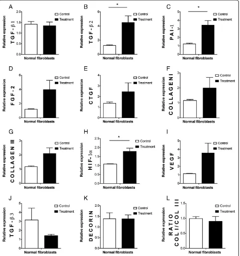
Wharton's jelly-derived MSCs enhance the expression of wound healing genes by paracrine signaling
Human WJ-MSCs upregulated the mRNA transcript ex-pression ofTGF-β2,hypoxia-inducible factor-1α, and plas-minogen activator inhibitor-1 genes (P≤0.05) in normal skin fibroblasts in our culture conditions. Other genes in-volved in re-epithelialization, neovascularization and/or remodeling –including VEGF,fibroblast growth factor-2, connective tissue growth factor, collagen I and collagen
III – were not changed. Decorin and TGF-β3 also re-mained unaffected (Figure 1).
Normal skin fibroblasts proliferate faster when treated with Wharton's jelly-derived MSC conditioned medium
In our culture conditions, WJ-MSC-CM accelerated nor-mal skin fibroblasts proliferation (P≤0.001), as measured by a Ki67 proliferation assay (Figure 2A,B,C).
Wharton's jelly-derived MSC conditioned medium does not induce apoptosis in normal skin fibroblasts
We did not find any significant modulation in the num-ber of apoptotic fibroblasts treated with WJ-MSC-CM using a terminal transferase TdT-mediated dUTP biotin end-labeling assay, suggesting that WJ-MSC-CM does not appear to affect normal skin fibroblast apoptosis under our culture conditions (Figure 2D). An increased apoptosis rate could imply a delayed wound healing pro-cess, whereas a much decreased apoptosis rate could im-ply enhanced risk of keloid scars.
Wharton's jelly-derived MSC conditioned medium promotes normal skin fibroblast migration and wound closure
To further examine whether the enhanced proliferation of fibroblasts treated with WJ-MSC-CM may also promote wound closure, a wound scratch assay was performed.
In our culture conditions, WJ-MSC-CM-treated nor-mal skin fibroblasts coapted wound borders faster than normal skin fibroblasts from the control group (that is, those treated with WJ-MSC nonconditioned medium), suggesting that WJ-MSC-CM enhanced fibroblast gration. This observation is important, because cell mi-gration is an essential step during wound healing. This fact highlights that healing promotion is partly due to faster migration, and consequently acceleration of wound closure, in human normal skin fibroblasts (Figure 3A,B).
Wharton's jelly-derived MSC conditioned medium pro-motes wound healing and repair in a mouse model
We next examined whether the in vitro wound healing promoting observed effects with WJ-MSC-CM might be translated into anin vivo wound healing model. BALB-c mice with WJ-MSC-CM-treated wounds showed enhanced wound healing rates compared with the control mice (P≤0.05) (Figure 4A,B, quantified in 4C). Increased and complete re-epithelialization, higher cellularity in newly formed granulation tissue, and less random and more organized extracellular matrix were observed in the CM-treated wounds, suggesting that WJ-MSC-CM promoted wound healing and repairin vivoin mice.
proliferative cells were found in the WJ-MSC-CM-treated wounds (P≤0.05 andP≤0.01, respectively) (Figure 5E,F). A microscopic wound cross-section showed a higher
number of positive proliferating nuclei (black arrows, BrdU-positive cells) (P≤0.01) in the WJ-MSC-CM-treated wounds (Figure 5D) compared with the control wounds Figure 1Upregulation of wound healing genes by human Wharton's jelly-derived mesenchymal stem cell conditioned medium in human normal skin fibroblasts. (A-L)mRNA transcript expression relative to 18S after 7 days of culture of human normal skin fibroblasts with Wharton's jelly-derived mesenchymal stem cell conditioned medium (WJ-MSC-CM; treatment group) or nonconditioned medium (control group) from five different patients (but four patients for FGF-2 and three patients for collagen I, collagen III and decorin). Overall, WJ-MSC-CM enhanced a wound healing promoting phenotype in human normal skin fibroblasts in our culture conditions. *P≤0.05. COL, collagen; CTGF, connective tissue growth factor; FGF-2, fibroblast growth factor-2; HIF-1α, hypoxia-inducible factor-1α; PAI-1, plasminogen activator inhibitor-1;
(Figure 5C), as well as general increased cellularity, matrix remodeling and overall wound repair. Higher magnifica-tion images corresponding to the aforemenmagnifica-tioned detailed micrographs of both control and treated wounds were shown in Figure 5A and 5B, respectively (Additional file 3 supports and complements Figure 5).
Discussion
The results of this study suggest that WJ-MSCs enhance wound healing and normal wound repair by paracrine
signaling mechanisms. Under our culture conditions, hu-man WJ-MSC-CM upregulated the gene expression of wound healing factors in human normal skin fibroblasts, and promoted fibroblast proliferation and migration to co-apt wound borders in vitro. Accordingly, WJ-MSC-CM accelerated the re-epithelialization rate and promoted wound repairin vivoin a full-thickness excisional mouse wound healing model.
Cutaneous wound repair is a complex orchestrated process that is activated upon injury and includes the
Figure 3Wharton's jelly-derived mesenchymal stem cell conditioned medium accelerates wound closurein vitro. (A),(B)Scratch wound assay was performed to examine migration properties of Wharton's jelly-derived mesenchymal stem cell conditioned medium (WJ-MSC-CM)-treated and untreated normal skin fibroblasts. The treated group showed significantly enhanced migration rates and coapted wound borders faster than the control group. *P≤0.05.
(See figure on previous page.)
Figure 2Wharton's jelly-derived mesenchymal stem cell conditioned medium increases normal skin fibroblast proliferation, but does not affect apoptosis.Cell proliferation was examined using Ki67 staining.(A)Wharton's jelly-derived mesenchymal stem cell conditioned medium (WJ-MSC-CM)-treated normal fibroblasts showed enhanced proliferative rates compared with the control group (quantified in(B)).(C)
multicellular overlapping and coordinated phases of inflammation, angiogenesis and formation of granulation tissue, re-epithelialization, and fibroproliferation or matrix formation and remodeling [14,15]. Decreased proinflam-matory cytokines, compromised neovascularization and/ or impairment in leukocyte recruitment might disturb and delay wound healing [16]. Despite the current use and availability of a wide array of wound dressings, ointments and devices, wound healing still remains a clinical chal-lenge, especially in older patients, diabetic patients, heavy smokers or burned patients [17–21]. There is therefore a need for new strategies to promote or at least coadjuvantly help in wound healing and repair.
Skin MSCs have been reported to populate the normal skin niche, remain quiescent and become active after in-jury, aiding in wound closure [14,15]. MSC paracrine sig-naling has been suggested to be the main underlying mechanism for MSCs enhanced wound repair effects [2,22,23]. BM-MSCs have been reported to promote wound healing, but their isolation requires an invasive and artificial method [24,25]. Harvesting adipose-derived stem
cells also requires a surgical procedure. Subsequently, we focused on a more advantageous MSC source, the umbil-ical cord-derived Wharton’s jelly [5,25,26]. The harvest of WJ-MSCs is not painful or invasive, as the cells are isolated naturally with no extra surgery and are dissected from dis-carded umbilical cords after birth. Indeed, caprine WJ-MSCs have already been shown to promote wound repair with minimal scarring [6], but to our knowledge there are no reports of human WJ-MSC treatment on human skin wounds. WJ-MSCs represent a very efficient stem cell source with reported immunoprivileged, anticancer and antifibrotic characteristics in animal models [25–28]. Due to their reported immunoprivileged properties and univer-sal and ever-lasting availability [5], WJ-MSC allotransplan-tion as an off-the-shelf therapy may represent an appropriate treatment strategy in the already compromised patients who suffer of recalcitrant cutaneous wounds [14].
promoting genes, including re-epithelialization, neovas-cularization and fibroproliferation inducing genes, in hu-man normal skin fibroblasts. Besides altering skin fibroblast gene expression, MSC signaling has also been shown to positively regulate cell survival, proliferation and migration [23,29]. Under our culture conditions, al-though apoptotic changes were not found in skin fibro-blasts treated with WJ-MSC-CM, significantly enhanced proliferation and migration were observed. This is import-ant, as cellular dynamics and cell migration constitute an essential step during cutaneous healing. WJ-MSC-CM ac-celerated wound closure both in vitro and in an in vivo mouse wound healing model, suggesting that human WJ-MSC-CM may promote wound repair.
Other researchers have also reported enhanced wound healing rates in mouse models with other human MSC sources, such as BM-MSCs [23,30] and umbilical cord blood-derived MSCs [31]. Blood of the umbilical cord has been reported to yield fewer MSCs than the cord it-self [31], however, and Wharton’s jelly may therefore emerge as a more appropriate stem cell source. To the best of our knowledge, no published studies regarding the use of human WJ-MSCs in human wounds have been reported, so this work is novel, and could prompt us to test whether the same results would be encoun-tered first in a more appropriate but more costly experi-mental model, such as the red duroc pig. Secondly, if the results appeared to be promising, the next step would be to translate them into phase I or phase II clinical trials for further evaluation and development. Indeed, pilot clinical studies have so far indicated that MSCs in ge-neral are safe in vivo, and they currently represent the most widely used stem cells in the clinical setting [26]. Accordingly, in the particular case of WJ-MSCs, 15 dia-betic patients received systemically WJ-MSCs with no documented relevant safety concerns [32]. Furthermore, many MSC clinical applications to manage intestinal fis-tulae, acute artery ischemic disease in diabetic patients, and periodontal defects have been reported, among others [26]. Anecdotally, one case report about autologous BM-MSCs used to help to heal a scapular recalcitrant wound in a Russian burned patient has also been published [33], but no reports regarding the use of WJ-MSCs have yet
been reported. Although WJ-MSCs secrete a more angio-genic secretome in comparison with BM-MSCs [34], the isolation of WJ-MSCs is more efficient, and WJ-MSCs have a higher capacity of proliferation and are less senes-cent than adipose-derived stem cells [35], proper studies comparing the wound healing abilities of different MSC sources are still lacking in the literature. On the other hand, the direct effects of human WJ-MSCs on wound healing, regeneration and repair still remain unknown. A new preclinical and clinical research arena investigating the potential of WJ-MSCs in wounds and wound healing has just been born and may hold promise for future me-dical therapies.
Conclusions
Human WJ-MSCs promoted wound healing by para-crine signaling in our culture conditionsin vitro, and in anin vivo preclinical animal model. If the reported im-munoprivileged character and safety of WJ-MSCs ob-served in experimental and first clinical models is further scientifically proven, WJ-MSCs might represent a feasible, universal and off-the-shelf technology to enhance normal wound healing to improve patient survival and quality of life.
Additional files
Additional file 1: Figure S1.Showing flow-cytometry markers and mesenchymal differentiation of human WJ-MSCs. Flow cytometry analysis of established human WJ-MSCs (successfully grown on plastic plates) showing markers used to characterize MSCs (a,b,c,d). Cells were able to differentiate into osteocytes (e), chondrocytes (f) and adipocytes (g). Images shown after alizarin red (e), safranin O (f) and oil red (g) staining, respectively. Additional file 2: Table S1.Showing primer sequences for real-time PCR. List of SYBR® Green gene primer sequences used in real-time PCR for the present work. *TGF-β, transforming growth factor-β; CTGF, connective tissue growth factor; PAI-I, plasminogen activator inhibitor-I; HIF-1-α, hypoxia inducible factor-1-α; VEGF, vascular endothelial growth factor; FGF-2, fibroblast growth factor-2.
Additional file 3: Figure S2.Showing WJ-MSC-CM promoted cell proliferation in anin vivowound healing model in BALB-c mice. (A) BrdU staining in the control group. (B) BrdU staining in the treatment group. (C), (D) Higher magnification (40×) of the above microscopic images were included for nonconditioned medium-treated (C is magnification of the marked area in A) and WJ-MSC-CM-treated normal skin fibroblasts (D is magnification of the marked area in B) to examine in further detail the (See figure on previous page.)
increase in cell number or stained nuclei in the WJ-MSC-CM-treated wounds, compared with controls. Brown nuclei correspond to cells that incorporated BrdU into their DNA. (Supplemental figure in support of Figure 5).
Abbreviations
BM-MSC:bone marrow-derived mesenchymal stem cell; BrdU: bromodeoxyuridine; CM: conditioned medium; DAPI: 4′ ,6-diamidino-2-phenylindole; DMEM: Dulbecco’s modified Eagle’s medium; FBS: fetal bovine serum; IL: interleukin; MSC: mesenchymal stem cell; PBS: phosphate-buffered saline; PCR: polymerase chain reaction; TGF-β: transforming growth factor beta; VEGF: vascular endothelial growth factor; WJ-MSC: Wharton’s jelly-derived mesenchymal stem cell.
Competing interests
The authors declare that they have no competing interests or any potential conflict of interest in any of the techniques or instruments mentioned in this manuscript.
Authors’contributions
AIA participated in the conception and design, administrative support, collection and/or assembly of data, data analysis and interpretation, manuscript writing and final approval of the manuscript. SA-N contributed to the conception and design, administrative support, collection and/or assembly of data, data analysis and interpretation, manuscript writing and final approval of the manuscript. PHB participated in the conception and design, administrative support, collection and/or assembly of data, data analysis and interpretation, manuscript writing and final approval of the manuscript. MA-S contributed to administrative support, collection and/or assembly of data and final approval of the manuscript. CB provided administrative support and helped in collection and/or assembly of data and final approval of the manuscript. EH carried out provision of study material or patients, administrative support, helped in collection and/or assembly or data and contributed to final approval of the manuscript. CHT provided financial and administrative support and study material, helped in collection of data and approved final version of the manuscript. MGJ carried out conception and design, financial support, administrative support, provision of study material or patient collection and/or assembly of data, data analysis and interpretation, manuscript writing and final approval of the manuscript. All authors read and approved the final manuscript.
Acknowledgements
The National Institutes of Health grant RO1 GM087285-01, the Canadian Institutes of Health Research #123336 funding, the CFI Leader’s Opportunity Fund (Project #25407), the Physician’s Services Incorporated Foundation– Health Research Grant Program, the Canadian Forces Health Services, and The Healing Foundation/BBA AB Wallace Memorial 2012 Award supported this work.
Author details
1Plastic Surgery Department and Burn Unit, Vall d’Hebron University Hospital,
Universitat Autònoma de Barcelona, Passeig de la Vall d'Hebron 119-129, 08035 Barcelona, Spain.2Ross Tilley Burn Centre and Sunnybrook Research
Institute, Sunnybrook Health Sciences Centre, University of Toronto, 2075 Bayview Avenue, Toronto, ON M4N 3M5, Canada.3Gynecology and
Obstetrics Department, Sunnybrook Health Sciences Centre, University of Toronto, 2075 Bayview Avenue, Toronto, ON M4N 3M5, Canada.4Canadian
Forces Health Services; Trauma, Emergency and Critical Care Program, Sunnybrook Health Sciences Centre, University of Toronto, 2075 Bayview Avenue, Toronto, ON M4N 3M5, Canada.
Received: 31 October 2013 Revised: 12 January 2014 Accepted: 18 February 2014 Published: 24 February 2014
References
1. Caspersen CJ, Thomas GD, Boseman LA, Beckles GL, Albright AL:Aging, diabetes, and the public health system in the United States.Am J Public Health2012,102:1482–1497.
2. Khosrotehrani K:Mesenchymal stem cell therapy in skin: why and what for?Exp Dermatol2013,22:307–310.
3. Liu S, Yuan M, Hou K, Zhang L, Zheng X, Zhao B, Sui X, Xu W, Lu S, Guo Q: Immune characterization of mesenchymal stem cells in human umbilical cord Wharton’s jelly and derived cartilage cells.Cell Immunol2012, 278:35–44.
4. Hanson SE, Bentz ML, Hematti P:Mesenchymal stem cell therapy for nonhealing cutaneous wounds.Plast Reconstr Surg2010,125:510–516. 5. Nekanti U, Rao VB, Bahirvani AG, Jan M, Totey S, Ta M:Long-term
expansion and pluripotent marker array analysis of Wharton’s jelly-derived mesenchymal stem cells.Stem Cells Dev2010, 19:117–130.
6. Azari O, Babaei H, Drakhsahnfar A, Nematollahi-Mahani SN, Poursahebi R, Moshrefi M:Effects of transplanted mesenchymal stem cells isolated from Wharton’s jelly of caprine umbilical cord on cutaneous wound healing: histopathological evaluation.Vet Res Commun2011, 35:211–222.
7. Moodley Y, Atienza D, Manuelpillai U, Samuel CS, Tchongue J, Ilancheran S, Boyd R, Trounson A:Human umbilical cord mesenchymal stem cells reduce fibrosis of bleomycin-induced lung injury.Am J Pathol2009, 175:303–313.
8. Du T, Cheng J, Zhong L, Zhao XF, Zhu J, Zhu YJ, Liu GH:The alleviation of acute and chronic kidney injury by human Wharton’s jelly-derived mesenchymal stromal cells triggered by ischemia–reperfusion injury via an endocrine mechanism.Cytotherapy2012,14:1215–1227.
9. Tsai PC, Fu TW, Chen YM, Ko TL, Chen TH, Shih YH, Hung SC, Fu YS: The therapeutic potential of human umbilical mesenchymal stem cells from Wharton’s jelly in the treatment of rat liver fibrosis.Liver Transpl 2009,15:484–495.
10. Xu Y, Huang S, Ma K, Fu X, Han W, Sheng Z:Promising new potential for mesenchymal stem cells derived from human umbilical cord Wharton’s jelly: sweat gland cell-like differentiative capacity.J Tissue Eng Regen Med 2012,6:645–654.
11. Choi M, Lee HS, Naidansaren P, Kim HK, O E, Cha JH, Ahn HY, Yang PI, Shin JC, Joe YA:Proangiogenic features of Wharton’s jelly-derived mesenchymal stromal/stem cells and their ability to form functional vessels.In t J Biohem Cell Biol2013,45:560–570.
12. Shohara R, Yamamoto A, Takikawa S, Iwase A, Hibi H, Kikkawa F, Ueda M: Mesenchymal stromal cells of human umbilical cord Wharton’s jelly accelerate wound healing by paracrine mechanisms.Cytotherapy2012, 14:1171–1181.
13. Kita K, Gauglitz GG, Phan TT, Herndon DN, Jeschke MG:Isolation and characterization of mesenchymal stem cells from the sub-amniotic human umbilical cord lining membrane.Stem Cells Dev2010,19:491–501. 14. Arno A, Smith AH, Blit BH, Al-Shehab M, Gauglitz GG, Jeschke MG:Stem cell
therapy: a new treatment for burns?Pharmaceuticals2011,4:1355–1380. 15. Blit PH, Arno AI, Jeschke MG:Stem cells and tissue engineering in wounds
and burns.InStem Cells Handbook.Edited by Snell S. New York: Springer; 2013:399–410.
16. Amini-Nik S, Glancy D, Boimer C, Whetstone H, Keller C, Alman BA:Pax7 expressing cells contribute to dermal wound repair, regulating scar size through aβ-catenin mediated process.Stem Cells2011,29:1371–1379. 17. Falanga V:Wound healing and its impairment in the diabetic foot.Lancet
2005,366:1736–1743.
18. Bielefeld KA, Amini-Nik S, Alman BA:Cutaneous wound healing: recruiting developmental pathways for regeneration.Cell Mol Life Sci 2013,70:2059–2081.
19. Jorgensen LN, Kallehave F, Christensen E, Siana JE, Gottrup F:Less collagen production in smokers.Surgery1998,123:450–455.
20. Menke NB, Ward KR, Witten TM, Bonchev DG, Diegelmann RF:Impaired wound healing.Clin Dermatol2007,25:19–25.
21. Wasiak J, Cleland H, Campbell F, Spinks A:Dressings for superficial and partial thickness burns.Cochrane Database Syst Rev2013,3:CD002106. 22. Li H, Fu X:Mechanisms of action of mesenchymal stem cells in
cutaneous wound repair and regeneration.Cell Tissue Res2012, 348:371–377.
23. Hocking AM, Gibran NS:Mesenchymal stem cells: paracrine signaling and differentiation during cutaneous wound repair.Exp Cell Res2010, 316:2213–2229.
25. Bongso A, Fong CY:The therapeutic potential, challenges and future clinical directions of stem cells from the Wharton’s jelly of the human umbilical cord.Stem Cell Rev2013,9:226–240.
26. Hass R, Kasper C, Bohm S, Jacobs R:Different populations and sources of human mesenchymal stem cells (MSC): a comparison of adult and neonatal tissue-derived MSC.Cell Commun Signal2011,9:12. 27. Kim DW, Staples M, Shinozuka K, Pantcheva P, Kang SD, Borlongan CV:
Wharton’s jelly-derived mesenchymal stem cells: phenotypic characterization and optimizing their therapeutic potential for clinical applications.Int J Mol Sci2013,14:11692–11712.
28. Taghizadeh RR, Cetrulo KJ, Cetrulo CL:Wharton’s Jelly stem cells: future clinical applications.Placenta2011,32:S311–S315.
29. Rodriguez-Menocal L, Salgado M, Ford D, Van Badiavas E:Stimulation of skin and wound fibroblast migration by mesenchymal stem cells derived from normal donors and chronic wound patients.Stem Cells Transl Med 2012,1:221–229.
30. Wu Y, Chen L, Scott PG, Tredget EE:Mesenchymal stem cells enhance wound healing through differentiation and angiogenesis.Stem Cells2007, 25:2648–2659.
31. Luo G, Cheng W, He W, Wang X, Tan J, Fitzgerald M, Li X, Wu J:Promotion of cutaneous wound healing by local application of mesenchymal stem cells derived from human umbilical cord blood.Wound Rep Regen2010, 18:506–513.
32. Hu JYX, Wang Z, Wang F, Wang L, Gao H, Chen Y, Zhao W, Jia Z, Yan S, Wang Y:Long term effects of the implantation of Wharton’s jelly-derived mesenchymal stem cells from the umbilical cord for newly-onset type 1 diabetes mellitus.Endocr J2013,60:347–357.
33. Rasulov MF, Vasilchenkov AV, Onishchenko NA, Krasheninnikov ME, Kravchenko VI, Gorshenin TL, Pidtsan RE, Potapov IV:First experience of the use bone marrow mesenchymal stem cells for the treatment of a patient with deep skin burns.Bull Exp Biol Med2005,139:141–144. 34. Balasubramanian S, Venugopal P, Sundarrai J, Zakaria Z, Majumdar AS, Ta M:
Comparison of chemokine and receptor gene expression between Wharton’s jelly and bone marrow-derived mesenchymal stromal cells. Cythotherapy2012,14:26–33.
35. Christodoulou I, Kolisis FN, Papaevangeliou D, Zoumpourlis V:Comparative evaluation of human mesenchymal stem cells of fetal (Wharton’s jelly) and adult (adipose tissue) during prolonged in vitro expansion: considerations for cytotherapy.Stem Cells Int2013,2013:246134.
doi:10.1186/scrt417
Cite this article as:Arnoet al.:Human Wharton’s jelly mesenchymal stem cells promote skin wound healing through paracrine signaling.
Stem Cell Research & Therapy20145:28.
Submit your next manuscript to BioMed Central and take full advantage of:
• Convenient online submission
• Thorough peer review
• No space constraints or color figure charges
• Immediate publication on acceptance
• Inclusion in PubMed, CAS, Scopus and Google Scholar
• Research which is freely available for redistribution