Patrick Mulligan, Bethwel Raore, Shuling Liu1, Jeffrey J. Olson
Department of Neurosurgery, Emory University School of Medicine, 1Department of Biostatistics, Rollins School of Public Health,
Emory University, Atlanta, Georgia, United States of America
Neurological and functional outcomes of subdural
hematoma evacuation in patients over 70 years of age
Introduction
Subdural hematoma (SDH) is a common disease pathology amongst elderly patients. Data collected from the US census bureau shows that the incidence of SDH almost doubles from age 65 to 75, and continues to increase to an incidence of 286/100000 in people over age 80.[1] Despite this, studies on the management of acute and chronic SDH in the elderly are limited. The paucity of data is particularly apparent for chronic SDH, and currently there are no treatment guidelines for its management. This may largely be due to the general perception that chronic subdural hematomas are
relatively benign pathology with minimal morbidity and mortality.[2] However, previous research has questioned this paradigm, and suggests that chronic subdural hematomas might confer long‑term increases in both morbidity and mortality in this population.[3,4]
While acute subdural hematomas have been studied more in depth, the guidelines pertaining to management are still based solely on class C evidence gleaned from case series, case studies, retrospective reviews, and expert opinion.[5] However, multiple studies do suggest that age is closely associated with increased morbidity and mortality after acute SDH;[6‑8] although, more recent evidence has not showed a correlation.[9,10] Regardless, the role of age in both acute and chronic subdural hematoma management and outcome remains ill defined.
Previous research on subdural hematomas (especially chronic subdural hematomas) within the elderly typically focus on the type of surgical treatment,[11‑14] the role of anticoagulant[15,16] and epidemiology.[1,17] However, the long‑term outcome of elderly patients and the role
Original Article
ABSTRACT
Background: Subdural hematoma (SDH) is a common disease entity treated by neurosurgical intervention. Although the incidence increases in the elderly population, there is a paucity of studies examining their surgical outcomes. Objectives: To determine the neurological and functional outcomes of patients over 70 years of age undergoing surgical decompression for subdural hematoma. Materials and Methods: We retrospectively reviewed data on 45 patients above 70 years who underwent craniotomy or burr holes for acute, chronic or mixed subdural hematomas. We analyzed both neurological and functional status before and after surgery. Results: Forty‑five patients 70 years of age or older were treated in our department during the study period. There was a significant improvement in the neurological status of patients from admission to follow up as assessed using the Markwalder grading scale (1.98 vs. 1.39; P =0.005), yet no improvement in functional outcome was observed as assessed by Glasgow Outcome Score. Forty‑one patients were admitted from home, however only 20 patients (44%) were discharged home, 16 (36%) discharged to nursing home or rehab, 6 (13%) to hospice and 3 (7%) died in the postoperative period. Neurological function improved in patients who were older, had a worse pre‑operative neurological status, were on anticoagulation and had chronic or mixed acute and chronic hematoma. However, no improvement in functional status was observed. Conclusion: Surgical management of SDH in patients over 70 years of age provides significant improvement in neurological status, but does not change functional status.
Key words: Elderly, evacuation, outcome, subdural hematoma
Address for correspondence:
Dr. Patrick Mulligan, Department of Neurosurgery, Emory University School of Medicine, 1365‑B Clifton Road, Suite 6200, Atlanta, GA 30322. E‑mail: pmullig@learnlink.emory.edu
Access this article online
Quick Response Code:
Website:
www.ruralneuropractice.com
DOI:
of conservative management versus surgery cannot be determined based on the limited data available.[3,4,18] In this study, we evaluate the neurologic and functional outcome of patients age 70 or older who underwent craniotomy or burr hole drainage for acute subdural hematoma (aSDH), chronic subdural hematoma (cSDH) or mixed acute and chronic subdural hematoma (a/cSDH).
Materials and Methods
This retrospective review includes all patients age 70 or older who underwent surgery for aSDH, cSDH, or a/cSDH between July 2010 and March 2012 in the Department of Neurosurgery at Emory University Hospital. Data was collected from the patient’s hospital records, previous hospital and clinic notes, and follow‑up clinic notes and/or documented phone conversations. All follow‑up visits were scheduled for 4 to 6 weeks from the time of discharge and most patients were followed up at the Emory Neurosurgery clinic. A small minority of patients followed up with their primary care physician, and data was pulled from their follow‑up clinic documentation. In total, the follow‑up data was available in all but 5 patients.
Inclusion criteria were based on imaging diagnosis of subdural hematoma, age 70 or older and initial surgical drainage of subdural hematoma; either via burr hole or craniotomy. The decision to perform burr hole drainage or craniotomy was based on the patient’s physical exam, neurologic exam, medical status and image findings.
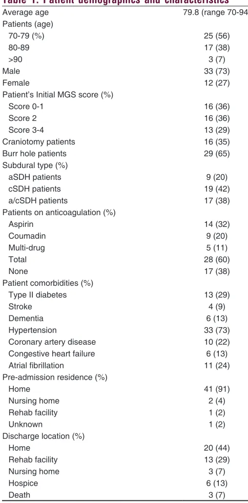
Hospital records were analyzed for patient demographics (age, gender), chronic or acute subdural hematoma, burr hole or craniotomy treatment, presence of comorbidities, use of anticoagulation therapy, platelet count and international normalized ratio (INR) at time of surgery, place of residence prior to admission and discharge placement [Table 1]. The neurological status of all patients was determined by reviewing the documented neurological exam within the chart and classified in terms of the Markwalder grading system (MGS) [Table 2].[19] Patients were assigned an MGS score on admission, post‑operative day 2, post‑operative day 7, and follow‑up visit.
Functional status was also quantified according to a pre‑operative and follow‑up Glasgow outcome score (GOS) [Table 3].[20] Pre‑operative Glasgow outcome scores were based on the patient’s neurological and functional status at the time of admission and presumptive functional outcome with no treatment.
Post‑operative Glasgow outcome scores were based on functional status at follow‑up visits, including physical exam findings, type of residence and daily assistance needed and family reports of daily function.
Statistical analysis was performed using SAS® Analytics Pro (SAS Institute Inc, Cary, North Carolina, USA). A proportional odds model was utilized for both MGS and GOS scores and adjusted for baseline scores. Differences between different MGS or GOS groups of patients for categorical variables were assessed Table 1: Patient demographics and characteristics
Average age 79.8 (range 70-94)
Patients (age)
70-79 (%) 25 (56)
80-89 17 (38)
>90 3 (7)
Male 33 (73)
Female 12 (27)
Patient’s Initial MGS score (%)
Score 0-1 16 (36)
Score 2 16 (36)
Score 3-4 13 (29)
Craniotomy patients 16 (35)
Burr hole patients 29 (65)
Subdural type (%)
aSDH patients 9 (20)
cSDH patients 19 (42)
a/cSDH patients 17 (38)
Patients on anticoagulation (%)
Aspirin 14 (32)
Coumadin 9 (20)
Multi-drug 5 (11)
Total 28 (60)
None 17 (38)
Patient comorbidities (%)
Type II diabetes 13 (29)
Stroke 4 (9)
Dementia 6 (13)
Hypertension 33 (73)
Coronary artery disease 10 (22)
Congestive heart failure 6 (13)
Atrial fibrillation 11 (24)
Pre-admission residence (%)
Home 41 (91)
Nursing home 2 (4)
Rehab facility 1 (2)
Unknown 1 (2)
Discharge location (%)
Home 20 (44)
Rehab facility 13 (29)
Nursing home 3 (7)
Hospice 6 (13)
Death 3 (7)
using the Chi‑square test. Differences for continuous measurements were analyzed by simple t‑tests. Statistical significance was determined using a P ≤ 0.05. Results are expressed as a mean ± SE.
Results
A total of 45 patients, 70 years of age or older presented with SDH during the study window. The mean age was 79.8 (range from 70 to 94 years), with 33 men (73%) and 12 women (27%). Forty‑two patients (93%) had at least one significant comorbidity, including cardiovascular disease, dementia, renal disease or diabetes mellitus. Overall, there was a significant improvement in the neurological status of patients from admission to follow‑up (MGS score: 1.98 Vs. 1.39; P = 0.005), yet no improvement in functional outcome was observed (GOS score: 3.55 vs. 3.53; P = 0.96) [Figure 1].
In total, 41 patients were admitted from home, 1 from a rehab facility, 2 from a nursing home and in 1 patient their residence on admission was unknown. Upon discharge there was a marked decrease in the number of patients returning home, at just 20 patients (44%). There was a rise in the number of patients discharged to long‑term rehab facilities (13 patients, 29%), and nursing homes (3 patients, 7%), along with 6 patients that were discharged to hospice. In total, 3 patients (7%) died during their hospital course, 2 from respiratory failure and one from congestive heart failure.
Table 2: Markwalder grading system of neurological status
MGS score Neurological status
0 Neurologically intact
1 Alert and oriented; mild symptoms such as headache; absent or mild neurological deficit, such as reflex asymmetry
2 Drowsy or disoriented with variable neurological deficit, such as hemiparesis
3 Stuporous but responding appropriately to noxious
stimuli; severe focal signs such as hemiplegia 4 Comatose with absent motor responses to painful
stimuli; decerebrate or decorticate posturing MGS: Markwalder grading system
Table 3: Glasgow outcome scale of functional status
GOS score Functional status
5 Resumption of normal life; there may be minor neurologic and/or psychological deficits 4 Able to work in a sheltered environment and
travel by public transportation
3 Dependent for daily support by reason of mental or physical disability or both
2 Unresponsive for weeks or months or until death
1 Death
GOS: Glasgow outcome score
Patients that died or were discharged to hospice were older than patients discharged to home or rehab facilities/nursing homes (83.5 vs. 78.9 years, P = 0.05). Furthermore, all patients that were discharged to hospice or died during admission initially presented from home. There was also no significant difference in the neurological status on admission of patients that were discharged home or to a rehabilitation/nursing home compared to those that died or went to hospice (MGS 2.0 vs. 1.8, P = 0.75). Furthermore, no difference in admission functional status was observed between the two groups (GOS 3.54 vs. 3.55, P = 0.96) [Figure 2].
Patient outcome was also assessed compared to their initial neurological status as determined by MGS score. Patients presenting with an MGS score of 0‑1 (mean MGS:0.93) on admission had an average MGS score of 0.85 on follow‑up examination (P = 0.41), demonstrating no statistical improvement in neurological function at follow‑up. In contrast there was significant neurological improvement in patients that initially presented with an MGS score of 2 (follow‑up MGS score: 1.41; P = 0.012) and in those patients that presented with an MGS score of 3‑4 (mean admission MGS score: 3.23; follow‑up MGS score: 2; P = 0.014) [Figure 3]. Interestingly, patients
Figure 1: Change in neurological and functional status from admission
to follow up
Figure 2: Admission neurological and functional status of patients
with an admission MGS score of 2 saw a modest improvement in their functional status on follow‑up exam as determined by GOS score (Admission GOS score: 3.53; Follow‑up GOS score: 3.33, P = 0.0002). However, no functional improvement was observed in patients with an admission MGS score 0‑1 (Admission GOS score: 4.0625; Follow‑up GOS score: 4.00; P = 0.96) or MGS 3‑4 (Admission GOS score: 2.92; Follow‑up GOS score: 3.17; P = 0.066).
There was a significant difference in neurological outcome based on age. Patients aged 70‑79 showed modest though statistically insignificant improvements in neurological status (admission MGS: 1.80; follow‑up MGS: 1.50; P = 0.50), while patient’s age 80 or older actually showed greater improvement in neurological function (admission MGS: 2.20; follow‑up MGS: 1.21; P = 0.01) [Figure 4]. Of note, patients age 80 and older presented with worse neurological function on admission but had better neurological function than the 70‑79‑year‑old group at follow‑up. There was however no significant change in functional status in either the 70‑79 year group (admission GOS: 3.80; follow‑up GOS: 3.80; P = 0.0790) or 80 years and older group (admission GOS: 3.21; follow‑up GOS: 3.16; P = 0.9389).
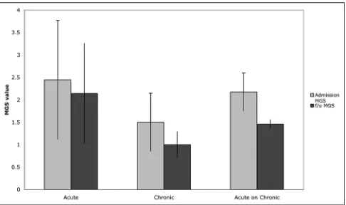
A total of 9 patients presented with aSDH, 19 patients with cSDH, and 17 patients presenting with a/cSDH. There was no statistically significant change in neurological function from admission to follow‑up in the aSDH group (admission MGS: 2.44; follow‑up MGS: 2.14; P = 0.8993), or the cSDH group (admission MGS: 1.5; follow‑up MGS: 1; P = 0.2244), although significant neurological improvement was seen in the a/cSDH group (admission MGS: 2.17; follow‑up MGS: 1.46; P = 0.0006) [Figure 5]. However the improvement in neurological status in the a/cSDH group did not translate into a statistically significant improvement of functional outcome from admission to follow‑up (admission GOS: 3.31; follow‑up GOS: 3.38; P = 1.00). In addition, no change in functional status from admission to follow‑up was seen in the aSDH group (admission GOS: 3.56; follow‑up GOS: 2.89; P = 0.1314) or cSDH group (admission GOS: 3.78; follow‑up GOS:4; P = 0.8199).
Of the twenty‑eight patients that underwent burr hole drainage, 17 had cSDH and 11 a/cSDH. Seventeen patients underwent craniotomy (9 aSDH, 6 a/cSDH, 1 cSDH). Patients undergoing burr hole drainage tended to have a better neurological exam on admission than patients that underwent craniotomy (burr hole admission MGS: 1.79; craniotomy admission MGS: 2.31; P = 0.08). Furthermore, burr hole patients showed greater improvement in
neurological outcome from admission to follow‑up visit (admission MGS: 1.79; follow‑up MGS: 1.00; P = 0.0049) while patients undergoing craniotomy experienced a smaller, and statistically insignificant improvement in neurological function (admission MGS: 2.31; follow‑up MGS: 2.07; P = 0.7588) [Figure 6]. However, there was no significant change in functional status from admission to follow‑up in either the burr hole patients (GOS: 3.55; follow‑up GOS: 3.86; P = 0.8638) or patients that underwent craniotomy (admission GOS: 3.53; follow‑up GOS: 2.93; P = 0.0657).
Figure 3: Neurological outcomes based on admission neurological
status. A comparison of mean pre-op and follow-up MGS score
Figure 4: Affect of age on neurological status from admission to
follow up
Twenty‑eight patients (62%) were on anticoagulation therapy at the time of admission, including 5 patients (11%) on multi‑drug therapy. The mean INR values on admission were significantly different for the anticoagulation patients vs. no therapy, 1.21 and 1.07, respectively (P =0.021). There was no statistical difference in the mean platelet value of the anticoagulation group compared to the group on no therapy (193.9 vs. 223.1, P = 0.27). Anticoagulated patients had a worse, though statistically insignificant, neurological exam on admission than patients not on anticoagulation therapy (anticoagulation MGS: 2.17; no therapy MGS: 1.65; P = 0.07). However, the anticoagulation cohort showed statistically significant improvement in neurological status at follow‑up (admission MGS: 2.17; follow up MGS: 1.43; P =0.0173), while patients on no anticoagulation saw a more modest, and not statistically significant improvement (admission MGS: 1.65; follow up MGS: 1.33; P =.5411) [Figure 7]. The improvement in neurological status of patients on anticoagulation however did not translate into an improvement in functional outcome (admission GOS: 3.52; follow‑up GOS: 3.46; P = 0.3235). Similarly, no improvement in functional status from admission to follow‑up was observed in patients not on anticoagulation (admission GOS: 3.59; follow‑up GOS: 3.65; P = 0.6219).
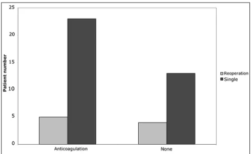
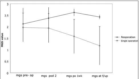
A total of 9 patients (20%) required additional surgery for recurrent SDH during their hospital course. Only 56% of patients requiring additional surgery (n = 5) were on anticoagulation therapy at the time of admission [Figure 8]. Furthermore, anticoagulation use did not increase the risk for reoperation compared to patients not on anticoagulants (X2 = 0.0059, P = 0.94). Interestingly, the group requiring additional surgery had a lower mean INR (1.06 vs. 1.16, P = 0.02), although mean platelet count was not statistically different (189.9 vs. 204.5, P = 0.60). There was also no significant difference in neurological status on admission between the patients receiving one operation and those requiring reoperation. (MGS: 2.13 vs. MGS: 1.98; P = 0.69). Reoperation was however associated with worsening of neurological function on follow‑up, though statistically insignificant (admission MGS: 2.13; follow‑up MGS: 2.43; P = 0.3665), while patients requiring only one operation saw a significant improvement in neurological function (admission MGS: 1.98; follow‑up MGS: 1.18; P = 0.0011). Furthermore, the decline in neurological status of patients undergoing a reoperation compared to patients with one operation was apparent as early as one week after the initial operation (reoperation MGS: 2.625; single operation MGS: 1.583; P = 0.0339) and this difference increased with time (reoperation MGS at follow‑up: 2.43; single operation MGS follow‑up: 1.18; P = 0.0077) [Figure 9]. There was a small, but statistically insignificant decline
in functional status at follow‑up in patients requiring additional surgery, and minimal improvement in the group undergoing just one operation (data not shown). Lastly, of the 9 patients that died or were discharged to hospice, only 1 underwent additional surgery during their stay.
Discussion
Subdural hematoma represents a common neurosurgical problem amongst elderly patients, yet surgical outcome
Figure 6: Affect of burr hole vs. craniotomy on neurological outcome
Figure 7: Affect of anticoagulation use on neurological outcome. The mean INR values on admission were significantly different for the anticoagulation patients vs. no therapy, 1.21 and 1.07 respectively
(P = 0.021)
remains an understudied field of research in this population. In this study we assessed the neurological and functional outcomes of 45 patients, aged 70 or older that were surgically treated for subdural hematomas.
While an overall improvement of neurological function was seen in our patients, this did not translate into functional improvements after discharge. Importantly, the initial GOS score assigned to patients was based on functional status at admission and not their baseline function before subdural hematoma onset. Thus, while there was no difference in the functional status from admission to follow‑up, this likely represents either an overall decline from the patient’s baseline before SDH onset or more likely a lower baseline functional status in this population. The discrepancy between neurological and functional outcome likely has several underlying factors. First, there was a trend of patients presenting from home and later discharged to rehab facilities despite returning to their neurological baseline or even improving. This suggests precautionary measures by the healthcare team given the known recurrence of subdural hematomas[11,14,17,21] and desire to have patients closely monitored because of other medical comorbidities, despite returning to their neurological baseline. This is in line with previous studies showing the morbidity of subdural hematomas in elderly patients, beyond just neurological deficits.[22] It must be noted that most patients were followed up 4 to 6 weeks after discharge, and thus it is uncertain what functional gains they may have made long term. However, previous studies suggest that the presence of subdural hematoma likely confers a morbidity and mortality risk well beyond 6 weeks post discharge.[3,4]
Interestingly, patients over 80 years old had an overall greater improvement of neurological function than those aged 70‑79. This is in contrast to past research,[23,24] but more recent studies have shown a similar trend to our results.[10,22,25] In our study, patients over 80 initially presented with worse neurological function. Given the greater neurological improvement overall, this may represent less ability to compensate for the initial
neurological insult compared to younger patients. However, it is possible that improved treatment for subdural hematomas and greater life expectancy may reduce the significance of age on SDH outcome. Thus, while patients over 80 initially presented at a lower baseline, they may have had more function to regain with the benefit of better treatment and perhaps better health than was observed in older studies.
Another paradoxical result of our study was that patients on anticoagulation therapy saw greater improvement in neurological function. Similar to the group of patients over 80 years, patients on anticoagulation therapy presented with worse neurological function. The mean INR at time of surgery of patients taking anticoagulants was statistically higher, however their mean INR was still relatively low at 1.21. Furthermore, only 1 patient had an INR greater than 2 on admission. Given the general therapeutic range for warfarin therapy is an INR of 2‑3, it is likely that many of these patients were on subtherapeutic doses of warfarin. Thus our data suggest that subtherapeutic levels of anticoagulation, and not just supratherapeutic levels, increase the risk of SDH. One likely mechanism is increased risk of transient ischemic attack or stroke leading to an increased risk of falling. Unfortunately, platelets function assays and bleeding times were not available for most of our patients, and its therefore uncertain if anti‑platelet medicines like aspirin were actually providing an intended therapeutic coagulopathy.
The mortality rate in our cohort was 7%. It is difficult to compare our mortality rate to other studies because we have included acute, acute on chronic, and chronic SDH. However, mortality rates in studies of cSDH among the elderly range from 1.2 to 16.7%,[4,25] while rates for aSDH range from 35 to 88%.[6,10,24] Interestingly, patients that died during their hospital stay or transferred to hospice had equivocal neurological and functional statuses on exam compared to patients that were discharged to home or a rehab facility. This suggests patient outcome cannot necessarily be determined by their presentation on admission. Furthermore, three patients died from either respiratory failure or congestive heart failure. As in previous reports,[2,26] chronic subdural hematoma likely predisposed our patients to greater morbidity and mortality from underlying medical conditions, as opposed to being the actual cause of death. Of note, 3 of the patients discharged to hospice were followed up in clinic. One had a worse neurological status while 2 had significant improvement.
Our rate of reoperation was 20%, whereas previous studies have reported recurrence rates ranging from 5.5 to 25%.[13,17,21] Surprisingly, there was no significant difference in the neurological status at presentation,
Figure 9: Changes in neurological status over time in patients
percentage of patients on anti‑coagulation, or ages of patients that required reoperation compared to those that did not. As with our mortality data, this suggests that a patient’s initial presentation does not predict their risk for SDH recurrence. However, reoperation did prove detrimental to long‑term neurological status as there was no change from admission to follow up, while patients undergoing a single operation saw marked gains in neurological function. These results are similar to a recent study of cSDH in elderly patients in which subdural recurrence (and presenting GCS score) were more predicative of patient outcome than age.[27] Yet despite worsening neurologic status within the reoperation group, none of the patients died, and only 1 of the 6 patients that went to hospice in this series underwent a reoperation for SDH. Therefore, reoperation for SDH increased morbidity but did not affect mortality during our study period.
Conclusion
Our results show that there is a role for surgical evacuation of subdural hematoma in the elderly, especially in patients 80 years and older, those patients on anticoagulation therapy, and those undergoing burr hole drainage. The perioperative course of SDH is often unpredictable and complications often arise from other medical comorbidities. While this study may challenge the opinion of some neurosurgeons regarding patient outcome, the results show that despite a possible tenuous hospital course, elderly patients frequently experience significant neurological improvement after surgery for subdural hematoma.
References
1. Frontera JA, Egorova N, Moskowitz AJ. National trend in prevalence, cost, and discharge after subdural hematoma from 1998‑2007. Crit Care Med 2011;39:1619‑25.
2. Gelabert‑Gonzalez M, Iglesias‑Pais M, Garcia‑Allut A, Martinez‑Rumbo R. Chronic subdural haematoma: Surgical treatment and outcome in 1000 cases. Clin Neurol Neurosurg 2005;107:223‑9. 3. Jones S, Kafetz K. A prospective study of chronic subdural haematomas
in elderly patients. Age Ageing 1999;28:519‑21.
4. Miranda LB, Braxton E, Hobbs J, Quigley MR. Chronic subdural hematoma in the elderly: Not a benign disease. J Neurosurg.2011;114:72‑6. 5. Bullock MR, Chesnut R, Ghajar J, Gordon D, Hartl R, Newell DW,
et al. Surgical management of traumatic brain injury author group:
Surgical management of acute subdural hematomas. Neurosurgery 2006;58 Suppl 3:S16‑24.
6. Cagetti B, Cossu M, Pau A, Rivano C, Viale G. The outcome from acute subdural and epidural intracranial haematomas in very elderly patients. Br J Neurosurg 1992;6:227‑31.
7. Luerssen TG, Klauber MR, Marshall LF. Outcome from head injury related to patient’s age. A longitudinal prospective study of adult and pediatric head injury. J Neurosurgery 1988;68:409‑16.
8. Taussky P, Widmer HR, Takala J, Fandino J. Outcome after acute traumatic subdural and epidural haematoma in Switzerland: A single‑centre experience. Swiss Med Wkly 2008;138:281‑5.
9. Baechli H, Nordmann A, Bucher HC, Gratzl O. Demographics and prevalent risk factors of chronic subdural haematoma: Results of a large single‑center cohort study. Neurosurg Rev2004;27:263‑6.
10. Taussky P, Hidalgo ET, Landolt H, Fandino J. Age and salvageability: Analysis of outcome of patients older than 65 years undergoing craniotomy for acute traumatic subdural hematoma. World Neurosurg 2011;78:306‑11.
11. Ducruet AF, Grobelny BT, Zacharia BE, Hickman ZL, DeRosa PL, Anderson K, et al. The surgical management of chronic subdural hematoma. Neurosurg Rev2012;35:155‑69.
12. Ernestus RI, Beldzinski P, Lanfermann H, Klug N. Chronic subdural hematoma: Surgical treatment and outcome in 104 patients. Surg Neurol 1997;48:220‑5.
13. Horn EM, Feiz‑Erfan I, Bristol RE, Spetzler RF, Harrington TR. Bedside twist drill craniostomy for chronic subdural hematoma: A comparative study. Surg Neurol2006;65:150‑3.
14. Mondorf Y, Abu‑Owaimer M, Gaab MR, Oertel JM. Chronic subdural hematoma‑ craniotomy versus burr hole trepanation. Br J Neurosurg 2009;23:612‑6.
15. Gonugunta V, Buxton N. Warfarin and chronic subdural haematomas. Br J Neurosurg2001;15:514‑7.
16. Torihashi K, Sadamasa N, Yoshida K, Narumi O, Chin M, Yamagata S. Independent predictors for recurrence of chronic subdural hematoma: A review of 343 consecutive surgical cases. Neurosurgery 2008;63:1125‑9. 17. Nayil K, Ramzan A, Sajad A, Zahoor S, Wani A, Nizami F, et al. Subdural hematomas: An analysis of 1181 Kashmiri patients. World Neurosurg 2012;77:103‑10.
18. Ramachandran R, Hegde T. Chronic subdural hematomas‑ causes of morbidity and mortality. Surg Neurol 2007;67:367‑72.
19. Markwalder TM, Steinsiepe KF, Rohner M, Reichenbach W, Markwalder H. The course of chronic subdural hematomas after burr‑hole craniostomy and closed‑system drainage. J Neurosurg 1981;55:390‑6.
20. Jennett B, Bond M. Assessment of outcome after severe brain damage. Lancet1975;1:480‑4.
21. Neils DM, Singanallur PS, Wang H, Tracy P, Klopfenstein J, Dinh D, et al. Recurrence‑free chronic subdural hematomas: A retrospective analysis of the instillation of tissue plaminogen activator in addition to twist drill or burr hole drainage in the treatment of chronic subdural hematomas. World Neurosurg 2011;78:145‑9.
22. Lau D, El‑Sayed AM, Ziewacz JE, Jayachandran P, Huq FS, Zamora‑Berridi GJ, et al. Postoperative outcomes following closed head injury and craniotomy for evacuation of hematoma in patients older than 80 years. J Neurosurg 2012;116:234‑45.
23. Amacher AL, Bybee DE. Toleration of head injury by the elderly. Neurosurgery1987;20:954‑8.
24. Howard MA 3rd, Gross AS, Dacey RG Jr, Winn HR. Acute subdural
hematomas: An age‑dependent clinical entity. J Neurosurg 1989;71:858‑63. 25. Mori K, Maeda M. Surgical treatment of chronic subdural hematoma in 500 consecutive cases: Clinical characteristics, surgical outcome, complications, and recurrence rate. Neurol Med Chir (Tokyo)2001;41:371‑81.
26. Rohde V, Graf G, Hassler W. Complications of burr‑hole craniostomy and closed‑system drainage for chronic subdural haematomas. A retrospective analysis of 376 patients. Neurosurg Rev 2002;25:89‑94.
27. De Araujo Silva DO, Matis GK, Costa LF, Kitamura MA,
de Carvalho Jr EV, de Moura Silva M, et al. Chronic subdural hematomas and the elderly: Surgical results from a series of 125 cases: Old “horses” are not to be shot! Surg Neurol Int 2012;3:150.
How to cite this article: Mulligan P, Raore B, Liu S, Olson JJ.
Neurological and functional outcomes of subdural hematoma evacuation
in patients over 70 years of age. J Neurosci Rural Pract 2013;4:250-6. Source of Support: The authors report no financial funding or other
forms of support for this study. Conflict of Interest: The authors report