CrossMark
Published by DiscoverSys
INTRODUCTION
Stroke is the second largest cause of death after ischemic heart, and causes the largest damage worldwide. Clinically, stroke can be divided into three types, namely ischemic stroke (80%), intra-cerebral bleeding (15%) and sub-arachnoids bleeding (5%).1 Study on stroke using trial animal such as Wistar rat is useful to study the patho-physiological aspects of each type of stroke before application for prevention and therapy of stroke in human. There is a limitation of study using animal testing, where animal with stroke is exposed only with single disease, while human with stroke in general is exposed to a complex risk factors and comorbid.2
In the late of 1970’s, study of ischemic stroke using animal testing was developed in order to know the mechanism by which the damage of brain cells occurred, as the basic of the develop-ment of pre-clinical studies and to develop the new method of stroke therapy. Previous study using animal testing was done to know the risk factors, restoration of brain cells, to study the new neuro-protective drug, and to develop strategy for re-canalization.3
There are several types of animals that commonly used for testing such as primates, pigs, goats, catchs, rats, and Mongolian rats. The use of primates and mammals is very important to obtain expected results, however, the use of smaller animals is still being done with the following reasons : it is suit-able to study ischemic stroke, they have variation in physiological properties, a small number of sample is appropriate enough for statistical analysis, and the cost is cheap. Rat is commonly used as a model for study of ischemic stroke.4 The stimulation of ischemic stroke is done through mechanical (intra-luminal monofilament and electro-coagulation) or pharmacological approach (intra-luminal throm-bin and topical endotelin).5-9
Stroke occurs as a result of the decrease of blood flow, and consequently may disturb neuron func-tion and the death of the cells.10 Rat is frequently used as a model for stroke due to several reasons. Its patho-physiology is similar to human and the lesion can be made according to the purpose. In addition, the procedure is relatively simple and it is not invasive, the cost is cheap, and monitoring of physiological parameters and analysis of brain
ABSTRACT
Background: Rat is frequently used as a model for ischemic stroke, because it has strains with homogeny gene, and ethically has been widely accepted. Model for ischemic stroke on animal testing are global and focal ischemic. Mechanical or pharmacological approach may be used to stop the blood flow of brain artery. The purposes of making ischemic stroke model on animal testing are to study how risk factors cause brain cells damage, thus can be used as consideration for preventing ischemic stroke, and to study pathophysiological aspects and therapeutic plan (rechanalization, neuroprotection, and neurorestoration). The blood flow in brain artery may be stopped by intraluminal filament, photochemical, tromboemboli and endotelin injection, electro coagulation on brain blood vein.
Methods: In this study, method to stimulate ischemic stroke on Wistar rat was done through conventional method, which is
by binding (ligation) common carotid artery, external carotid artery, and internal carotid artery on proximal middle cerebral artery. Twenty five Wistar rats were used in this experiment. This experiment was conducted in Institute of Bioscience, Brawijaya University, Malang.
Results: From 25 Wistar rats that subjected to the artery binding method, 2 rats (8%) showed neurological deficit level 1, one rat (4%) with level 2, 17 rats (68%) with level 3, and 5 rats (20%) with no symptom of neurological deficit.
Conclusion: This simple method successfully stimulate ischemic stroke on 80% of treated rats, suggested that this method can be considered as one of methods for stimulating ischemic stroke on Wistar rat for animal testing.
Keywords: Wistar rat, ligation of brain artery, ischemic stroke, neurological deficit
Cite This Article: Adnyana, I.M.O., Sudewi, A.A.R., Samatra, D.P.G.P., Suprapta, D.N., Aulanni’am, A. 2017. A simple method to stimulate ischemic stroke in Wistar rat for animal testing. Bali Medical Journal 6(1): 156-160. DOI:10.15562/bmj.v6i1.430
*Correspondence to: I Made Oka
Adnyana, Depertment of Neurology, Faculty of Medicine, Udayana University, Jl. PB. Sudirman Denpasar Bali, Indonesia
Received: 2017-1-25 Accepted: 2017-02-23 Published: 2017-2-28
1Depertment of Neurology, Faculty
of Medicine, Udayana University, Jl. PB. Sudirman Denpasar Bali, Indonesia. 2Laboratory
of Biopesticide, Faculty of Agriculture, Udayana University.
3Institute of Bioscience, Brawijaya
University, Malang, Indonesia
A simple method to stimulate ischemic
stroke in Wistar rat for animal testing
I Made Oka Adnyana,1* Anak Agung Raka Sudewi,1 Dewa Putu Gde Purwa
Samatra,1 Dewa Ngurah Suprapta,2 Aulanni’am Aulanni’am3
Volume No.: 6
Issue: 1
First page No.: 156
P-ISSN.2089-1180
E-ISSN.2302-2914
tissues can be done as intended. Genetically, the strains of rat is homogeny, and ethically is widely accepted.11
To date, there are two methods or models have been developed to stimulate stroke in rats. The first model is based on the size of ischemic, namely global ischemic and focal ischemic. Global isch-emic is an ischisch-emic developed by stopping all four brain-serving arteries i.e. two charotic arteries and two left and right vertebral arteries. Focal ischemic resulted from the stopping of one of the brain artery, viz. middle cerebral artery.10,12 The second model is based on the duration of occlusion, viz. permanent occlusion and temporary occlusion. Permanent occlusion occurred permanently with a damage on cerebral cortex, striatum/globus palidua, nucleus thalamus, and hippocampus. This type of occlusion is used to study the new thrombolytic medicine. Temporary occlusion occurs in a certain time, and the damage may occur on cortex and striatum areas. This occlusion is often used to study the effects of antioxidants.12,13
Both of above mentioned methods hardly applied in Indonesia due to the limitation of supporting tools and chemicals, as well as the cost is relatively high. Based on these reasons this study was done to develop a simple method to stimulate ischemic stroke in animal testing (Wistar rat) by stopping common carotid, external carotid, and internal carotid arteries on the basal of middle cerebral artery.
MATERIALS AND METHODS
Stimulation of ischemic strokeWistar rats on ages of 3-3.5 months with the body weight of 200-250 g were used in this experiment. The procedure for animal testing in this study was
done based on letter released by Ethical Clearance Committee of Brawijaya University, Malang Number 651-KEP-UB/2016. The rats were reared for a week in a cage of Institute of Bioscience, Brawijaya University, Malang. The rats were fasted along the night, and prior to the treatment they were anesthetized intravenally with 10 mg ketamine through vena caudles. Middle cere-bral artery was bound according to the method developed previously with slight modification.14 Operation space was cleaned up using scissor, and the skin around the petrosum of scapula was incised and the neck muscle was displaced to reach common carotid artery. Dissection was done on bifurcation carotid artery and carotid glomus around nervus vagus. Occipital artery, the branch of external carotid artery was carefully displaced from carotid artery. Internal carotid artery was carefully displaced on distal part, an entrance into intracranial. Internal carotid artery was then branched into pterigopalatinum nearby proximal, an entrance into head. Location of middle cerebral artery is about 17-20 mm from bifurcation common carotid artery. After all the arteries have been identified, then occlusion was done by binding common carotid artery, exter-nal carotid artery, and interexter-nal carotid artery on proximal of middle cerebral artery. Occlusion was done for 2 h, and then the binding was released for reperfusion for 7 days. Treatment to the Wistar rats with medicine or antioxidants can be done after reperfusion.
Evaluation of Neurological Deficit
The result of this method is determined by neuro-logical scores. The rat was lifted up as high as one meter by holding the tail. When both of the forelimbs are straight, means the rat is normal, however when flexion on forelimbs occurred on contra lateral lesion it indicated that infarction has occurred. Then, the rat was laid on the floor while holding the tail. Assessment of neurological deficit was done according to the criteria developed by Bederson15 as shown in Table 1.
RESULTS
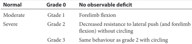
Results of experiment showed that 80% of Wistar rats subjected to the artery binding method showed neurological deficit with deficit levels varied from 1 to 3 (Table 2). Most (68%) of them showed neuro-logical deficit level 3, one rat (4%) with level 2, and two rats (8%) with level 1, while five rats (20%) did not show neurological deficit. Symptom of rat with neurological deficit level 1 indicated by the occur-rences of flexi on right forelimb as shown in Fig. 2. In this method, no side effects was observed. Table 1 Neurologic Examination Grading System
Normal Grade 0 No observable deficit
Moderate Gtade 1 Forelimb flexion
Severe Grade 2 Decreased resistance to lateral push (and forelimb flexion) without circling
Grade 3 Same behaviour as grade 2 with circling
Table 2 Neurological deficit level of Wistar rats subjected to the artery binding method
No Neurological deficit level Number of rats Percentage (%)
1 0 (normal) 5 20
2 1 2 8
3 2 1 4
DISCUSSION
To date there are five models or methods that have been developed to result in ischemic stroke condition on trial animal namely filament method, photochemical method, thromboemboli method, endothelin method, and Tamura method. However, each of these five methods has its respective advan-tages and disadvanadvan-tages.
Intraluminal filament method was firstly devel-oped by Koizumi.6 This method is done by putting a suture filament through internal carotid artery to
block blood flow into middle cerbral artery. This method is often used, because it is relatively simple and it is not invasive. The infarct occurred in the cortex cerebri of rat is assessed by using magnetic resonance imaging (MRI). Complications that frequently occurred are sub-arachnoids bleeding, spontaneous hyperthermia when lasting more than 2 h, and mechanical endothelial disorder. This method uses 4.0 nylon filament suture under the help of microscope through external carotid artery and then stop the middle cerbral artery so that the stop is accurate.16 Advantages of this method include craniotomy is not necessary, duration of ischemic can be controlled, and the changes occurred after ischemic, and can make reperfusion. Disadvantages of this method is that intraluminal filament is not the same as thrombosis in the human, and cannot assess combined therapy of recanalization with recombi-nant tissue plasminogen activator (rTPA).17 Other filament that also used is Poly-L-Lysine(PLL)coated filament. The success of this filament is very low, only 13-14% with the death rate at 21-31%. In addition, a complication of subarachnoid bleeding is occurred.18 Other filament type method is Flame/heat-blunted occluders. The success rate of this method is only 46%, with subarachnoid bleeding complication (40%), premature reperfusion (24%). This method is not recommended to study neuroprotection.19,20
Photochemical method is done by using photo-sensitive injection with laser light or filtered non-laser light that can pass through barrier of brain blood, so that singlet oxygen and free radicals are formed, and resulted in endothelial disorder and formation of micro thrombus. This method is used to assess neuroprotective medicines.21 This method offer several advantages, i.e the neuron disorder can be generated on intended location because the phothochemical is directly toward intended area, and can be used for animals with various size. However, several limitations sujected to this method. Craniotomy is needed to use this method, and cannot appropriately assess the effect of neuroprotective medicine, because duration of reperfusion cannot be predicted, and occlusion occurred permanently.17,22
Other method is thromboemboli method which is developed by injecting freez blood to stimulate embolic condition. This method was first used on dog, and then it was used for rat as thrombolytic as rTPA.23,24 Advantage of this method is that the resulted lump is similar to that of human, and it is suitable to study combined medicines for trom-bolytic and neuroprotective. Disadvantages of this method is that it is difficult to make reperfusion and difficult to assess occurrences of a complex change.17
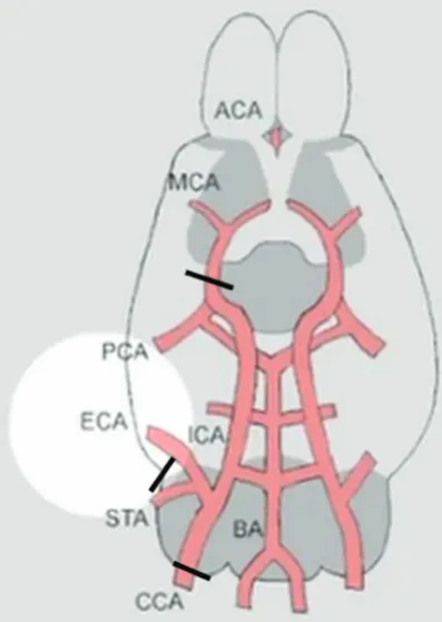
Figure 1 Schematic of brain arteries of Wistar rat. = places for binding arteries
Endothelin method is a method developed by injecting natural peptide, endotelin-1 into artery, that act as vasoconstrictor, which constrict blood vessel and result in a decrease in brain blood circulation, and finally causes ischemic lesion. Following this method, its advantages include no lesion occur around injection site, low mortality even in old animal, less invasive, lesion can be made with intended anatomy, and can be done without anesthesia. While the disad-vantage is the result is very much depending on the dose and duration of occlusion, so that the resulted infarct is not clear.9,25
The last method is Tamura method that has been developed by Tamura.7 This method uses electroco-agulation on cerebral artery. This method is devel-oped using micro clip or ligation with nylon thread. This method is alternative to study neuro-protector medicine. Advantage of this method is that occlu-sion can be made correctly on a artery that are going to be stopped. The disadvantage are craniot-omy is needed, and level of neurological deficit is often not consistent.7,26
Presently, the common method applied to stimulate ischemic stroke is intraluminal filament method facilitated with MRI to assess the size of infarct. Due to the limited access to get filament and MRI in Indonesia, a simple method was devel-oped by binding brain artery and successfully showed clinical symptom, which is paresis on contralateral side of body. This symptom indicated that lesion has occurred on brain with the success level by 80%. These rats are ready for further use to study medicines or antioxidants. Advantage of this method is that ischemic stroke in Wistar rat can be stimulated without sophisticated tools with the level of success relatively high. Its disadvan-tage is that the infarct resulted from this method is not as wide as intraluminal filament method, because the binding was not done directly in the middle cerebral artery, but in the internal carotid close to branching toward middle cerebral artery. Furthermore, since the rats in this study are still under treatment with antioxidants and have not yet subjected to craniotomy, the occurrences of infarct has not evaluated yet.
CONCLUSION
Application of a simple method through binding of brain artery successfully stimulated ischemic stroke on Wistar rat indicated by clinical symptom, a pare-sis on contralateral side of body with the level of success by 80%. This method can be implemented as an altenative method to stimulate ischemic stroke for animal testing.
REFERENCES
1. Lopez AD, Mathers CD, Ezzati M, Jamison DT, Murray JL. Global and regional burden of disease and risk factor. 2001: systematic analysis of population health and data.
Lancet. 2006;367:1747-57.
2. Krafft PR, Bailey EL, Lekic T, Rolland WB, Altay O, Tang J, et al. International Journal of stroke. 2012;7:398-406. 3. Bacigaluppi M, Comi G, Hermann DM. Animal Model of
Ischemic Stroke. Part Two: Modeling Cerebral Ischemia.
The Open Neurology Journal. 2010;4:34-8.
4. Carmichael ST. Rodent models of ischemic stroke: size, mechanisms, and purpose. NeuroRx. 2005;2(3):396-409. 5. Longa EZ, Weinstein PR, Carlson S, Cummins R.
Reversible middle cerebral artery occlusion without crani-otomy in rats. Stroke. 1989;20:84-91.
6. Koizumi J, Yoshida Y, Nakazawa T, Ooneda G. Experimental studies of ischemic brain edema: 1 A new experimental model of cerebral embolisms in rats in wich recirculation can be introduced in the ischemia area. Jpn Stroke J. 1986; 8: 1-8.
7. Tamura A, Graham GI, McCulloh J, Teasdale GM. Focal cerebral ischemia in the rat: 1: Description of technique and early neuropathological consequences following mid-dle cerebral artery occlusion. J Cereb Blood Flow Metab. 1981;1: 53-60.
8. Orset C, Haelewyn B, Vivien K, Vivien D, Young AR. Rodent model of thromboembloc stroke. In: Dirnagl U, ed. Rodent Model of stroke. Springer Protocols Neuromethods 47. New York: Humana Press; 2010. p. 55-70.
9. Marcae IM, Robinson MJ, Graham DI, Reid JL, McCulloh J. Endothelin induced reduction blood flow: dose depen-dency, time course and neuropathological consequences.
J Cereb Blood Flow Metab. 1993;13:276-84.
10. Mohr JP, Gautier JC, Hier D, Stein RW. Middle cerebral artery. In: Barnettt, et al, eds. Stroke, vol 1. Pathophysiology, Diagnosis and Management. New York: Churchill Livingston; 1986. p. 377-450.
11. Sicard K, Fisher M. Animal Model of focal brainn ischemia.
Experimental & Translational Stroke Medicine. 2009;1:1-6. 12. Marcae IM. Preclinical stroke research-advantage and
disadvantage of the most common rodent models
of focal ischemia. British Journal of Pharmacology. 2011;164:1062-1078.
13. Satriotomo I. Pre Conference: 1st National Conference of Neuroscience Indonesia; 2013 April; Indonesia Brain Research Center (IBRC), Surya University. Jakarta; 2013. 14. Ahmad A, Khan MM, Javed H, Raza SS, Ishrat T,
Khan MB, et al. Edaravone ameliorates stress associated cholinergic dysfunction and limits apoptosis response following focal cerebral ischemia in rat. Mol Cell Biochem. 2012;367:215-225.
15. Bederson JB, Pitts LH, Tsuji M, Nishimura MC, Davis RL, Bartkowski H. Rat middle cerebral artery occlusion: evalu-ation of the model and development of a neurologic exam-ination. Stroke. 1986;17:472-76.
16. Durukan A, Tatlisumak T. Animal Model of ischemic stroke. In Fisher M eds. Handbook of Clinical neurology (3rd series). Stroke. Part I, Basic and Epidemiological Aspect. 2009; 92:450-64.
17. Herson PS., Traystman R. Animal model of stroke: transla-tional potential at present and in 2050. Future Neurol. 2014 9(5):541-51.
18. Spatt NJ, Fernandez J, Chen M, Rewell S, Cox S, vaan Ray L, Hogan L, Howells DW. Modification of the method of thread manufacture improves stroke induction rate and reduce mortality after thread occlusion of the middle cerebral artery in young or aged rats. J Nuerosci Methods. 2006;155:285-90.
19. Tsuchiya D, Hong S, Kayama T, Panter SS, Weinstein PR. Effect of suture clip application upon blood flow and infarct volume after permanent and temporary middle cerebral artery occlusion in mice. Brain Res. 2003;970:131-39. 20. Schimid-Elsaesser R, Zausinger S, Hungerhuber E,
intraluminal thread model of focal cerebral ischemia: evidence of inadvertent premature reperfusion and sub-arachnoid hemorrhage in rats by lase-Doppler flowmetry.
Stroke. 1998;29:2162-70.
21. Lozano JD, Abulifia DP, Danton GH, Watson BD, Dietrich WD. Characterization of thromboembolic photo-chemical model repeated stroke in mice. J Neurosci meth-ods. 2007;162:244-54.
22. Wang CX, Yang T, Shuaib A. An improved version of embolic model of brain ischemic injury in the rat.
J Neurosci Method. 2001:190;147-51.
23. Kudo M, Aoyama A, Ichimori S, Fukunaga A. An Animal model of cerebral infarction: homologous blood clot emboli rats. Stroke. 1982;13:505-08.
24. Albers GW. Antithrombotic agent in cerebral ischemia.
Am J Cardiol. 1995;75:348-88.
25. Ansari S, Azari H, Cadwell KJ, Regenhardt RW, Hedna VS, Waters MF, et al. Endothelin-1 induced middle cere-bral artery occlusion model for ischemic stroke with lasser dopler flowmetry guidance in rat. Journal of Visual Expreriments. 2013;72:1-6.
26. Liu S, Zhen G, Meloni B, Campbell K,Win R. Rodent stroke model guidelines for preclinical stroke trials (1st edition).
J Exp Stroke Trans Med. 2009;2(2):2-27.