Functions after Initiation of Mitosis
Meera Govindaraghavan,a,bAlisha A. Lad,aStephen A. Osmania,b
‹Department of Molecular Geneticsaand Molecular, Cellular and Developmental Biology Program,bThe Ohio State University, Columbus, Ohio, USA
The G2-M transition inAspergillus nidulansrequires the NIMA kinase, the founding member of the Nek kinase family.
Inactiva-tion of NIMA results in a late G2arrest, while overexpression of NIMA is sufficient to promote mitotic events independently of
cell cycle phase. Endogenously tagged NIMA-GFP has dynamic mitotic localizations appearing first at the spindle pole body and then at nuclear pore complexes before transitioning to within nuclei and the mitotic spindle and back at the spindle pole bodies at mitotic exit, suggesting that it functions sequentially at these locations. Since NIMA is indispensable for mitotic entry, it has been difficult to determine the requirement of NIMA for subaspects of mitosis. We show here that when NIMA is partially inacti-vated, although mitosis can be initiated, a proportion of cells fail to successfully generate two daughter nuclei. We further define the mitotic defects to show that normal NIMA function is required for the formation of a bipolar spindle, nuclear pore complex disassembly, completion of chromatin segregation, and the normal structural rearrangements of the nuclear envelope required to generate two nuclei from one. In the remaining population of cells that enter mitosis with inadequate NIMA, two daughter nuclei are generated in a manner dependent on the spindle assembly checkpoint, indicating highly penetrant defects in mitotic progression without sufficient NIMA activity. This study shows that NIMA is required not only for mitotic entry but also se-quentially for successful completion of stage-specific mitotic events.
I
dentification of cell cycle specific mutations in model organismshas been instrumental in the discovery of proteins required for progression through all stages of the cell cycle. The pioneering
work of Ron Morris (1) enabled the isolation of numerous highly
conserved genes required for mitotic progression utilizing the
model filamentous fungusAspergillus nidulans (2).A. nidulans
undergoes both sexual and asexual development to produce dor-mant ascospores and conidiospores, respectively. Because of their ease of production, conidiospores (conidia) are most often used as
inoculum forA. nidulanscell cycle analysis. Conidia are
uninucle-ated dormant cells that upon exposure to suitable growth condi-tions first undergo isotropic growth, during which the first mitosis is often completed. After a single site for polarized growth is es-tablished, germ tube extension occurs, during which the two nu-clei transition the cell cycle and parasynchronously undergo mitosis to generate germlings with four nuclei in a common cyto-plasm. Typically only after the third synchronous mitotic division
does septation occur (3–5).
InA. nidulans, the never in mitosis A (NIMA) and cell division cycle 1 (CDK1) kinases are essential for mitotic entry but not for
short-term germling growth (6). Like Cdk1 (7–13) NIMA was
identified through a genetic screen to isolate temperature-sensi-tive (ts) cell cycle mutants, some of which caused an arrest in
interphase (never in mitosis [nim]mutants) at the restrictive
tem-perature (1). TwonimAalleles contain point mutations causing
amino acid substitutions in the catalytic domain of the kinase (nimA5[Y91N] andnimA7[E41G]), while thenimA1allele has a change in the C-terminal regulatory domain (L304P) just
down-stream of the catalytic domain (14). Cells carrying these
temper-ature-sensitive nimA alleles, when incubated at the restrictive
temperature, arrest with a single G2nucleus, duplicated spindle
pole bodies (SPBs), and cytoplasmic microtubule architecture
(15). When restored to the permissive temperature, these cells
synchronously enter mitosis, indicating that mitotic entry is
con-tingent upon NIMA activation (16, 17). NIMA is the founding
member of the Nek family of NIMA-related kinases conserved through all eukaryotes, which have diverse roles in mitosis as well
as ciliogenesis (18,19). InSchizosaccharomyces pombe, the NIMA
ortholog Fin1 plays a central role in the G2-M transition by
miti-gating the inhibition of Cdk1 activity by the Pom1 kinase as well as by establishing a positive feedback of Polo kinase activity to cause
a switch-like increase in Cdk1 activity (20,21). However, inA.
nidulans, although NIMA is required for entry into mitosis, it is not required for mitotic activation of Cdk1 kinase activity via
tyrosine dephosphorylation (17). On the other hand, mitotic
ac-tivation of Cdk1 is required for the final complete mitotic activa-tion of NIMA during entry into mitosis, most likely via
phosphor-ylation of NIMA by mitotic Cdk1 (22). Moreover, through the use
of temperature-sensitivenimAmutants, NIMA function has been
shown to be required for the nuclear localization of cyclin B, which is critical for Cdk1-cyclin B-mediated phosphorylation of
mitotic substrates (23). It has been shown that the dramatic
mi-totic targeting of NIMA to SPBs (24) and nuclear pore complexes
(NPCs) at the initiation of mitosis also requires mitotic activation
of Cdk1 (25,26).
Emphasizing its functional conservation, overexpression of
NIMA causes mitotic chromatin condensation not only inA.
ni-dulansbut also, strikingly, in fission yeast,Xenopus, and human
cells (27, 28). NIMA can also promote the phosphorylation of
histone H3S10, a mark of mitotic chromatin (25). Using a forward
Received4 September 2013Accepted31 October 2013
Published ahead of print1 November 2013
Address correspondence to Stephen A. Osmani, [email protected].
Supplemental material for this article may be found athttp://dx.doi.org/10.1128 /EC.00231-13.
Copyright © 2014, American Society for Microbiology. All Rights Reserved.
doi:10.1128/EC.00231-13
on September 8, 2020 by guest
http://ec.asm.org/
genetic screen, mutations in two NPC proteins, SONA and SONB,
were identified as suppressors of thenimA1temperature-sensitive
mutation, suggesting that NIMA may regulate these nuclear pore
proteins (23,29). Consistent with that expectation, NIMA is
re-quired and sufficient to promote NPC disassembly, one of the
earliest mitotic events, not only inA. nidulans(26,30–33) but also
vertebrate systems (34), and NIMA and related human kinases
can phosphorylate the NPC protein Nup98in vitro(34).
Since NIMA function is essential for all aspects for mitosis, it has been difficult to assess whether NIMA is required for specific mitotic events subsequent to the start of mitosis. However, the analysis of cells that are mutated in the gene encoding the ana-phase-promoting complex (APC) subunit BIME in addition to
carrying animA5mutant allele (nimA5 bimE7) suggested the
pos-sibility that specific mitotic events might require NIMA function.
The absence ofbimEfunction abrogates the G2-M-mediated
ar-rest innimA5cells, promoting premature mitotic entry causing
abnormal spindle formation and nuclear envelope (NE) invagina-tions potentially due to the initiation of mitosis in the absence of
normal NIMA function (35).
Our studies presented here using cells with partial NIMA func-tion provide further strong evidence that in addifunc-tion to being required for mitotic entry, NIMA is also required to regulate spin-dle pole body functions, nuclear pore complex permeability, and NE dynamics for successful mitotic generation of daughter nuclei.
MATERIALS AND METHODS
Standard conditions were used for propagating and generatingA. nidu-lansstrains, as described in reference36with minor alterations. The ge-notypes of strains used in this study are provided in Table S1 in the sup-plemental material. Live-cell confocal imaging was performed using a 60⫻, 1.49-numerical aperture (NA) TIRF objective lens on a Nikon 484 Eclipse TE 2000-U (Nikon, Inc.) microscope equipped with an UltraView ERS spinning-disk 485 confocal system (PerkinElmer Inc.), and images were captured using a Hamamatsu ORCA-AG 486 camera. Temperature-controlled experiments were carried out using Bioptechs Delta T dishes and heating equipment from Bioptechs, Inc., PA, USA. The cells were grown at the required temperature in humidity chambers for 4.5 h. The objective and the stage were calibrated for the correct temperature, and then the imaging dish was transferred to the temperature-controlled setup on the microscope. For confocal imaging at room temperature, conidio-spores were germinated in 35-mm glass-bottom petri dishes (MatTek Cultureware). Images and movies were processed and signal intensity inside the nucleus measured using ImageJ version 1.46m (http://rsbweb .nih.gov/ij/). All images are maximum-intensity projections unless indi-cated otherwise. Rotational analysis was done using the Projector 4D pl-ugin on ImageJ. Statistical analyses were done using MS Excel, except for box plots, which were generated on Stata software. For the box plots, the ends of the whiskers extend to 1.5 times the height of the box or, if there is no value in that range, to the minimum and maximum values. If the data are distributed normally, approximately 95% of the data are expected to lie between the whiskers. For analysis of mitosis in the presence of unpo-lymerized tubulin, cells were germinated in the presence of 2.4g/ml benomyl at the required temperature for 4.5 h before imaging at the same temperature.
RESULTS
Using live-cell spinning disk 4D confocal microscopy, we exam-ined the first mitosis in germinating uninucleated conidia in which the function of the mitotic NIMA kinase was partially in-hibited to identify the earliest defects caused and avoid the possi-bility of looking at cumulative effects of previous mitotic failures.
We employed thenimA7mutation because this is a tightnimA
allele encoding a mutation in the catalytic domain that facilitated temperature-controlled imaging at 35°C under conditions that allowed some cell growth and mitotic progression. We examined
nimA7strains that carried fluorescently tagged proteins locating to the kinetochore, nucleus, and NE by live-cell microscopy. These
studies revealed that 46% (n⫽54) of cells with partial NIMA
function fail to complete the first mitosis successfully (strains MG190, MG229, MG227, and MG228). Of these 46%, 11% un-dergo mitotic failure due to an inability to form a bipolar spindle, while 35% exhibit defects downstream of bipolar spindle forma-tion. Below we report in more detail how normal NIMA function is required for different aspects of mitosis.
Cells with reduced NIMA function have defects in forming a bipolar spindle.InA. nidulans, the kinetochores are clustered near the spindle pole body at the nuclear periphery during inter-phase. At mitotic entry, nuclear spindle microtubules are initially formed from duplicated, unseparated SPBs in a monopolar fash-ion. Subsequently, the separation of the SPBs results in the
bipo-larization of the spindle. We generatednimA7strains that carried
green fluorescent protein (GFP)-Tub (TubA) (37) and the
kinet-ochore marker Ndc80-mCherry Red (Ndc80-CR) (38) to monitor
spindle formation and kinetochore segregation, respectively. In
nimA7cells grown at the permissive temperature of 22°C, the kinetochores are in a single focus during interphase, as expected. During mitosis, the kinetochores become spread along the spindle and then divide equally, regenerating SPB-associated foci within
daughter nuclei upon successful completion of mitosis (Fig. 1A).
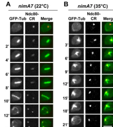
When NIMA is partially inactivated by germinating the same strain at the semipermissive temperature of 35°C, we find that
11% (n⫽54) of the cells form a monopolar spindle. These
spin-dles appear to be arrested at the monopolar stage due to an inabil-ity to separate the duplicated SPBs, as indicated by the presence of
clustered unseparated kinetochores (Fig. 1B). The monopolarity
of the spindle was further confirmed innimA7cells carrying
GFP-Tub and an SPB marker, theA. nidulansortholog of
Schizosaccha-FIG 1Monopolar spindle formation in cells with partial NIMA function.
nimA7cells carrying GFP-Tub and the kinetochore protein Ndc80-CR (strain MG190) were followed through mitosis at 22°C (A) or at 35°C (B). Bars, 5m.
on September 8, 2020 by guest
http://ec.asm.org/
romyces pombe Sad1 (annotated as AN6868 at The Aspergillus
Genome Database [AspGD]) (39), at the semipermissive
temper-ature (data not shown), where 14% of SPBs (n⫽28) were seen to
remain as a single focus for the duration of mitosis.
Cells with reduced NIMA function show defects in anaphase completion, defective segregation of the nucleolus, and abnor-mal NE dynamics during mitosis.In wild-type (WT) cells, mi-totic entry is accompanied by chromatin condensation and
spin-dle formation. During anaphase (Fig. 2A, 3=), the bipolar spindle
segregates the chromatin and subsequent chromatin
decondensa-tion occurs during the formadecondensa-tion of two G1nuclei (Fig. 2A). In
35% of thenimA7cells (n⫽54), we find that a bipolar spindle is
established; however, mitosis is not completed successfully, as chromatin fails to segregate completely upon spindle elongation (Fig. 2B, 8=, arrowhead) and collapses back into a single mass (Fig.
2B). InnimA7cells that form a bipolar spindle carrying
Ndc80-CR, GFP-Tub, and the spindle pole body marker GCP3-GFP (33,
40), the kinetochores could be seen to segregate, giving rise to two
Ndc80 foci in an apparently normal fashion (Fig. 2C,
arrow-heads). Surprisingly, however, the segregation of Ndc80 was not maintained, and the kinetochores collapsed back to a single focus indicative of a defect in anaphase completion. Remarkably, the failure to segregate the kinetochores was accompanied by the
sep-arated SPBs also fusing back to give a single focus (Fig. 2C, 14=) at
mitotic exit. Together with the examination ofnimA7strains
car-rying the SPB marker Sad1 or TinA (41) (data not shown), we find
that 12% (n⫽50) ofnimA7cells showed mitotic bipolar spindle
formation and SPB separation followed by their reassociation into closely paired unresolvable foci, demonstrating that the pheno-type is reproducible using different SPB markers. The population ofnimA7cells showing this phenotype is likely to be an underes-timate, since clear observation of this phenotype is limited by the orientation of the mitotic apparatus. Collectively, these results show that partial inactivation of NIMA impairs successful ana-phase completion.
To monitor NE dynamics, a recently identified inner nuclear membrane (INM) marker that contains a C-terminal
transmem-brane domain with similarity to the S. pombe INM protein
Bqt4 (S. A. Osmani and M. Chemudupati, unpublished data)
(an-notated at AspGD as AN0162 [http://www.aspergillusgenome.org
/cgi-bin/locus.pl?dbid⫽ASPL0000057939]) was utilized. During WT mitosis, the NE, marked by CR-AN0162, restricts at two
points, giving rise transiently to 3 compartments (Fig. 3A,
ar-rows). The middle compartment contains the nucleolus, and the two compartments at either end encompass the segregated
chro-matin of the forming daughter nuclei (42). Interestingly, 30%
(n⫽27) of cells with reduced NIMA function show a defect in
executing the NE double pinch (Fig. 3B). Moreover, a significant
proportion ofnimA7cells (65%,n⫽17 failed first mitoses) show
the formation of an apparent intranuclear ring-like structure of NE (Fig. 3B, arrowhead), a phenomenon not observed after
nor-mal mitosis. We refer to this NE configuration as “theta” (),
owing to its resemblance to the Greek letter. Intriguingly, we found, using a tagged nucleolar marker protein fibrillarin
(Fib-CR) (42), that the nucleolus is present within the ring-like
struc-ture in thenuclei observed (Fig. 3D). Z sections through the
nucleus (Fig. 3E) and three-dimensional (3D) rotations (see
Movie S1 in the supplemental material) revealed that the
nucleo-lus was positioned in an aberrant location within the nuclei,
causing a protrusion which changed the normal oval shape of interphase nuclei. Notably this configuration was dynamic, with the nucleolar protrusion changing to become more or less prom-inent through time. In contrast to the normal nucleolar segrega-tion observed in WT cells at the same temperature (see Fig. S1 in
the supplemental material) (42), we also observed that some
nimA7nuclei apparently managed to segregate their nucleolus
into two (Fig. 3C, 5=to 6=), only to then have the two nucleoli
collapse back into one (Fig. 3C, 7=). The data indicate that
success-ful generation of daughter nuclei via mitotic NE double restric-tions, coupled with nucleolar segregation, requires normal NIMA functions.
Cells with reduced NIMA function are unable to undergo normal nuclear pore complex disassembly during mitosis.InA. nidulans, around half of the nuclear pore complex (NPC) pro-teins, representing the peripherally located components, disperse
from NPCs during mitosis, abolishing nuclear transport (26,30).
Complete NIMA inactivation prevents initiation of mitosis and
FIG 2Failure of nuclear division in cells with partial NIMA function subse-quent to bipolar spindle formation. (A and B) Histone H1-chRFP and GFP-Tub were used to follow mitosis in WT (strain MG300) (A) andnimA7(strain MG298) (B) cells at 35°C. In panel A, the arrowhead points to chromatin segregation in anaphase. In panel B, the arrowhead points to chromatin that is attempting to segregate but eventually collapses into a single nucleus after failed anaphase. (C)nimA7cell carrying GFP-Tub, GCP3-GFP and Ndc80-CR (strain MG190) followed through mitosis at the semipermissive temperature of 35°C. Arrowheads indicate kinetochore foci as they first become segregated (6 to 10 min) and then return back together (10 to 14 min). The GCP3-GFP foci at separated SPBs at 10 min also collapse back together by 14 min. Bars, 5m.
on September 8, 2020 by guest
http://ec.asm.org/
thus prevents downstream NPC disassembly, while ectopic ex-pression of NIMA promotes partial nuclear pore complex
disas-sembly, even out of cell cycle phase (26). Therefore, NIMA has
been proposed to initiate mitosis at least in part by promoting NPC disassembly, which allows tubulin and mitotic regulators to access the nucleoplasm. The mitotic failure in cells with partial NIMA function might therefore be due to a defect in
disassem-bling the NPCs. To test this, we generated strains having WTnimA
or thenimA7mutant allele, in which the location of the NPC
protein Nup49-CR can be followed in addition to GFP-Tub. Nup49 is a peripheral nucleoporin that locates to the NPCs during
interphase but disperses from them at mitosis (26,43). Most
sur-prisingly, in cells with reduced NIMA function, the majority of the Nup49-CR signal remained at the nuclear periphery during mito-sis, when it should be dispersed as the mitotic spindle was formed (Fig. 4A, 3=). This dramatic defect, which was seen in allnimA7
cells at 35°C, indicates that in addition to being sufficient to pro-mote NPC disassembly, NIMA function is actually required for NPC disassembly during mitosis after nuclei are triggered to
un-dergo the G2-M transition. To further define the status of NPCs in
mitoticnimA7cells, we visualized nuclear transport by following
NLS (nuclear localization sequence)-DsRed, a marker protein
that is actively transported into interphase nuclei (44). During
FIG 3Defective NE dynamics and nucleolar segregation in cells with partial NIMA function. (A and B) Mitotic dynamics of the NE marker protein CR-AN0162 and GFP-Tub in WT (A) (strain MG224) andnimA7(B) (strain MG227) cells. The arrows in panel A indicate the normal NE restriction points during telophase. The arrowhead in panel B at 26 min shows the formation of the “theta”-shaped nuclear structure (see the text for details). (C) Mitotic dynamics of the nucleolus marked by fibrillarin-CR and GFP-AN162 in a
nimA7nucleus (strain MG294) that fails to generate two daughter nuclei after first mitosis even though the nucleolus first appears to be segregated into two. (D) The nucleolar protein fibrillarin-CR (strain MG294) is apparently envel-oped by GFP-AN0162 (arrow). (E) Z sections through a nimA7nucleus marked with GFP-AN162 reveal an NE protrusion and the abnormal shape of the nucleus (see also Movie S1 in the supplemental material). Bars, 5m.
FIG 4Cells with reduced NIMA function have defects in mitotic NPC disas-sembly. (A) Mitosis in animA7cell carrying GFP-Tub and the peripheral nucleoporin Nup49-CR (strain MG321) at the semipermissive temperature. During spindle formation, unlike in WT cells, most Nup49 remains associated at the NE. (B) Mitosis in animA7cell carrying the heterochromatin binding protein HP1-CR and GFP-Tub (strain MG378) at the semipermissive temper-ature. When spindles form, HP1-CR is released from nuclei. (C and D) Nu-clear transport marked by NLS-DsRed and spindle formation (GFP-Tub) fol-lowed in mitotic WT cells (C) (strain MG273) andnimA7cells (D) (strain MG229). (E) Behavior of NLS-DsRed innimA7⌬mad2cells (strain MG313) during mitosis in the presence of benomyl. Arrows indicate the nuclear shadow revealed by cytoplasmic GFP-Tub. Bars, 5m.
on September 8, 2020 by guest
http://ec.asm.org/
interphase, NLS-DsRed is exclusively nuclear (Fig. 4C). During mitosis, partial NPC disassembly results in the dispersal of
NLS-DsRed until active transport is reestablished in daughter G1nuclei
(Fig. 4C). However, when NIMA is partially functional, NLS-DsRed does not disperse from nuclei upon mitotic entry, as marked by formation of the mitotic spindle, but continues to
re-main within nuclei (Fig. 4D, 2=and 4=). This indicates that in
mitoticnimA7cells, both partial NPC disassembly and the
open-ing of nuclear pores are defective.
It has been proposed that NPC disassembly functions to allow the entry of tubulin into the nucleus, thus promoting spindle
for-mation. However, since spindles were able to form innimA7cells
even though NLS-DsRed fails to escape nuclei, we wanted to con-firm whether tubulin is able to enter the nucleus during mitotic entry in these cells. To this end, we quantitated the change in tubulin signal inside the nucleus using single confocal sections
through the middle of WT andnimA7 nuclei (Fig. 5). In WT
interphase nuclei, unpolymerized fluorescently tagged tubulin is
excluded from the nucleus, revealing a nuclear shadow in late G2
(Fig. 5A, arrow). Upon mitotic entry, this mitotic shadow fills in as tubulin equilibrates across the NE concurrent with the dispersal of
NLS-DsRed from the nucleus (Fig. 5AtoC). Interestingly in cells
with partial NIMA function, nuclear GFP-Tub increases sharply
at the G2-M transition; however, the nuclear signal of NLS-DsRed
does not decrease precipitously as it does in the WT cells (Fig. 5D
toF). This shows that when NIMA is partially inactive, tubulin is
able to access the mitotic nucleoplasm and hence form the spindle even though NLS-DsRed is unable to escape from the mitotic nucleus in the normal fashion. This might suggest that tubulin is actively transported into these nuclei at mitosis. One alternative explanation, however, is that in the absence of normal NIMA function, the nuclear pores are insufficiently opened during
mi-tosis, such that␣-tubulin dimers (GFP-TubA-BenA [127 kDa]
[39]) can diffuse across the NE, while proteins of larger molecular
size and/or shape, such as the tetrameric NLS-DsRed (190 kDa)
(45), are prevented from escaping the nucleus. To test this idea, we
examined the behavior of heterochromatic protein 1 (HP1) (also
termed HepA inA. nidulans) (46), which is nuclear during
inter-phase and disperses during mitosis. We find that HP1-CR, which is expected to be 53 kDa, does indeed diffuse from mitotic nuclei innimA7cells (Fig. 4B). This indicates that partial inactivation of NIMA allows only incomplete NPC disassembly, such that small proteins like tubulin and HP1 can diffuse across nuclear pores while larger protein complexes, such as NLS-DsRED, remain trapped within mitotic nuclei.
Intriguingly, in nimA7cells that form a bipolar spindle,
al-though NLS-DsRed does not disperse at mitotic entry, it does escape the nucleus transiently at a point corresponding to spindle elongation, suggesting that perhaps the mechanical force of
spin-dle elongation compromises NE integrity (Fig. 4D, 6=). To test
whether the release of NLS-DsRed is caused by mechanical forces acting on the NE driven by the elongating spindle, we germinated
nimA7cells withmad2deleted and carrying GFP-Tub and NLS-DsRed in the presence of benomyl, a microtubule-depolymerizing drug. The absence of Mad2 prevents the engagement of the spin-dle assembly checkpoint (SAC)-mediated mitotic arrest in the
presence of unpolymerized tubulin (32, 43), allowing nuclei to
enter and then exit mitosis without a mitotic spindle. As in WT
cells, during G2, GFP-Tub is excluded from the nucleus, revealing
a nuclear shadow in thenimA7⌬mad2cells treated with benomyl
(Fig. 4E, arrow). Entry into mitosis was marked by the diffusion of
soluble GFP-Tub into the nucleus, as expected (37), but
NLS-DsRed remained nuclear at this time. In contrast, in WT cells with normal NIMA function, NLS-DsRed is known to be released upon
mitotic entry under identical conditions (43). Importantly, we
found that NLS-DsRed still dispersed briefly in the absence of
spindle formation in mitoticnimA7cells (Fig. 4E, 5=) before then
being reimported into the G1nucleus. In G1, GFP-Tub is once
again excluded from the nucleus (Fig. 4E, 11’, arrow). The data
show that in the absence of forces exerted by spindle microtu-bules, NLS-DsRed is able to disperse briefly in cells with partial
FIG 5Tubulin can enter the mitotic nucleus in cells with reduced NIMA function. (A and D) Single Z-slice images of GFP-Tub and NLS-DsRed in WT andnimA7cells at 35°C during the G2-M transition. Arrows mark the shadow
of the nucleus revealed by the absence of GFP-tubulin. Bars, 5m. (B and E) Schematic representation of the location of tubulin and NLS-DsRed in inter-phase versus mitosis in WT cells (B) andnimA7cells (E). Nuclear pore com-plex disassembly in mitosis is indicated by the dotted line enveloping the nu-cleus. The signal intensity in the yellow-dotted area was measured. (C and F) Changes in signal intensities of tubulin (green) and NLS-DsRed (red) with time during the G2-M transition of WT (C) (n⫽4, strain MG273) ornimA7
(F) (n⫽6, strain MG229) cells.
on September 8, 2020 by guest
http://ec.asm.org/
NIMA function, indicating that the mitotic permeability proper-ties of the NE are being modified in a manner independent of the mitotic spindle (see Discussion).
Insufficient NIMA function causes a delay in mitotic pro-gression.The mitotic defects seen in cells with partial NIMA
func-tion suggest thatnimA7cells may be delayed in mitotic
progres-sion, as indicated by comparing the times of mitosis for WT and
nimA7cells inFig. 1and2. To quantitate this, we calculated the average time that spindle microtubules are detectable in WT and
nimA7cells from time lapse analysis of mitosis at the
semipermis-sive temperature fornimA7. Cells with partial NIMA function
show the presence of spindle microtubules for more than double the time on average that WT cells do and also display more vari-ability in time in mitosis (see Fig. S2A and B in the supplemental material). There was no correlation between the time spent in mitosis and whether the mitosis was successful or not (data not shown). In order to determine whether the delay in mitosis seen in
cells with partial NIMA function is specific to this allele ofnimA,
we followed mitosis in strains with the two other available
tem-perature-sensitivenimAalleles,nimA1andnimA5. We find that
spindle microtubules are similarly present for a prolonged time
period innimA1andnimA5cells compared to WT cells (see Fig.
S2C in the supplemental material), demonstrating that partial in-activation of NIMA results in prolonged mitosis independent of the allele used.
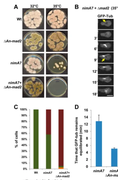
Cells with partial NIMA function are dependent on the spin-dle assembly checkpoint for their survival.The spindle assembly checkpoint functions to monitor mitotic errors and impose a de-lay in mitosis so that these errors can be corrected before anaphase is triggered. Given that partial NIMA function results in multiple mitotic defects, as well as a delay in mitosis, it is possible that the
completion of mitosis innimA7cells might be dependent on the
SAC. To test this, we generated strains carryingnimA7that have a
deletion of a gene essential for SAC function,mad2(32). To
ex-amine whether the deletion ofmad2affects the growth ofnimA7
cells, we compared the colony growth of thenimA7⌬mad2
mu-tant and the single mumu-tants at the semipermissive temperature.
The double mutants lackingmad2with partial NIMA function are
unable to form any visible colonies at this temperature (Fig. 6A),
highlighting the requirement of the SAC for the survival ofnimA7
cells with partial NIMA functions. Using live-cell microscopy and
NLS-DsRed and GFP-Tub as markers, we find thatnimA7⌬mad2
cells show a highly penetrant inability to complete their first mi-tosis successfully, where nuclei enter mimi-tosis but fail to generate
daughter nuclei (Fig. 6BandC). Because GFP-Tub is excluded
from G2nuclei but diffuses into the nucleus during mitosis and is
reexported back out of the nucleus during G1, nuclear tubulin can
be used as a marker of mitosis. We find that mitoticnimA7⌬mad2
cells have tubulin within the nucleus for an average of 5 min,
compared to a longer 12 min innimA7cells (Fig. 6BandD).
However, surprisingly, we see that even after tubulin is exported from the nucleus, astral microtubules continue to extend from the
cytoplasmic side ofnimA7 ⌬mad2nuclei (Fig. 6B). Taken
to-gether, these data show that completion of mitoses in cells with partial NIMA function is dependent on SAC function.
To ascertain whether the two nuclei generated innimA7cells
after an apparently successful first mitosis are normal, we followed them into their second mitosis (see Fig. S3 in the supplemental material). Surprisingly, we find that in such cells, daughter nuclei can behave differently from each other. As shown in the example
in Fig. S3B in the supplemental material, although both nuclei enter mitosis forming spindles, one nucleus (n2) attempts ana-phase, while the other (n1) does not. The nucleus that does not attempt anaphase also does not disperse NLS-DsRed and likely forms a monopolar spindle, whereas nucleus n2 disperses
NLS-DsRed briefly (at 6=). However, since nucleus n1 is actively
trans-porting, much of the NLS-DsRed dispersed from n2 is available for import into nucleus n1. Thus, the two nuclei undergo asyn-chronous mitoses in an autonomous manner, in sharp contrast to the parasynchronous mitoses seen in all WT cells.
The generation of two nuclei after first mitosis in 54% of
nimA7cells depends on SAC function, indicating that the SAC is functional. However, the remaining 46% of the first mitoses fail to generate daughter nuclei, although nuclei exit mitosis, suggesting that perhaps SAC function is not completely normal in these
nimA7cells. To test whether SAC-mediated mitotic arrest can be appropriately engaged in the absence of normal NIMA function,
we followed mitosis innimA7cells at the semipermissive
temper-ature in the presence of unpolymerized GFP-tubulin. Mitotic ini-tiation can be followed by the entry of GFP-Tub into the nucleus,
FIG 6Cells with reduced NIMA function depend on the spindle assembly checkpoint (SAC) for survival. (A) Colony growth of strains of the indicated genotypes at 35°C after 72 h. Strains: WT, R153;nimA7, CDS790;⌬An-mad2, CDS629;nimA7⫹⌬An-mad2, MG381. (B) Mitosis as followed using NLS-DsRed and GFP-Tub in animA7cell that also lacksmad2(strain MG313). Arrows point to the exclusion of GFP-Tub from the nucleus in G2and then G1.
Bar, 5m. (C) Quantitation of the number of cells that complete the first mitosis successfully versus those which do not in WT (n⫽15),nimA7(n⫽
26), andnimA7⌬mad2(n⫽40) strains. Unequal division refers to a mitosis resulting in daughter nuclei of different sizes. (D) Quantitation of the time that GFP-Tub is seen equilibrated across the NE (n⫽11 for each).
on September 8, 2020 by guest
http://ec.asm.org/
which in WT cells is followed immediately by the dispersal of
NLS-DsRed from the nucleus (Fig. 7AandC; see Fig. S4A in the
supplemental material). However, in 50% of thenimA7cells (n⫽
30), entry of GFP-Tub into the nucleus was not accompanied by dispersal of NLS-DsRed (data not shown), while in the other half of the population, NLS-DsRed dispersed after an average delay of
18 min (Fig. 7BandC; see Fig. S4A in the supplemental material).
This suggests that the delay in mitosis imposed by the SAC allows somenimA7cells to accumulate enough NIMA function so as to eventually affect NLS dispersal. To compare the time of mitotic
SAC arrest between WT andnimA7cells, mitotic exit could be
monitored by the reimport of NLS-DsRed, as this is an M-G1
transition-regulated event. The actual time of mitotic SAC arrest could therefore be measured as the time between entry of GFP-Tub into nuclei (start of mitosis) and the nuclear reimport of
NLS-DsRed (mitotic exit) (Fig. 7). In WT cells, mitotic entry in
the presence of the microtubule-depolymerizing drug benomyl results in a SAC-mediated mitotic arrest, as previously
docu-mented, for an average of 40.6⫾10 min (Fig. 7AandD; see Fig.
S4B in the supplemental material) (43). Importantly, mitosis in
cells with partial NIMA function in the absence of spindles was
similarly prolonged to an average of 46⫾13 min (Fig. 7BandD;
see Fig. S4B in the supplemental material) in a manner dependent
on the SAC (Fig. 4E). The data indicate that the SAC is engaged
normally during first mitosis in the absence of spindles innimA7
cells at the semipermissive temperature.
DISCUSSION
Initiation of mitosis in the absence of sufficient NIMA function.
When NIMA is partially inhibited by incubating strains having the
nimA7temperature-sensitive allele at semipermissive tempera-tures, nuclear division can be initiated. Because NIMA is not re-quired for activation of mitotic Cdk1 kinase activity, this is pre-sumably because low levels of NIMA7 activity are sufficient to trigger mitotic entry, since CDK1 is expected to be mitotically activated. However, in the absence of normal NIMA function, penetrant defects in specific mitotic events ensue. Therefore, the
analysis ofnimA7cells at the semipermissive temperature enabled
the uncoupling of NIMA’s requirement for mitotic initiation from its functions during mitotic progression and proved to be a powerful approach to uncover the requirement for NIMA in
reg-ulating specific mitotic events (Fig. 8).
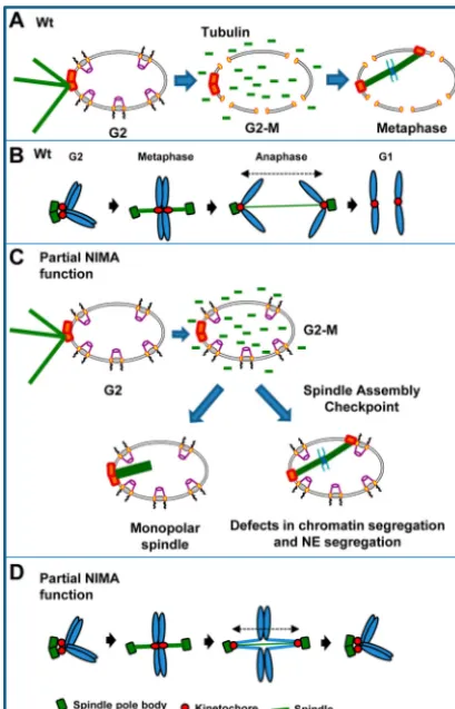
NIMA-mediated regulation of spindle pole body separation.
Partial inactivation of NIMA leads to monopolar spindle forma-tion implicating NIMA in the separaforma-tion of the duplicated spindle
pole bodies during mitotic entry (Fig. 8AandC). This provides
strong evidence for the functional significance of the localization
of NIMA-GFP to SPBs as the first step in promoting the G2-M
transition (24). The formation of monopolar spindles is likely not
due to a defect in SPB duplication, since SPB duplication has been shown to occur in the absence of NIMA function and bipolar
spindles form immediately upon return ofnimA mutants to a
permissive temperature (15,47,48). The human NIMA-related
kinase Nek2 regulates the removal of centrosomal linker proteins
C-Nap1, rootletin, and-catenin through phosphorylation, while
the Nek9-Nek6/Nek7 phosphorylation cascade regulates the
func-tion of the BIMC kinesin Eg5 during centrosome separafunc-tion (19,
49–52). Along similar lines, a temperature-sensitive mutant of the
S. pombeNIMA orthologfin1was identified in a screen for spindle architecture defects where the microtubule nucleation of one of
FIG 7Cells with partial NIMA function are capable of engaging the SAC-medi-ated mitotic arrest in response to unpolymerized GFP-Tub. (A) Mitosis in a WT cell (strain MG273) in the presence of benomyl, a microtubule-depolymerizing drug. Mitotic entry is indicated by the filling in of the GFP-Tub nuclear shadow (arrow) followed immediately by the dispersal of NLS-DsRed throughout the cy-toplasm. (B) Mitosis in animA7cell (strain MG229) in the presence of benomyl at the semipermissive temperature (35°C). The dispersal of NLS-DsRed is seen after a time lag following the entry of GFP-Tub into the nucleus. Bars, 5m. (C) Quantitation of the time interval between the entry of GFP-Tub into the nucleus and the dispersal of NLS-DsRed in WT (n⫽25) versusnimA7(n⫽41) cells at 35°C. The difference in time interval between nuclear entry of GFP-Tub and the release of NLS-DsRed between WT andnimA7cells is highly significant (P⫽0.00 by unpairedttest). (D) The length of SAC-mediated mitotic arrest in the absence of spindles is quantitated as the time interval between the entry of GFP-Tub into the nucleus and the reimport of NLS-DsRed at mitotic exit for WT (n⫽25) cells andnimA7(n⫽41) cells at the semipermissive temperature. The difference in mitotic arrest time between WT andnimA7cells is not significant (P⬎0.01 using unpairedttest).
on September 8, 2020 by guest
http://ec.asm.org/
the spindle pole bodies was affected (53). Moreover, Fin1 localizes
to the spindle pole body from G2to mitotic exit and regulates SPB
function via phosphorylation during the G2-M transition (20,54).
Our functional analysis of NIMA, revealing its role in regulating microtubule-organizing centers during mitosis, further demon-strates that this function is highly conserved.
NIMA-mediated regulation of mitotic NPC disassembly and NE permeability.Dispersal of peripheral nucleoporins (Nups) can be induced by NIMA overexpression in a cell
cycle-indepen-dent manner (26). Our data showing that the peripheral Nup49
does not disperse at mitosis in cells with partial NIMA function provide strong evidence that NIMA is not only sufficient but also required for partial mitotic disassembly of NPCs in cells that have
transitioned from G2into mitosis. Furthermore, our data revealed
that in the absence of normal NIMA function, nuclear pores are opened incompletely compared to those of WT cells during
mi-totic progression (Fig. 8AandC). This allowed HP1-CR and
GFP-tubulin to traverse the mitotic NPCs at mitotic entry, but not the larger NLS-DsRED tetramer. Because mitotic nuclear pore
com-plex disassembly happens in a hierarchical manner (55), it is
pos-sible that partial NIMA function is sufficient to promote only the early steps of NPC disassembly. Alternatively, there might be a more general defect in the extent of dispersal of all peripheral Nups. In either scenario, our findings indicate that the distinctive
location of NIMA at nuclear pore complexes during mitosis (24,
26) is reflective of its function to open NPCs to promote mitosis, a
function recently shown to be conserved in mammalian cells (34).
Interestingly, although in nimA7cells NLS-DsRed does not
disperse at mitotic entry it does disperse, albeit momentarily, co-incident with spindle elongation, even though there is no detect-able dispersal of Nup49. This suggested that perhaps mechanical forces driven by spindle elongation might be responsible for NE tearing and transient release of NLS-DsRed. Intriguingly,
how-ever, we found that NLS-DsRed disperses innimA7mitotic cells
even when microtubules are depolymerized. This indicates that
subsequent to the partial disassembly of the NPCs at the G2-M
transition, permeability across the NE is further increased at a later time point in mitosis. In support of this idea, the dispersal of proteasomal components, which are expected to be in large com-plexes, has been observed to occur later than NPC disassembly in WTA. nidulanscells but can be made to occur earlier when the mitotic nuclear pores are more dramatically disassembled and
further opened in the absence ofnup37andelys(Yi Xiong and Berl
Oakley, personal communication). Furthermore, NE breakdown
in anaphase has been demonstrated in the fission yeast
Schizosac-charomyces japonicasin a manner independent of the mitotic
spin-dle, similar to the case for ournimA7cells (56). This suggests the
existence of a second mechanism, in addition to partial NPC
dis-assembly, inA. nidulansregulating the permeability properties of
the NE late in mitosis that is potentially important for normal mitotic progression.
Functions for NIMA in successful segregation of SPBs, kinet-ochores, and chromosomes.Some cells with partial NIMA func-tion fail to divide their nuclei after establishment of a bipolar spindle, with the kinetochores and chromatin transiently segre-gating but then collapsing back together. This potentially reflects defects in sister chromatin separation, which is known to involve
cohesin and topoisomerase II (57,58), and potentially defects in
chromosome condensation and the regulation of condensin (59).
Such defects would help explain how kinetochores can apparently be successfully segregated but still be attached via unresolved sister
chromatid cohesion and/or catenation (Fig. 8D). Then, upon
mi-totic spindle dissolution, sister chromatids might collapse back
together, effectively reassociating sister kinetochores (Fig. 8D).
Importantly, it has been shown that chromosomes can act like springs to counteract the motive force of the mitotic spindle,
which would also support this model (60). Because separated
SPBs also collapse back together in somenimA7cells, this suggests
that some connection is maintained between the segregated kinet-ochores and the separated SPBs before kinetkinet-ochores are pulled
back together via unresolved sister chromatids (Fig. 8D).
It is possible that some defects during mitosis caused by lack of NIMA function might reflect secondary effects of insufficient NIMA activity toward SPBs and/or NPCs earlier during mitosis.
FIG 8Schematic representation of the mitotic defects seen in cells with partial NIMA function. (A) In WT cells at the semipermissive temperature, mitotic entry is accompanied by the partial disassembly of the nuclear pore complexes, which allows the entry of unpolymerized tubulin into the nucleus and the formation of the metaphase spindle. (B) Schematic representation of the be-havior of the spindle pole body, kinetochores, and chromatin in WT cells. (C) In cells with partial NIMA function (nimA7cells, 35°C), the disassembly of nuclear pore complexes at the G2-M transition is incomplete. However, this
incomplete NPC disassembly is sufficient to allow tubulin into the nucleus and spindle formation. In the absence of sufficient NIMA, about half of the cells fail to complete mitosis successfully either due to an inability to establish a bipolar spindle or due to defects in anaphase completion and NE dynamics. The suc-cessful mitoses innimA7cells are dependent on the function of the spindle assembly checkpoint. (D) Schematic representation of the behavior of the spindle pole body, kinetochores, and the chromatin in some cells with partial NIMA function. (The parts of the cartoons in panels A and C depicting the NE and nuclear pore complexes were adapted from reference63with permission.)
on September 8, 2020 by guest
http://ec.asm.org/
Although we do not discount this possibility, because NIMA has dynamic mitotic location on the mitotic apparatus after it has located to SPBs and NPCs, it is also possible, if not likely, that later mitotic defects do reflect deficiencies in NIMA function at these later mitotic locations.
Full NIMA activity is required for normal NE dynamics and G1nuclear structure.DuringA. nidulansmitosis, the NE
under-goes restriction and abscission at two points to generate two
daughter nuclei coupled with segregation of the nucleolus (42).
NIMA has been implicated in regulation of these complex mitotic NE dynamics. Expression of a truncated version of TINC, a yeast two-hybrid-interacting protein of NIMA, results in defects in the normal dynamics of the NE, causing large masses of chromatin to be enveloped within a single NE as well as premature loss of NIMA
in mitotic cells (61). In our study, we found that in cells with
partial NIMA, the NE fails to complete its normal double restric-tions and abscissions, causing failure to successfully generate two nuclei during mitotic exit. This further implicates NIMA in the regulation of NE dynamics during mitotic exit. We also observed marked defects in interphase nuclear structure after mitotic
fail-ure innimA7nuclei. In these nuclei, the nucleolus was pushed
out from the nucleus, forming a protrusion of the normal ovoid nuclei. This indicates that the normal spatial constraints acting upon the nucleolus to position it within interphase nu-clei are defective in postmitotic cells formed with insufficient NIMA function. Interestingly, recent work also points to a po-tential function for NIMA with SONC, a chromatin-associated protein encoded by a gene identified as an extragenic
suppres-sor of thenimA1mutant, in regulating global chromatin
orga-nization during mitosis (62). Taking this together with our
functional analysis of NIMA during mitosis, it is becoming clearer that NIMA is important for execution of several key aspects of mitotic nuclear restructuring that can affect subse-quent interphase nuclear architecture when impaired.
The spindle assembly checkpoint and NIMA.Colony growth
and successful mitoses at 35°C ofnimA7cells are completely
de-pendent on the function of the SAC, indicating that the SAC is functional in cells with partial NIMA function. However, in cells with partial NIMA function that undergo mitotic failure, their SAC-mediated mitotic delay is far shorter than the mitotic arrest seen in WT cells treated with microtubule poisons that prevents
spindle formation (43). This suggests that the SAC innimA7cells
may be defective. Importantly, however, we find thatnimA7cells
undergo cell cycle delay the same as WT cells during drug-induced mitotic SAC arrest. Therefore, the core SAC machinery is func-tional in the absence of normal levels of NIMA function. The
inability of some nimA7 cells to complete mitosis successfully
might therefore stem from defects downstream of SAC satisfac-tion or possibly be due to ineffective engagement of the check-point in the absence of normal NIMA function due to the unique mitotic defects cause by partial NIMA function.
In conclusion, by utilizing a temperature-sensitive allele of NIMA at a semipermissive temperature, we have been able to query what mitotic events require NIMA activity after mitosis has been initiated. Our findings indicate that NIMA plays complex roles during mitosis by functioning as a spatially regulated rheo-stat-like regulator as well as an on/off switch at the initiation of mitosis.
ACKNOWLEDGMENTS
We thank Jian-Qiu Wu (Molecular Genetics, The Ohio State University) for assistance with the live-cell confocal microscopy, Colin P. De Souza and Mahesh Chemudupati (Molecular Genetics, The Ohio State Univer-sity) for strains, and Sridhar Vedachalam (Cornell UniverUniver-sity) for techni-cal assistance in generating the box plots. We are also grateful to the past and present members of the Osmani lab for their feedback and support.
This study was funded by NIH grant GM042564 to S.A.O. and a Pelotonia Graduate Fellowship to M.G.
REFERENCES
1.Morris NR.1975. Mitotic mutants of Aspergillus nidulans. Genet. Res.
26:237–254.http://dx.doi.org/10.1017/S0016672300016049.
2.Morris NR, Enos AP.1992. Mitotic gold in a mold: Aspergillus genetics and the biology of mitosis. Trends Genet.8:32–37.http://dx.doi.org/10 .1016/0168-9525(92)90022-V.
3.Rosenberger RF, Kessel M.1967. Synchrony of nuclear replication in individual hyphae of Aspergillus nidulans. J. Bacteriol.94:1464 –1469. 4.Clutterbuck AJ.1970. Synchronous nuclear division and septation in
Aspergillus nidulans. J. Gen. Microbiol.60:133–135.http://dx.doi.org/10 .1099/00221287-60-1-133.
5.Fiddy C, Trinci AP.1976. Mitosis, septation, branching and the duplica-tion cycle in Aspergillus nidulans. J. Gen. Microbiol.97:169 –184.http: //dx.doi.org/10.1099/00221287-97-2-169.
6.Osmani SA, Ye XS.1996. Cell cycle regulation in Aspergillus by two protein kinases. Biochem. J.317:633– 641.
7.Nurse P, Thuriaux P, Nasmyth K. 1976. Genetic control of the cell division cycle in the fission yeast Schizosaccharomyces pombe. Mol. Gen. Genet.146:167–178.http://dx.doi.org/10.1007/BF00268085.
8.Hartwell LH, Culotti J, Pringle JR, Reid BJ.1974. Genetic control of the cell division cycle in yeast. Science183:46 –51.http://dx.doi.org/10.1126 /science.183.4120.46.
9.Hartwell LH, Mortimer RK, Culotti J, Culotti M.1973. Genetic control of the cell division cycle in yeast. V. Genetic analysis of cdc mutants. Genetics74:267–286.
10. Hartwell LH.1971. Genetic control of the cell division cycle in yeast. IV. Genes controlling bud emergence and cytokinesis. Exp. Cell Res.69:265– 276.
11. Culotti J, Hartwell LH.1971. Genetic control of the cell division cycle in yeast. III. Seven genes controlling nuclear division. Exp. Cell Res.67:389 – 401.
12. Hartwell LH.1971. Genetic control of the cell division cycle in yeast. II. Genes controlling DNA replication and its initiation. J. Mol. Biol.59:183– 194.
13. Hartwell LH, Culotti J, Reid B.1970. Genetic control of the cell-division cycle in yeast. I. Detection of mutants. Proc. Natl. Acad. Sci. U. S. A.
66:352–359.http://dx.doi.org/10.1073/pnas.66.2.352.
14. Pu RT, Osmani SA.1995. Mitotic destruction of the cell cycle regulated NIMA protein kinase of Aspergillus nidulans is required for mitotic exit. EMBO J.14:995–1003.
15. Osmani SA, May GS, Morris NR.1987. Regulation of the mRNA levels of nimA, a gene required for the G2-M transition in Aspergillus nidulans. J. Cell Biol.104:1495–1504.http://dx.doi.org/10.1083/jcb.104.6.1495. 16. O’Connell MJ, Osmani AH, Morris NR, Osmani SA.1992. An extra
copy of nimEcyclinB elevates pre-MPF levels and partially suppresses mu-tation of nimTcdc25 in Aspergillus nidulans. EMBO J.11:2139 –2149. 17. Osmani AH, McGuire SL, Osmani SA.1991. Parallel activation of the
NIMA and p34cdc2 cell cycle-regulated protein kinases is required to ini-tiate mitosis in A. nidulans. Cell67:283–291.http://dx.doi.org/10.1016 /0092-8674(91)90180-7.
18. Quarmby LM, Mahjoub MR.2005. Caught Nek-ing: cilia and centrioles. J. Cell Sci.118:5161–5169.http://dx.doi.org/10.1242/jcs.02681. 19. Fry AM, O’Regan L, Sabir SR, Bayliss R.2012. Cell cycle regulation by
the NEK family of protein kinases. J. Cell Sci.125:4423– 4433.http://dx .doi.org/10.1242/jcs.111195.
20. Grallert A, Chan KY, Alonso-Nunez ML, Madrid M, Biswas A, Alvarez-Tabares I, Connolly Y, Tanaka K, Robertson A, Ortiz JM, Smith DL, Hagan IM.2013. Removal of centrosomal PP1 by NIMA kinase unlocks the MPF feedback loop to promote mitotic commitment in S. pombe. Curr. Biol.23:213–222.http://dx.doi.org/10.1016/j.cub.2012.12.039. 21. Grallert A, Connolly Y, Smith DL, Simanis V, Hagan IM.2012. The S.
on September 8, 2020 by guest
http://ec.asm.org/
pombe cytokinesis NDR kinase Sid2 activates Fin1 NIMA kinase to con-trol mitotic commitment through Pom1/Wee1. Nat. Cell Biol.14:738 – 745.http://dx.doi.org/10.1038/ncb2514.
22. Ye XS, Xu G, Pu RT, Fincher RR, McGuire SL, Osmani AH, Osmani SA.1995. The NIMA protein kinase is hyperphosphorylated and activated downstream of p34cdc2/cyclin B: coordination of two mitosis promoting kinases. EMBO J.14:986 –994.
23. Wu L, Osmani SA, Mirabito PM.1998. A role for NIMA in the nuclear localization of cyclin B in Aspergillus nidulans. J. Cell Biol.141:1575–1587. 24. Shen KF, Osmani SA.23 October 2013. Regulation of mitosis by the
NIMA kinase involves TINA and its newly discovered partner An-WDR8 at spindle pole bodies. Mol. Biol. Cell.http://dx.doi.org/10.1091/mbc.E13 -07-0422.
25. De Souza CP, Osmani AH, Wu LP, Spotts JL, Osmani SA.2000. Mitotic histone H3 phosphorylation by the NIMA kinase in Aspergillus nidulans. Cell102:293–302.http://dx.doi.org/10.1016/S0092-8674(00)00035-0. 26. De Souza CP, Osmani AH, Hashmi SB, Osmani SA. 2004. Partial
nuclear pore complex disassembly during closed mitosis in Aspergillus nidulans. Curr. Biol.14:1973–1984.http://dx.doi.org/10.1016/j.cub.2004 .10.050.
27. O’Connell MJ, Norbury C, Nurse P.1994. Premature chromatin con-densation upon accumulation of NIMA. EMBO J.13:4926 – 4937. 28. Lu KP, Hunter T.1995. Evidence for a NIMA-like mitotic pathway in
vertebrate cells. Cell 81:413– 424. http://dx.doi.org/10.1016/0092-8674 (95)90394-1.
29. De Souza CP, Horn KP, Masker K, Osmani SA. 2003. The SONB-(NUP98) nucleoporin interacts with the NIMA kinase in Aspergillus ni-dulans. Genetics165:1071–1081.
30. Osmani AH, Davies J, Liu HL, Nile A, Osmani SA.2006. Systematic deletion and mitotic localization of the nuclear pore complex proteins of Aspergillus nidulans. Mol. Biol. Cell17:4946 – 4961.http://dx.doi.org/10 .1091/mbc.E06-07-0657.
31. De Souza CP, Osmani SA.2007. Mitosis, not just open or closed. Eu-karyot. Cell6:1521–1527.http://dx.doi.org/10.1128/EC.00178-07. 32. De Souza CP, Hashmi SB, Nayak T, Oakley B, Osmani SA.2009. Mlp1
acts as a mitotic scaffold to spatially regulate spindle assembly checkpoint proteins in Aspergillus nidulans. Mol. Biol. Cell20:2146 –2159.http://dx .doi.org/10.1091/mbc.E08-08-0878.
33. Liu HL, De Souza CP, Osmani AH, Osmani SA.2009. The three fungal transmembrane nuclear pore complex proteins of Aspergillus nidulans are dispensable in the presence of an intact An-Nup84-120 complex. Mol. Biol. Cell20:616 – 630.http://dx.doi.org/10.1091/mbc.E08-06-0628. 34. Laurell E, Beck K, Krupina K, Theerthagiri G, Bodenmiller B, Horvath
P, Aebersold R, Antonin W, Kutay U.2011. Phosphorylation of Nup98 by multiple kinases is crucial for NPC disassembly during mitotic entry. Cell144:539 –550.http://dx.doi.org/10.1016/j.cell.2011.01.012. 35. Osmani AH, O’Donnell K, Pu RT, Osmani SA.1991. Activation of the
nimA protein kinase plays a unique role during mitosis that cannot be bypassed by absence of the bimE checkpoint. EMBO J.10:2669 –2679. 36. Pontecorvo G, Roper JA, Hemmons LM, Macdonald KD, Bufton AW.
1953. The genetics of Aspergillus nidulans. Adv. Genet.5:141–238.http: //dx.doi.org/10.1016/S0065-2660(08)60408-3.
37. Ovechkina Y, Maddox P, Oakley CE, Xiang X, Osmani SA, Salmon ED, Oakley BR.2003. Spindle formation in Aspergillus is coupled to tubulin movement into the nucleus. Mol. Biol. Cell14:2192–2200.http://dx.doi .org/10.1091/mbc.E02-10-0641.
38. Yang L, Ukil L, Osmani A, Nahm F, Davies J, De Souza CP, Dou X, Perez-Balaguer A, Osmani SA.2004. Rapid production of gene replace-ment constructs and generation of a green fluorescent protein-tagged cen-tromeric marker in Aspergillus nidulans. Eukaryot. Cell3:1359 –1362. http://dx.doi.org/10.1128/EC.3.5.1359-1362.2004.
39. Arnaud MB, Cerqueira GC, Inglis DO, Skrzypek MS, Binkley J, Chi-bucos MC, Crabtree J, Howarth C, Orvis J, Shah P, Wymore F, Binkley G, Miyasato SR, Simison M, Sherlock G, Wortman JR. 2012. The Aspergillus Genome Database (AspGD): recent developments in comphensive multispecies curation, comparative genomics and community re-sources. Nucleic Acids Res.40:D653–D659.http://dx.doi.org/10.1093/nar /gkr875.
40. Xiong Y, Oakley BR.2009. In vivo analysis of the functions of gamma-tubulin-complex proteins. J. Cell Sci.122:4218 – 4227.http://dx.doi.org /10.1242/jcs.059196.
41. Osmani AH, Davies J, Oakley CE, Oakley BR, Osmani SA.2003. TINA interacts with the NIMA kinase in Aspergillus nidulans and negatively
regulates astral microtubules during metaphase arrest. Mol. Biol. Cell14:
3169 –3179.http://dx.doi.org/10.1091/mbc.E02-11-0715.
42. Ukil L, De Souza CP, Liu HL, Osmani SA.2009. Nucleolar separation from chromosomes during Aspergillus nidulans mitosis can occur with-out spindle forces. Mol. Biol. Cell20:2132–2145.http://dx.doi.org/10 .1091/mbc.E08-10-1046.
43. De Souza CP, Hashmi SB, Yang X, Osmani SA.2011. Regulated inac-tivation of the spindle assembly checkpoint without functional mitotic spindles. EMBO J.30:2648 –2661.http://dx.doi.org/10.1038/emboj.2011 .176.
44. Suelmann R, Sievers N, Fischer R.1997. Nuclear traffic in fungal hyphae: in vivo study of nuclear migration and positioning in Aspergillus nidulans. Mol. Microbiol.25:757–769.http://dx.doi.org/10.1046/j.1365-2958.1997 .5131873.x.
45. Toews MW, Warmbold J, Konzack S, Rischitor P, Veith D, Vienken K, Vinuesa C, Wei H, Fischer R.2004. Establishment of mRFP1 as a fluo-rescent marker in Aspergillus nidulans and construction of expression vectors for high-throughput protein tagging using recombination in vitro (GATEWAY). Curr. Genet. 45:383–389. http://dx.doi.org/10.1007 /s00294-004-0495-7.
46. Reyes-Dominguez Y, Bok JW, Berger H, Shwab EK, Basheer A, Gall-metzer A, Scazzocchio C, Keller N, Strauss J.2010. Heterochromatic marks are associated with the repression of secondary metabolism clusters in Aspergillus nidulans. Mol. Microbiol.76:1376 –1386.http://dx.doi.org /10.1111/j.1365-2958.2010.07051.x.
47. Osmani SA, Pu RT, Morris NR.1988. Mitotic induction and mainte-nance by overexpression of a G2-specific gene that encodes a potential protein kinase. Cell 53:237–244. http://dx.doi.org/10.1016/0092-8674 (88)90385-6.
48. Oakley BR, Morris NR.1983. A mutation in Aspergillus nidulans that blocks the transition from interphase to prophase. J. Cell Biol.96:1155– 1158.http://dx.doi.org/10.1083/jcb.96.4.1155.
49. Fry AM, Mayor T, Meraldi P, Stierhof YD, Tanaka K, Nigg EA.1998. C-Nap1, a novel centrosomal coiled-coil protein and candidate substrate of the cell cycle-regulated protein kinase Nek2. J. Cell Biol.141:1563– 1574.http://dx.doi.org/10.1083/jcb.141.7.1563.
50. Bahe S, Stierhof YD, Wilkinson CJ, Leiss F, Nigg EA.2005. Rootletin forms centriole-associated filaments and functions in centrosome cohe-sion. J. Cell Biol.171:27–33.http://dx.doi.org/10.1083/jcb.200504107. 51. Bahmanyar S, Kaplan DD, Deluca JG, Giddings TH, Jr, O’Toole ET,
Winey M, Salmon ED, Casey PJ, Nelson WJ, Barth AI.2008.-Catenin is a Nek2 substrate involved in centrosome separation. Genes Dev.22:91– 105.http://dx.doi.org/10.1101/gad.1596308.
52. Bertran MT, Sdelci S, Regue L, Avruch J, Caelles C, Roig J.2011. Nek9 is a Plk1-activated kinase that controls early centrosome separation through Nek6/7 and Eg5. EMBO J.30:2634 –2647.http://dx.doi.org/10 .1038/emboj.2011.179.
53. Grallert A, Hagan IM.2002. Schizosaccharomyces pombe NIMA-related kinase, Fin1, regulates spindle formation and an affinity of Polo for the SPB. EMBO J.21:3096 –3107.http://dx.doi.org/10.1093/emboj/cdf294. 54. Krien MJ, West RR, John UP, Koniaras K, McIntosh JR, O’Connell MJ.
2002. The fission yeast NIMA kinase Fin1p is required for spindle function and nuclear envelope integrity. EMBO J.21:1713–1722.http://dx.doi.org /10.1093/emboj/21.7.1713.
55. Dultz E, Zanin E, Wurzenberger C, Braun M, Rabut G, Sironi L, Ellenberg J.2008. Systematic kinetic analysis of mitotic dis- and reassem-bly of the nuclear pore in living cells. J. Cell Biol.180:857– 865.http://dx .doi.org/10.1083/jcb.200707026.
56. Yam C, He Y, Zhang D, Chiam KH, Oliferenko S. 2011. Divergent strategies for controlling the nuclear membrane satisfy geometric con-straints during nuclear division. Curr. Biol.21:1314 –1319.http://dx.doi .org/10.1016/j.cub.2011.06.052.
57. Andrews CA, Vas AC, Meier B, Gimenez-Abian JF, Diaz-Martinez LA, Green J, Erickson SL, Vanderwaal KE, Hsu WS, Clarke DJ.2006. A mitotic topoisomerase II checkpoint in budding yeast is required for ge-nome stability but acts independently of Pds1/securin. Genes Dev.20:
1162–1174.http://dx.doi.org/10.1101/gad.1367206.
58. Remeseiro S, Losada A.2013. Cohesin, a chromatin engagement ring. Curr. Opin. Cell Biol.25:63–71.http://dx.doi.org/10.1016/j.ceb.2012.10.013. 59. Cuylen S, Haering CH.2011. Deciphering condensin action during
chro-mosome segregation. Trends Cell Biol.21:552–559.http://dx.doi.org/10 .1016/j.tcb.2011.06.003.
60. Stephens AD, Haggerty RA, Vasquez PA, Vicci L, Snider CE, Shi F,
on September 8, 2020 by guest
http://ec.asm.org/
Quammen C, Mullins C, Haase J, Taylor RM, 2nd, Verdaasdonk JS, Falvo MR, Jin Y, Forest MG, Bloom K.2013. Pericentric chromatin loops function as a nonlinear spring in mitotic force balance. J. Cell Biol.
200:757–772.http://dx.doi.org/10.1083/jcb.201208163.
61. Davies JR, Osmani AH, De Souza CP, Bachewich C, Osmani SA.2004. Potential link between the NIMA mitotic kinase and nuclear membrane fission during mitotic exit in Aspergillus nidulans. Eukaryot. Cell3:1433– 1444.http://dx.doi.org/10.1128/EC.3.6.1433-1444.2004.
62. Larson JR, Facemyer EM, Shen K-F, Ukil L, Osmani SA.8 November 2013. Insights into dynamic mitotic chromatin organization through the NIMA kinase suppressor SonC, a chromatin-associated protein involved in the DNA damage response. Genetics. http://dx.doi.org/10.1534 /genetics.113.156745.
63. De Souza CP, Osmani SA.2009. Double duty for nuclear proteins—the price of more open forms of mitosis. Trends Genet.25:545–554.http://dx .doi.org/10.1016/j.tig.2009.10.005.