Tanaffos (2008) 7(1), 58-62
©2008 NRITLD, National Research Institute of Tuberculosis and Lung Disease, Iran
Accuracy of Chest X-Ray in the
Diagnosis of Tracheobronchial Foreign
Bodies in Children
Hamid Reza Mansourian, Abdol-Reza Sadrearhami, Amir Abbas Shadman Yazdi
Department of Radiology, Shaheed Sadoughi University of Medical Sciences, YAZD – IRAN.
ABSTRACT
Background: Tracheobronchial foreign body (TFB) aspiration is a common cause of mortality and morbidity in early
childhood. The aim of this study was to evaluate the diagnostic value of chest x-ray in tracheobronchial foreign body
aspiration among a group of Iranian children.
Materials and Methods: We evaluated 32 children who underwent bronchoscopy for suspected airway foreign bodies. We
reviewed the patients' age, symptoms, duration of symptoms, prebronchoscopy posterior-anterior x-rays, type of foreign
body, and anatomic location of foreign body. Sensitivity, specificity, negative predictive value (NPV), positive predictive value
(PPV), and accuracy indices of chest x-ray for the diagnosis of TFB aspiration were measured in this study.
Results: The mean age of patients was 30 months (range 6–54 months), and 19(59.4%) were males. Among 32 patients
who underwent bronchoscopy, foreign body was found and removed in 26 (81.2%) of them. Foreign bodies observed were
the hull of nuts in 10 (38.5%), pomegranate seeds in 6 (23%), beans in 4 (15.4%), and some food products such as meat in
two (7.7%) children. Chest x-ray was normal in 12 patients (37.5%). The most common symptom was cough in 80% of
patients; followed by wheezing in 60%, tachypnea in 40%, dyspnea in 20%, and stridor in 5%. The sensitivity, specificity,
positive predictive value (PPV), negative predictive value (NPV), and accuracy of expiratory chest radiography in this study
were 65%, 50%, 85%, 25%, and 62.5%, respectively.
Conclusion: Chest x-ray is not specific for diagnosis of foreign body aspiration, and a normal chest x-ray does not always
rule out the diagnosis of foreign body aspiration in patients with a history suggestive of foreign body aspiration and positive
physical examinations. (Tanaffos 2008; 7(1): 58-62)
Key words: Foreign Bodies, Chest X-ray, Bronchoscopy
INTRODUCTION
Tracheobronchial foreign body (TFB) aspiration is a common cause of mortality and morbidity in early
Correspondence to: Mansourian HR
Address: Department of Radiology, Shaheed Sadoughi University of Medical Sciences, Yazd, Iran.
Email address: hr_mansourian2000@yahoo.com Received: 1 Oct 2007
Accepted:22 Jan 2008
atelectasis, which are serious complications of TFB aspiration (2).
In younger children, due to their distensible trachea, larger foreign bodies can be aspirated in comparison with older ones (3).
History and physical examination are the hallmarks for evaluation of these patients. Appropriate diagnostic methods can minimize unnecessary bronchoscopies in cases that mimic TFB aspiration sign and symptoms, and reduce the delay time when patients need bronchoscopy.
Chest x- ray has been used as the first imaging modality in children suspected for upper airway obstruction before bronchoscopy (4).
The most common reported chest x-ray findings are obstructive emphysema, mediastinal shift, pneumonia, and atelectasis (5-7).
The aim of this study was to evaluate the diagnostic value of chest x-ray for prediction of tracheobronchial foreign body aspiration in a group of Iranian children.
MATERIALS AND METHODS
This retrospective study evaluated 32 children treated for TFB aspiration in the Pediatric Ward of Shaheed Sadoughi affiliated Hospital, Yazd during a 4-year period.
All patients had chest radiographs taken before bronchoscopy. Foreign bodies were removed by a rigid bronchoscope (magnet type) while the patient was under general anesthesia.
After obtaining ethical committee’s approval, we analyzed completed bronchoscopy reports of children admitted to our department who underwent rigid bronchoscopy for removal of TFB.
Choking, wheezing, coughing, and acute dyspnea were the symptoms leading to bronchoscopy.
Patients with known chronic lung disease undergoing explorative bronchoscopy were not included in this study. Chest x-rays were taken in an expiratory posterior-anterior position.
For the posterior-anterior film, a 1-mm aluminum filter was used at a distance of 40 inches from the patient. The mili ampere per second settings ranged from 0.25-0.8 and the kilovolt potential settings ranged from 100-115.
An expert radiologist, who was unaware of the diagnosis made at the time of bronchoscopy, retrospectively reviewed each of the radiographs.
Statistical analysis was performed using SPSS Version 11.5. Sensitivity, specificity, negative predictive value (NPV), positive predictive value (PPV) and accuracy indices of chest x-ray for the diagnosis of TFB aspiration were measured in this study.
RESULTS
Among 86 children referred to our hospital, bronchoscopy was performed for 32 of them.
The mean age of patients was 30 months (range 6–54 months), 19(59.4%) were males and 13 (40.6%) were females.
The age of two children was between 6-12 months.
Duration of symptoms before the admission was between 15 to 60 minutes.
The interval between aspiration and bronchoscopy was between 12 hours to 7 days.
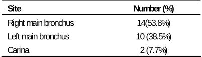
Foreign bodies were found and removed in 26 (81.2%) out of 32 patients who underwent bronchoscopy. The location of foreign bodies extracted by bronchoscopy is shown in Table 1.
Table 1. The location of bronchoscopically extracted foreign bodies
Site Number (%)
Right main bronchus 14(53.8%)
Left main bronchus 10 (38.5%)
Carina 2 (7.7%)
two (7.7%) cases.
The most common symptom in our patients was cough in 80%; followed by wheezing in 60%, tachypnea in 40%, dyspnea in 20%, and stridor in 5%. Chest x-ray was normal in 12 patients (37.5%).
Radiographic findings on expiratory posterior-anterior chest x-ray showed hyperaeration in 18 and atelectasis in 2 patients.
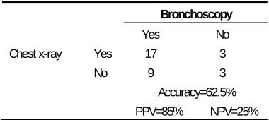
The sensitivity of expiratory chest radiography in this study was 65%, and the specificity was 50% (Table 2).
Table 2. Sensitivity and specificity of expiratory chest radiography for
detection of tracheobronchial foreign body
Bronchoscopy
Yes No
Yes 17 3
Chest x-ray
No 9 3
Accuracy=62.5% PPV=85% NPV=25%
DISCUSSION
Children are at risk of aspiration. This is because of the poor chewing ability of young children due to the lack of posterior dentition as well as the parents’ unawareness of the hazards of feeding nuts in young children (8).
In Ayed et al. report, watermelon seeds in summer and nuts in winter were the most commonly aspirated foreign bodies in Kuwaiti children (9). In our study, the most commonly encountered foreign body was the hull of nuts.
Foreign body aspiration can be difficult to diagnose especially in infants and small children. History of a choking or coughing spell can be usually obtained from the parents (9). Furthermore, history of acute choking/coughing or a witnessed aspiration episode, or the presence of wheezing or stridor is an important clue for the diagnosis of foreign body
aspiration.
The most common clinical presentation in our study was coughing, which occurred in 80% of our subjects with foreign body aspiration.
In a study conducted by Mantor et al., a choking spell was the most common clinical presentation which occurred in 88% of their subjects (10).
Plain chest x-ray is useful for detection of foreign bodies since it may show obstructive emphysema, atelectasis, pulmonary infiltration or radio-opaque foreign bodies (11-13). However, radiolucent aspirated objects on routine chest x-rays may not be seen (12).
Ayed et al. showed that the most common radiological sign in these subjects was obstructive emphysema (9).
Mu et al. also reported obstructive emphysema in 62% of their patients (14). Hyperaeration in our study was seen in 18 children.
Mu and his colleagues also reported that radiological examination was essential for diagnosis and localization of airway foreign bodies. Among their patients, 32.3% with bronchial foreign bodies had normal chest rays. In their study, 67% of x-rays were abnormal when the duration of symptoms was more than one day, and 44% were abnormal when the duration was less than one day (13).
Burton et al. found normal radiographs in 19% of their patients among which, 35% were symptomatic for longer than three days before bronchoscopy (15).
A normal chest x-ray reported in about 25%-35% of patients in different studies (2, 9, 16-18).
In our study, 37.5% of patients with foreign body had normal x-rays which is a higher rate than those of the above-mentioned studies. One possible reason for this might be the fact that all x-rays in our study were done in the first 24 hours post-aspiration.
61.3% for normal chest x-rays in their patients with a foreign body who referred early after aspiration (19).
Different studies reported that the majority of patients with foreign body aspiration are younger than 3 years of age (2, 11, 12). In our survey, all children were younger than 54 months.
Male to female ratio in many other studies was 2:1 (19-22). In our study this ratio was about 3:2. The reported sensitivity of chest x-ray in previous studies was 60-80% (21). Previous reports found specificities of 30–51% for pathological chest x-rays (21, 23).
Our study result is consistent with that of previous studies demonstrating the low sensitivity and specificity of chest x-ray in children (24, 25).
False negative in our study was common (28.1%) and we had a poor negative predictive value which is consistent with previous studies (2).
Patients with retained foreign bodies are at risk of infection and bronchiectasis that can lead to pulmonary resection. Therefore, negative x-ray should not preclude bronchoscopy in patients with characteristic signs and symptoms (2).
Early bronchoscopy is essential for reducing the complications of prolonged foreign body retention (8).
Indeed, in infants and children, it may be impossible or difficult to obtain technically adequate inspiratory/expiratory chest x-rays (26).
There were a number of limitations in this study. The sample size was small. We limited this study to children younger than 54 months, and our findings may not apply to older children.
In conclusion, chest x-ray is not specific for diagnosis of foreign body aspiration, and a normal chest x-ray does not always rule out the diagnosis of foreign body aspiration in patients with a history suggestive of foreign body aspiration and positive physical examinations.
REFERENCES
1. Black RE, Choi KJ, Syme WC, Johnson DG, Matlak ME. Bronchoscopic removal of aspirated foreign bodies in children. Am J Surg 1984; 148 (6): 778- 81.
2. Zerella JT, Dimler M, McGill LC, Pippus KJ. Foreign body aspiration in children: value of radiography and complications of bronchoscopy. J Pediatr Surg 1998; 33
(11): 1651- 4.
3. Humphries CT, Wagener JS, Morgan WJ. Fatal prolonged foreign body aspiration following an asymptomatic interval.
Am J Emerg Med 1988; 6 (6): 611- 3.
4. Walner DL, Ouanounou S, Donnelly LF, Cotton RT. Utility of radiographs in the evaluation of pediatric upper airway obstruction. Ann Otol Rhinol Laryngol 1999; 108 (4): 378-
83.
5. Tan HK, Brown K, McGill T, Kenna MA, Lund DP, Healy GB. Airway foreign bodies (FB): a 10-year review. Int J
Pediatr Otorhinolaryngol 2000; 56 (2): 91- 9.
6. Paşaoğlu I, Doğan R, Demircin M, Hatipoğlu A, Bozer AY. Bronchoscopic removal of foreign bodies in children: retrospective analysis of 822 cases. Thorac Cardiovasc Surg
1991; 39 (2): 95- 8.
7. Fontoba JEB, Gutierrez C, Lluna J, Vila JJ, Poquet J, Ruiz-Company S. Bronchial foreign body: should bronchoscopy be performed in all patients with a choking crisis? Pediatr
Surg Int 1997; 12 (2/3): 118-20.
8. Inglis AF Jr, Wagner DV. Lower complication rates associated with bronchial foreign bodies over the last 20 years. Ann Otol Rhinol Laryngol 1992; 101 (1): 61- 6.
9. Ayed AK, Jafar AM, Owayed A. Foreign body aspiration in children: diagnosis and treatment. Pediatr Surg Int 2003; 19
(6): 485- 8.
10.Mantor PC, Tuggle DW, Tunell WP. An appropriate negative bronchoscopy rate in suspected foreign body aspiration. Am
J Surg 1989; 158 (6): 622- 4.
11.Black RE, Johnson DG, Matlak ME. Bronchoscopic removal of aspirated foreign bodies in children. J Pediatr Surg 1994;
29 (5): 682- 4.
12.Cotton E, Yasuda K. Foreign body aspiration. Pediatr Clin
13.Mu LC, Sun DQ, He P. Radiological diagnosis of aspirated foreign bodies in children: review of 343 cases. J Laryngol
Otol 1990; 104 (10): 778- 82.
14.Mu L, He P, Sun D. The causes and complications of late diagnosis of foreign body aspiration in children. Report of 210 cases. Arch Otolaryngol Head Neck Surg 1991; 117 (8): 876- 9.
15.Burton EM, Brick WG, Hall JD, Riggs W Jr, Houston CS. Tracheobronchial foreign body aspiration in children. South
Med J 1996; 89 (2): 195- 8.
16.Aytaç A, Yurdakul Y, Ikizler C, Olga R, Saylam A. Inhalation of foreign bodies in children. Report of 500 cases.
J Thorac Cardiovasc Surg 1977; 74 (1): 145- 51.
17.Cataneo AJ, Reibscheid SM, Ruiz Júnior RL, Ferrari GF. Foreign body in the tracheobronchial tree. Clin Pediatr
(Phila) 1997; 36 (12): 701- 6.
18.Melaku G. Foreign body aspiration in children: experience from Ethiopia. East Afr Med J 1996; 73 (7): 459- 62.
19.Eren S, Balci AE, Dikici B, Doblan M, Eren MN. Foreign body aspiration in children: experience of 1160 cases. Ann
Trop Paediatr 2003; 23 (1): 31- 7.
20.Reilly J, Thompson J, MacArthur C, Pransky S, Beste D, Smith M, et al. Pediatric aerodigestive foreign body injuries are complications related to timeliness of diagnosis.
Laryngoscope 1997; 107 (1): 17- 20.
21.Hoeve LJ, Rombout J, Pot DJ. Foreign body aspiration in children. The diagnostic value of signs, symptoms and pre-operative examination. Clin Otolaryngol Allied Sci 1993; 18 (1): 55- 7.
22.Martinot A, Closset M, Marquette CH, Hue V, Deschildre A, Ramon P, et al. Indications for flexible versus rigid bronchoscopy in children with suspected foreign-body aspiration. Am J Respir Crit Care Med 1997; 155 (5): 1676-
9.
23.Ciftci AO, Bingöl-Koloğlu M, Senocak ME, Tanyel FC, Büyükpamukçu N. Bronchoscopy for evaluation of foreign body aspiration in children. J Pediatr Surg 2003; 38 (8): 1170- 6.
24.Silva AB, Muntz HR, Clary R. Utility of conventional radiography in the diagnosis and management of pediatric airway foreign bodies. Ann Otol Rhinol Laryngol 1998; 107
(10 Pt 1): 834- 8.
25.Svedström E, Puhakka H, Kero P. How accurate is chest radiography in the diagnosis of tracheobronchial foreign bodies in children? Pediatr Radiol 1989; 19 (8): 520- 2.