C A S E R E P O R T
Open Access
Irreversible fatal contrast-induced
encephalopathy: a case report
Wei Zhao, Jinping Zhang, Yun Song, Lili Sun, Meimei Zheng, Hao Yin, Jun Zhang, Wei Wang and Ju Han
*Abstract
Background:Contrast-induced encephalopathy (CIE) is a well-known complication of iodinated contrast agents during angiography and vascular interventions. It can manifest as hemiparesis, cortical blindness, speech changes, Parkinsonism, confusion, seizure, and coma. Most of the reported CIE cases have been transient and reversible. Irreversible fatal CIE cases have been rarely reported. All the fatal CIE cases reported involved the use of ionic high osmolar contrast agents. Here, we document a heretofore unreported fatal CIE after digital subtraction angiography (DSA) using iopamidol, which is a type of non-ionic monomer low osmolar contrast agent.
Case presentation:A 71-year-old woman was admitted to our Department of Neurology for tinnitus in the head. The cerebral magnetic resonance angiography (MRA) detected atherosclerotic cerebral arteries and bilateral stenosis of the middle cerebral arteries. The patient underwent DSA for further diagnostic work-up. The total amount of iopamidol used during the procedure was 110 ml. The patient experienced headache during the procedure, followed by dizziness with nausea and vomiting. Despite treatment with anti-oedema medications, her clinical status was gradually deteriorating and ended up with deep coma due to irreversible cerebral oedema which was confirmed by cerebral computed tomography (CT). Finally, the patient died 56 days after the procedure due to irreversible fatal cerebral oedema.
Conclusions: This report documents that iopamidol-induced encephalopathy may not always have a benign
outcome and can result in irreversible fatal cerebral oedema.
Keywords: Digital subtraction angiography, Iodinated contrast agents, Complications, Contrast-induced encephalopathy
Background
Contrast-induced encephalopathy (CIE) is a known but rare complication of angiography and endovascular in-terventions. The presentations may include hemiparesis, cortical blindness, speech changes, Parkinsonism, confu-sion, seizure, and coma [1, 2]. In most reported cases, the symptoms are reversible, and fatal encephalopathy following iodinated contrast administration has been rarely reported. Only 8 cases of autopsy-proven fatal cerebral oedema due to contrast neurotoxicity in the early stage of angiography have been reported [1, 3, 4]. All these reported fatal cases involved the use of high os-molar contrast agents. Iopamidol is a non-ionic monomer low osmolar contrast agent, which has been reported in cases of reversible contrast-induced encephalopathy [5–9].
Here, we describe a patient who suffered irreversible fatal encephalopathy after DSA using iopamidol.
Case presentation
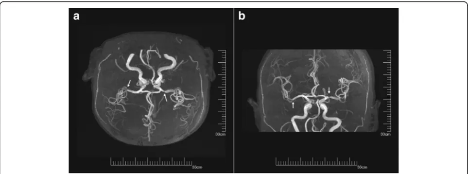
A 71-year-old woman with a history of hypertension, hyperlipemia, and angina was admitted to our Department of Neurology for tinnitus in the head. On physical examin-ation, bilateral hearing impairment was found. The cere-bral magnetic resonance imaging (MRI) detected signal changes consisted with multiple cerebral infarctions and bilateral demyelination in the centrum semiovale. And the cerebral MRA detected atherosclerotic cerebral arteries and bilateral stenosis of the middle cerebral arteries (Fig. 1a, b). For further diagnosis, the patient underwent DSA subsequently. The total amount of iopamidol (Bracco Imaging Italia S.r.L.) administered during the procedure was 110 ml. The DSA showed that the patient had bilat-eral embryonic posterior cerebral arteries, 40% stenosis of
© The Author(s). 2019Open AccessThis article is distributed under the terms of the Creative Commons Attribution 4.0 International License (http://creativecommons.org/licenses/by/4.0/), which permits unrestricted use, distribution, and reproduction in any medium, provided you give appropriate credit to the original author(s) and the source, provide a link to the Creative Commons license, and indicate if changes were made. The Creative Commons Public Domain Dedication waiver (http://creativecommons.org/publicdomain/zero/1.0/) applies to the data made available in this article, unless otherwise stated.
* Correspondence:hanjujack@163.com
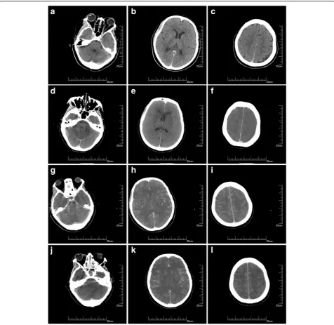
the left middle cerebral artery and tortuous vertebral ar-teries bilaterally. There was no obvious calcification of the aortic arch; angiography of the arch using 25 ml iopamidol was performed only once. Ten minutes after the aortic arch angiography, the patient experienced mild headache. The pain was bearable, and the patient could cooperate during the remainder of the procedure. The DSA was completed 20 min later. No haemorrhage or vasospasm was detected during the procedure. The headache was continuous, and the patient suffered nausea and vomiting. The immediate physical examination showed no obvious abnormal sign. The patient was treated with 8 mg ramose-tron and 10 mg dexamethasone. After 20 min of observa-tion, the symptoms were relieved. Her cerebral CT scan at the time was normal (Fig. 2a, b, c). Two hours later, the patient manifested dizziness with nausea and vomiting and was treated with 8 mg ondansetron and 20 mg di-phenhydramine. Meanwhile, compound sodium chloride injection was used to facilitate the elimination of the con-trast agent. The treatment alleviated her symptoms. Four hours after the procedure, the patient re-experienced diz-ziness; thus 5 mg dexamethasone was administered but re-sulted in no alleviation until after 11 h wherein dizziness was relived but her blood pressure was 183/92 mmHg. The patient was drowsy but could answer questions cor-rectly. The pupil were symmetric and reactive, and limb movements were preserved. To manage hypertension, 30 mg nimodipine tablets were used. Fourteen hours after the procedure, the patient fell asleep, but at 17 h, respira-tory failure progressed and oxygen saturation dropped to 88%. The patient was in a coma state, with sighing respir-ation. Anisocoria with non-reactive pupils developed, limb drop test was positive along with flexor plantar and Babinski sign was negative. Therefore, cerebral hernia was
considered. The patient was treated with 20% mannitol, nikethamide, lobeline and diprophylline, and was trans-ferred to the intensive care unit for further treatment after cardio-pulmonary resuscitation, endotracheal intubation and mechanical ventilation. Two days after the procedure, cerebral CT scan indicated diffuse cerebral oedema, loss of grey-white differentiation, effacement of the cerebral sulci and decrease in cerebrospinal fluid space (Fig.2d, e, f ). The patient was treated with dehydration, mechanical ventilation, and anti-infectious agent, but the diffuse cere-bral oedema did not improve. Nine days after the proced-ure, the third cerebral CT scan showed that the cerebral oedema had become much more severe, the ventricles had disappeared and there was hyperdense signal in the subarachnoid space, which was considered to be indicative of a pseudo-subarachnoid haemorrhage due to the severe cerebral oedema [10] (Fig. 2g, h, i). Fifteen days after the procedure, the cerebral CT scan detected unrelieved dif-fuse cerebral oedema, and the hyperdense signal in the subarachnoid space persisted (Fig.2j, k, l). None of these cerebral CT scans showed intracerebral haemorrhage or infarct in this patient. The patient remained in a continuous deep coma state, and the brainstem re-flexes had disappeared; she died 56 days after that sudden deterioration.
Discussion and conclusions
other 3 received aortography. All fatal cerebral oedema cases reported involved the use of ionic high osmolar contrast agents, and ionic high osmolar contrast agents are no longer used in routine angiography and inter-vention procedures. The case which we report here may be the first fatal cerebral oedema after DSA using iopamidol. This case highlights the potential for other types of iodinated contrast agents to induce fatal encephalopathy.
brain after DSA (immediately, 2 days, 9 days and 15 days after the procedure) indicated intracerebral haemorrhage or infarct. Therefore, the possibility of multiple embo-lisms was not considered in this case. The hyperdense signal in the subarachnoid space in the cerebral CT scans was considered to be due to the severe diffuse cerebral oedema. The hyperdense appearance results from a combination of loss of grey-white differenti-ation, narrowing and effacement of the subarachnoid spaces, and corresponding engorgement of superficial pial veins [10].
The patient experienced headache 10 minutes after the aortic arch angiography during the DSA procedure, and suffered nausea, vomiting and dizziness after the proced-ure. The symptoms were continuous, the patient was co-matose at 17 h with respiratory failure. Anisocoria with non-reactive pupils developed, the limb drop test was positive, and Babinski sign was negative. All these symp-toms supported the diffuse lesion of the brain, there was no sign of focal brain lesion. The cerebral CT scans de-tected diffuse unrelieved cerebral oedema after the se-vere deterioration. Therefore, cerebral hernia was considered due to severe cerebral oedema. The sudden respiratory failure was due to cerebral oedema leading to cerebral hernia. Hence the anoxic brain injury was not considered in this case. And the history, symptoms and cerebral CT scans did not support the diagnoses of sec-ondary viral encephalitis, secsec-ondary cerebral vein throm-bosis and metabolic causes.
The mechanism of CIE is controversial. The temporary disruption of the blood-brain barrier (BBB) after injec-tion of the iodinated contrast agent is widely accepted
[2,11–15]. Experimental studies have demonstrated that
contrast agents can penetrate the altered BBB and that this is dependent on the contact time, anions and dosage
[1,12,13,15]. Both the hyperosmolality and
chemotoxi-city of the contrast agents contribute to the disruption of the BBB. Hyperosmolality of the contrast medium is hypothesised to cause shrinkage of endothelial cells and disrupt tight junctions [12]. Other studies suggest that the alteration of the BBB is due to the physical or chem-ical effects of the contrast medium on the BBB instead of the hyperosmolality [14]. The expression of endothe-lin, which can be induced by radiocontrast agents, can increase human brain endothelial cell permeability and has been implicated in the pathophysiology of disorders associated with BBB injury [2,15].
Studies have indicated that opening of the BBB may be accompanied by brain oedema, resulting from the flux of proteins, electrolytes, and water across the abnormally permeable cerebral vessels into the extracellular space [4]. An idiosyncratic response to small doses of contrast agent, which may be related to the areas of incomplete-ness of their BBB, has been reported [1]. We postulate
that the idiosyncratic response to contrast agents may have contributed to the patient’s prolonged and progres-sive brain oedema. Contrast agents can produce direct neurotoxic effects on the neurons and astrocytes when they penetrate the altered BBB. Experimental studies have shown that ionised contrast agents can severely alter neuronal function when directly introduced into the nervous system [1,12,13,15]. We hypothesised that the direct neurotoxic effect of the contrast agent also contributed to the patient’s progressive and fatal brain oedema.
All types of iodinated contrast agents can induce the development of neurotoxicity, but the occurrence of fatal cerebral oedema is very rare. Unfortunately, there is no currently available effective treatment for such a severe fatal CIE. In the case reported by L. Junck and W.H. Marshall [4], the post mortem tissue iodine concentra-tions were the highest in the urine, serum and kidney. The use of continuous renal replacement therapy and continuous blood purification may be potential treat-ments for cases of fatal CIE.
In summary, although CIE has typically been associ-ated with benign outcomes in previous studies, we present a case of fatal cerebral oedema after DSA using iopamidol. This case illustrates the potential to cause se-vere complications, even fatal cerebral oedema, with all types of iodinated contrast agents. The doctors perform-ing angiography and interventions should be aware of this severe potentially harmful effect. The rare occur-rence of severe contrast-induced complications renders their prevention very difficult. Further studies are needed to define the risk factors and the mechanism of the iodinated contrast agent neurotoxicity, which may help minimise the occurrence of severe complications.
Abbreviations
BBB:Blood-brain barrier; CIE: Contrast-induced encephalopathy; CT: Computed tomography; DSA: Digital subtraction angiography; MRA: Magnetic resonance angiography; MRI: Magnetic resonance imaging
Acknowledgements None.
Funding None.
Availability of data and materials
All data generated and analysed during this study are included in this article.
Authors’contributions
WZ designed and wrote the manuscript. JPZ, YS, and LLS examined the patient. MMZ, HY, JZ and WW analysed the neuroimages. JH examined the patient, designed the case report and helped draft the manuscript. All authors read and approved the final manuscript.
Ethics approval and consent to participate
Consent for publication
A signed informed consent was obtained from the patient’s guardian for publication of this case report and accompanying neuroimages.
Competing interests
The authors declare that they have no competing interests.
Publisher’s Note
Springer Nature remains neutral with regard to jurisdictional claims in published maps and institutional affiliations.
Received: 13 December 2018 Accepted: 21 March 2019
References
1. Lalli AF. Contrast media reactions: data analysis and hypothesis. Radiology. 1980;134(1):1–12.https://doi.org/10.1148/radiology.134.1.6985735. 2. Leong S, Fanning NF. Persistent neurological deficit from iodinated contrast
encephalopathy following intracranial aneurysm coiling. A case report and review of the literature. Interv Neuroradiol. 2012;18(1):33–41.
3. Shrivastava S, Mohan JC, Chopra P. Fatal cerebral edema following angiocardiography: a case report. Int J Cardiol. 1985;8(4):490–1. 4. Junck L, Marshall WH. Fatal brain edema after contrast-agent overdose.
AJNR Am J Neuroradiol. 1986;7(3):522–5.
5. Parry R, Rees JR, Wilde P. Transient cortical blindness after coronary angiography. Br Heart J. 1993;70(6):563–4.
6. Kamata J, Fukami K, Yoshida H, Mizunuma Y, Moriai N, Takino T, et al. Transient cortical blindness following bypass graft angiography. A case report. Angiology. 1995;46(10):937–46.
7. Uchiyama Y, Abe T, Hirohata M, Tanaka N, Kojima K, Nishimura H, et al. Blood brain-barrier disruption of nonionic iodinated contrast medium following coil embolization of a ruptured intracerebral aneurysm. AJNR Am J Neuroradiol. 2004;25(10):1783–6.
8. Gellen B, Remp T, Mayer T, Milz P, Franz WM. Cortical blindness: a rare but dramatic complication following coronary angiography. Cardiology. 2003; 99(1):57–9.https://doi.org/10.1159/000068443.
9. Merchut MP, Richie B. Transient visuospatial disorder from angiographic contrast. Arch Neurol. 2002;59(5):851–4.
10. Hasan TF, Duarte W, Akinduro OO, Goldstein ED, Hurst R, Haranhalli N, et al. Nonaneurysmal "pseudo-subarachnoid hemorrhage" computed tomography patterns: challenges in an acute decision-making heuristics. J Stroke Cerebrovasc Dis. 2018;27(9):2319–26.https://doi.org/10.1016/j.jstrokecerebrovasdis. 2018.04.016.
11. Lantos G. Cortical blindness due to osmotic disruption of the blood-brain barrier by angiographic contrast material: CT and MRI studies. Neurology. 1989;39(4):567–71.
12. Junck L, Marshall WH. Neurotoxicity of radiological contrast agents. Ann Neurol. 1983;13(5):469–84.https://doi.org/10.1002/ana.410130502. 13. Torvik A, Walday P. Neurotoxicity of water-soluble contrast media. Acta
Radiol Suppl. 1995;399:221–9.
14. Wilson AJ, Evill CA, Sage MR. Effects of nonionic contrast media on the blood-brain barrier. Osmolality versus chemotoxicity. Investig Radiol. 1991; 26(12):1091–4.