Identifying key modulators of panic attacks in panic
disorder: A translational model
D.L.M. Paes
Abstract
Panic disorder (PD) has a prevalence of 4% in the general population, but its pathophysiology remains elusive. This research focuses on identifying molecular and genetic modulators of unpredictable, recurrent panic attacks which characterize PD. Prior studies revealed that the serotonin transporter, 5-HTT, is involved in fear behavior and that genetic variations might affect panic-related behavior. In order to test these genetic variations on a molecular level a translational model consisting of humans and rodents was used. As follow-up of a human study, 40 male mice (background C57BL/6), wild-type and heterozygous 5-HTT knock-out, were exposed to either 9% CO2 or room air to experimentally provoke fear-related behavior.
Furthermore, the acid-sensing ion channel ASIC1a is hypothesized to be a linking factor between fear detection and fear response. It was tested whether this ASIC1a is expressed in serotonergic neurons in the dorsal raphe nucleus, main source of serotonin, the pivotal neurotransmitter in panic responses. Localization of the ASIC1a channel was determined by use of immunofluorescence.
Analysis of the behavioral tests showed no significant difference between genotypes in both air and carbon dioxide condition. Carbon dioxide exposure however increased freezing time in both genotypes. ASIC1a has been indicated in the dorsal raphe and hippocampus in perfusion-fixated rat tissue, but could not yet be detected in the dorsal raphe of the fresh-frozen tissue of tested mice.
Introduction
literally of vital importance among many animals, the pivotal part that creates this panic response is located in the more preserved, ancient brain structures. Higher vertebrates like humans and rodents still possess lower structures like medulla and pons. Because of the preservation of these brain regions among species during years of evolution a translational model between humans and rodents might be reliable (1).
Neuronal signalling needs to be modulated very precisely. Hence, alterations among individuals due to genotypic differences may result in different signalling and thus behavior. In case of an insufficient stimulus detection or signalling in humans concerning panic behavior, panic disorder (PD) might develop. Sufferers from PD experience recurrent, unpredictable panic attacks (PAs) that are characterized by symptoms such as feelings of intense fear and physiological reactions like choking, dizziness and palpitations (2). Panic disorder is quite prevalent with 4% of patients in the general population (3). Panic attacks occur even in about 23% of the general population at least once in their lifetime (4). Panic behavior occurs when being exposed to very direct threats that occur from inside the body, for example by inhalation of an elevated concentration of carbon dioxide in an experimental manner (5). Given the fact that both patients and subjects react to elevated CO2-concentrations indicates that a similar mechanism is involved (6). Patients however
might be more sensitive to stimuli at the molecular level resulting in more severe reactions.
Genetic determinant: 5-HTT
In case of panic behavior there is convincing evidence that the serotonergic (5-HT) system is involved (7). Signalling at the synaptic level is dependent on factors such as the amount of neurotransmitter, 5-HT in this case, which again is dependent on the availability of conversing enzymes, (auto)receptors and 5-HT transporters that transport the 5-HT from the synapse back into the cell. The amounts and/or efficiency of the mentioned factors determine the speed, duration and amplitude of the signal. Several experiments have been executed in which the effect of 5-HT availability and use of agonists and antagonists of the 5-HT receptor was investigated (8). These studies concluded that 5-HT signalling does modulate the response of a PA.
more transporters and thus a quicker termination of the signalling compared to SS or SL genotypes. Possession of minimally one S-allele was shown to result in a decreased fear response compared to the LL-genotype (9). The polymorphism is not present in mice, but in order to create a translational model we investigated wild-type and heterozygous knock-out mice concerning the 5-HTT. Both wild-type and heterozygous knock-knock-out conditions are comparable to the polymorphism genotypes used in human studies since heterozygous mice will possess less transporters which is similar to the SS and SL genotypes in humans. By making use of behavioral tests, wild-type and heterozygous 5-HTT mice will be investigated by scoring several fear indicating parameters. We expect that heterozygous mice will show less fear-related behavior due to the longer lasting 5-HT signalling.
Molecular modulator: ASIC1a
It is known that serotonergic signalling plays a pivotal role when it comes to fear-related behavior. The initiation of the signalling however remains quite indistinct. It has however been shown that the amygdala, involved in fear-related behavior, expresses certain ion channels, ASIC1a (11,12). ASICs, acid-sensing ion channels, are ion channels through which sodium and calcium ions flow into the cell when exposed to an acid environment. This influx may then induce neuronal firing by depolarizing the membrane potential (13-15). The most abundant ASIC subtype, ASIC1a, (16) and its modulating effect on fear responses has been elucidated by Coryell et al. in 2007 (12). The chemoreceptive property of amygdale neurons seemed to be a linking factor of sensing stimuli and responses. Additionally, a lowered pH in midbrain 5-HT neurons leads to an increased firing rate of these neurons (17), which might be explained by pH-sensing ion channels such as ASIC1a. Raphe neurons are chemoreceptive as well and react by increasing the firing rate, thus an increased 5-HT signalling.
When taking the findings of mentioned prior studies together, it sounds plausible that 5-HT raphe neurons in the midbrain might express ASICs because of their chemosensing ability. Since raphe neurons are a pivotal source of 5-HT, which plays a major role in panic responses, we hypothesize that 5-HT neurons in the dorsal raphe nucleus express ASICs which allow them to be a linking factor between extracellular sensing and neuronal firing.
Material and Methods
Effect of 5-HTT genotype on fear-related behavior
mice (background C57BL/6), wildtype and heterozygous knock-out, kindly provided by Prof. Lesch (University of Würzburg), underwent the experiments. Mice were housed paired for 10 weeks with access to mouse chow and water ad libitum. Then, the animals were subjected to behavioral tests and at the age of 4 months were sacrificed by decapitation. Only male animals were used because of the influence of the estrous cycle on the response to CO2 exposure (18).
Both genotypes were tested by making use of the open field test (OFT) and aversion assay (AA) in a Plexiglass box (50x50x40cm). In the AA a septum including a passage was placed inside the box to create two separated chambers. The amount of leaking CO2
was negligible. During the OFT mice were exposed to either normal room air or 9% CO2 for 20 minutes. Scored parameters included total distance moved, time spent in centre and corners and time spent freezing of which the last is a correlate of panic (19). Freezing time was manually scored being blind regarding genotype and condition whereas other parameters were scored via video tracking EthoVision XT 7.5 software.
In the AA mice were placed in randomized manner in one of the chambers. Chambers contained either both normal air or one randomized side 9% CO2. The time spent per chamber was scored for 10 minutes, however compared to others studies we corrected for freezing time (20), since total times spent will increase by freezing even though it may not be the animal’s intention.
Immunohistochemical staining of ASIC1a
then repeated, this time in the dark. Samples were then placed conversely on a plastic plate before pipetting 300 µl Hoechst (1:500 in TBS) to determine cell positions per glass between the plate and glass. Hoechst incubated for half an hour in the dark after which the two washing steps in TBS were executed again. Thereafter the glasses were covered by mounting solution, 80% glycerol and 20% TBS, and a coverslip was mounted and secured on the glass. By means of an Olympus BX-51 microscope and cellSens imaging software stainings were examined.
Statistical analysis
Freezing times scored during the OFT and AA were statistically analyzed. Mice were grouped per genotype and condition. In case of the AA starting side and CO2-side were used as differentiating parameters also. The mean values of freezing times of the four groups (n=10) in both tests were compared by means of a one- and two-way ANOVA-test and LSD post-hoc test in the IBM SPSS Statistics 21-software, using a significance level of p<0.05.
Results
Behavioral test analysis
As can be seen in Figure 1, different conditions affected the amount of time freezing significantly independent of genotype (p<0.001) in OFT and AA. However, between genotypes, no significant difference in freezing times was found per condition.
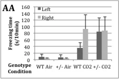
Figure 2. Freezing time in AA per genotype and condition in both left and right chamber. Means + SEM, corrected for starting side. Significant differences (p<0.05) among groups, but no effect of genotype and CO2- side.
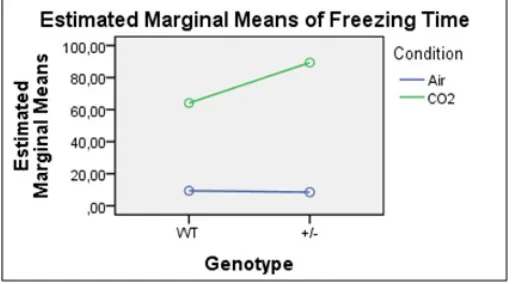
Figure 3. Chart of the Genotype*Condition interaction effect in AA. Genotype*Condition: p<0.475.
Genotype and CO2-side do not show significant differences in freezing time in the AA (Fig.
Immunofluorescent staining
Concerning the immunofluorescent staining promising results were achieved. Specific signals on cell bodies and dendrites in the hippocampus were found in fresh-frozen tissue as can be seen in Figure 4, which corresponds to a prior study (16).
Figure 4. Signal from cell bodies and sprouting fibers in the CA2 region of the hippocampus. Condition: Santa Cruz goat-α-mouse, blocking buffer, citrate buffer incubation and Alexa488. Magnification 20x, exposure time: 50ms, FITC-filter.
Figure 5. A Image of fluorescent signal Alexa 488 in hippocampus. Magnification 20x. Exposure time 50ms, FITC-filter. B Negative control. Exposure time 150 ms.
Figure 6. A Image of fluorescent signal Alexa 488 in dorsal raphe nucleus. Magnification 20x. Exposure time 100ms, FITC-filter. B Neg. Exposure time 150ms. Blur due to folded tissue.
Discussion
altered conformation of the channel in such a way that the antibody’s epitope becomes more approachable since the channels will open at a lowered pH (20). However, because mice subjected to the behavioral tests were sacrificed by decapitation and their brains were freshly frozen the protocol should be adjusted to produce similar results in fresh frozen tissue as has been seen in perfusion-fixated tissue.
Since the brains of the animals used in the behavioral part are fixated by freezing them freshly, the protocol has to be adjusted to this kind of fixation method. Future protocols will be based on trying to mimic the mechanisms of perfusion-fixation. When the protocol has led to specific staining of cells in the dorsal raphe nucleus, it additionally has to be replicated in a double-staining protocol including tryptophan hydroxylase, TPH2, antibodies in order to determine whether the stained cells are 5-HT neurons (22). This extra staining might lead to altered results or additional reactions to which the protocol might be adapted again.
References
1. Darwin, Charles. The origin of species by means of natural selection: or, the preservation of favored races in the struggle for life. Ed. William F. Bynum. AL Burt, 2009.
2. American Psychiatric Association. The Diagnostic and Statistical Manual of Mental Disorders: DSM 5. bookpointUS, 2013.
3. Weissman, Myrna M., et al. “The cross-national epidemiology of panic disorder.” Archives of General Psychiatry 54.4 (1997): 305-309.
4. Kessler, Ronald C., et al. “The epidemiology of panic attacks, panic disorder, and agoraphobia in the National Comorbidity Survey Replication.” Archives of general psychiatry 63.4 (2006): 415-424.
5. Perna, Giampaolo, et al. “Carbon dioxide/oxygen challenge test in panic disorder.” Psychiatry research 52.2 (1994): 159-171..
6. Schruers, K. R. J., et al. “Symptom profiles of natural and laboratory panic attacks.” Acta Neuropsychiatrica 16.2 (2004): 101-106.
7. Grove, Gregory, Jeremy D. Coplan, and Eric Hollander. “The neuroanatomy of 5-HT dysregulation and panic disorder.” The Journal of neuropsychiatry and clinical neurosciences 9.2 (1996): 198-207.
8. Schruers, Koen, et al. “Effects of tryptophan depletion on carbon dioxide provoked panic in panic disorder patients.” Psychiatry research 93.3 (2000): 179-187.
9. Schruers, Koen, et al. “Genetic moderation of CO2-induced fear by 5-HTTLPR genotype.” Journal of psychopharmacology 25.1 (2011): 37-42.
10. Lesch, Klaus-Peter, et al. “Association of anxiety-related traits with a polymorphism in the serotonin transporter gene regulatory region.” Science274.5292 (1996): 1527-1531.
11. Coryell, Matthew W., et al. “Acid-sensing ion channel-1a in the amygdala, a novel therapeutic target in depression-related behavior.” The Journal of neuroscience 29.17 (2009): 5381-5388.
14. Xiong, Zhi-Gang, et al. “Acid-sensing ion channels (ASICs) as pharmacological targets for neurodegenerative diseases.” Current opinion in pharmacology 8.1 (2008): 25-32.
15. Yermolaieva, Olena, et al. “Extracellular acidosis increases neuronal cell calcium by activating acid-sensing ion channel 1a.” Proceedings of the National Academy of Sciences of the United States of America 101.17 (2004): 6752-6757.
16. Zha, Xiang-ming, et al. “Acid-sensing ion channel 1a is a postsynaptic proton receptor that affects the density of dendritic spines.” Proceedings of the National Academy of Sciences 103.44 (2006): 16556-16561.
17. Severson CA, Wang W, Pieribone VA, Dohle CI, Richerson GB. Midbrain serotonergic neurons are central pH chemoreceptors. Nature neuroscience. 2003 Nov;6(11):1139-40. PubMed PMID: 14517544.
18. Brack, K. E., S. M. T. Jeffery, and T. A. Lovick. “Cardiovascular and respiratory responses to a panicogenic agent in anaesthetised female Wistar rats at different stages of the oestrous cycle.” European Journal of Neuroscience 23.12 (2006): 3309-3318.
19. Mongeluzi DL, Rosellini RA, Ley R, Caldarone BJ, Stock HS. The Conditioning of Dyspneic Suffocation Fear: Effects of Carbon Dioxide Concentration on Behavioral Freezing and Analgesia. Behavior Modification. 2003;27(5):620-36.
20. Ziemann, Adam E., et al. “The amygdala is a chemosensor that detects carbon dioxide and acidosis to elicit fear behavior.” Cell 139.5 (2009): 1012-1021.
21. Wemmie, John A., et al. “Acid-sensing ion channel 1 is localized in brain regions with high synaptic density and contributes to fear conditioning.” The journal of neuroscience 23.13 (2003): 5496-5502.