THEORETICAL STUDY ON THE HYDROGEN BONDING INTERACTIONS IN
PARACETAMOL-WATER COMPLEXES
MEIFANG XU
a,b, BOHAI ZHANG
a, QI WANG
a, YUAN YUAN
a, LE SUN
a, ZHENGGUO HUANG
a1*
aTianjin Key Laboratory of Structure and Performance for Functional Molecules; Key Laboratory of Inorganic-Organic Hybrid Functional Materials
Chemistry (Tianjin Normal University), Ministry of Education; College of Chemistry, Tianjin Normal University, Tianjin 300387, People’s Republic of China
bCollege of Mathematics Science, Tianjin Normal University, Tianjin 300387, People’s Republic of China
ABSTRACT
The paracetamol–water (PA–H2O) complexes formed by hydrogen bonding interactions were investigated at the MP2/6–311++G(d,p) level. Six PA–H2O
complexes possessing various types of hydrogen bonds (H–bonds) were characterized by geometries, energies, vibrational frequencies. Natural bond orbital (NBO), quantum theory of atoms in molecules (QTAIM) and the localized molecular orbital energy decomposition analysis (LMO–EDA) were performed to explore the nature of the hydrogen–bonding interactions in these complexes. The intramolecular H–bond formed between the methylene and carbonyl oxygen atom of paracetamol is retained in most of complexes. The H–bonds in PW1 and PW6 are stronger than other H–bonds, moreover, the researches show that both the hydrogen bonding interaction and structural deformation play important roles for the relative stabilities of PA–H2O complexes.
Keyword: paracetamol; hydrogen bonding interactions; natural bond orbital (NBO); quantum theory of atoms in molecules (QTAIM)
1. INTRODUCTION
Paracetamol (PA) is a widely used non-prescription drug, which can enhances the analgetic activity and reduces the nephrotoxicity 1. A randomized
controlled trial of chronic pain from osteoarthritis in adults found similar benefit from PA and diclofenac . In recommended doses, the side effects of PA are mild to non–existent, but acute overdoses of PA can cause potentially fatal kidney, brain and liver damage and, in rare individuals, a normal dose can do the same 2-6. The risk may be heightened by chronic alcohol abuse. PA
toxicity is the foremost cause of acute liver failure and accounts for most drug overdoses 7-12.
The infrared and Raman spectra of PA have been studied widely 13-17.
Joseph M. et al. have measured the resonant 2–photon ionization spectrum of jet–cooled PA, and analyzed the results in the light of theoretical calculations of the ground–state geometry and vibrational frequencies 18. The complexes
formed by PA with ethanol and acetone species have been studied by Y. Danten et al. 19. Two nearly isoenergetic conformers were distinctly found in a
supersonic molecular beam expansion and positively identified as the cis– and trans– isomers of PA by UV–UV hole–burning spectroscopy 20.
Weak interactions, especially hydrogen bonding interaction, play important roles in biological systems 21-26. For example, hydrogen bonding interaction
construct the structure of DNA and RNA (bonding between nitrogenous bases), the secondary structure of proteins (helix or pleated sheet) and the different branching patterns of sugar chains. For the pharmacological activity of PA, it is of importance to have information about the conformation of PA in solution, and hydrogen bonding interaction plays an important role on the conformations of PA since it is the major interaction in solution of PA. However, there is few literatures can be found so far. Therefore, we dedicated to study the hydrogen bonding interaction between PA and H2O solvent molecule by ab initio
method, and we hope that this study will be helpful to the further study on the solvent effects of PA. It is important to note that not all theoretical methods are reliable for the description of hydrogen bond (H–bond) because it is usually weak. Compared with density functional theory (DFT), MP2 is too time-consuming to apply to large biomolecular systems even with a medium-size basis set, however, it is a reliable method to descript H–bond because it treats electron correlation well. Therefore, here MP2 was used to study the hydrogen bonding interactions in PA–H2O complexes. The geometric parameters (bond
length and bond angle) of H–bond usually provide us preliminary information about the strength of H–bond. However, more technical tools are required to elucidate the nature of H–bond in PA–H2O complexes. The quantum theory of
atoms in molecules (QTAIM) 27, 28, natural bond orbital (NBO) analysis 29 and
the localized molecular orbital energy decomposition analysis (LMO–EDA)
30 methods meet this requirements since they have been proved to be very
useful tools in understanding of H–bond 31-36. Therefore, ab initio calculations
combined with QTAIM, NBO and LMO-EDA approaches were performed to investigate the hydrogen bonding interactions in PA–H2O complexes.
2. COMPUTATIONAL DETAILS
The PA and water monomers were optimized at MP2/6–311++G (d,p) level , then the PA–H2O complexes were constructed starting from the most stable
monomers and were fully optimized at the same level. Harmonic vibrational frequencies calculations were carried out to characterize the structures as minima and enable the evaluation of zero–point vibrational energies (ZPVE). To take into account the effects of the basis set superposition error (BSSE), the counterpoise corrections were implemented to insure that complexes and monomers are being computed with a consistent basis set. Then the interaction energies were calculated based on the ZPVE and BSSE corrections.
In order to analyze the properties of the H–bonds in complexes, QTAIM, NBO and LMO-EDA calculations were carried out. According to QTAIM, the first descriptor of X–H···Y H–bond is the existence of the bond critical point (BCP) at the H···Y bond. Moreover, some descriptors at BCPs have been used widely to characterize the bonding between the atoms, such as the electron density (ρb), the Laplacian of electron density (s2rb) and the total energy
density (Hb) 37. Both ρb and s2ρb at the H···Y BCP are good measures of the
strength of H–bond. According to the criteria established by Koch and Popelier
38, the s2ρ
b should fall in the range of 0.024–0.139 a.u., and the ρb is within
0.002–0.034 a.u.. Such criteria can used to distinguish hydrogen–bonding interactions from van der Waals interactions. The total electron energy density (Hb) is the sum of the potential energy density (Vb) and kinetic energy density
(Gb), which can also be used to characterize the interactions between atoms.
The local priority of Vb at the BCP results into a negative Hb, which means a
partial covalent character is attributed to the H–bonds. Meanwhile, the low and positive s2ρ
b at the BCP means typical closed–shell interactions. Therefore,
both Hb and s2ρb at the BCP can used to characterize the interaction 39-42: for
very strong H–bonds, s2ρ
b <0 and Hb <0; for weak or medium–strength H–
bonds, s2ρ
b >0 and Hb >0; for strong H–bonds, s2ρb >0 and Hb <0.
According to NBO theory 43, the formation of H–bond results into that
electron density from the lone pair nB of the H–acceptor delocalizes into the
unfilled anti–bonding orbital of the H–donor. Therefore, the occupancy of the anti–bond orbital increases, which leads to that the X–H bond is weakened and lengthened. The charge transfer (CT) effects between nB and
is estimated by second–order perturbation energies E(2), in other words, the E(2) lowering is responsible for the orbital interaction of H–bond, the larger E(2) values correspond to stronger CT interaction occurred in the H–bond. In LMO–EDA, total interaction energy DEMP2 is decomposed into five terms:
electrostatic energy (DEele), exchange energy (DEex), repulsion energy (DErep),
polarization energy (DEpol) and dispersion energy (DEdisp). The ab initio and
NBO calculations were carried out using Gaussian 09 44, QTAIM analysis was
performed using the wave functions obtained at the MP2/6–311++G(d,p) level by AIM2000 45, and the LMO–EDA was implement at the same level using the
3. RESULTS AND DISCUSSION
The PA and water monomers were optimized at the MP2/6–311++G(d,p) level, and the molecular graphs are presented in Figure 1. As shown in Figure 1, water molecule can donate/accept proton to form H–bond, in which the hydroxyl and oxygen atom act as H–donor/acceptor, respectively. PA has several possible proton–donor/acceptor sites to form H–bonds. The imino group in the benzene ring and the phenolic hydroxyl are the main H–donor sites of PA, while the methylene can also form weak H–bonds with water in
some complexes. The main H–acceptors of PA are the oxygen atoms of the hydroxyl and carbonyl groups. Moreover, the oxygen atom of the carbonyl groups usually accepts one proton to form intramolecular H–bond with the methylene.
3.1 Structures
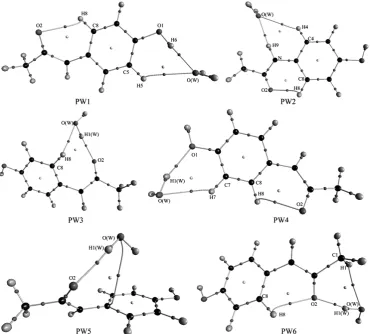
All molecular graphs of optimized PA–H2O complexes are shown in
Figure 2, and the structural parameters ofH–bonds are listed in Table 1. As shown in Figure 2, different types of
Figure 1. Molecular graphs of paracetamol (PA) and water (W) monomers. Large circles correspond to attractors attributed
to atomic positions: white, H; blue, N; gray, C; red, O. Small circles are attributed to critical points: red, bond critical point; yellow, ring critical point.
Figure 2. Molecular graphs of PA–H2O complexes. Large circles correspond to attractors attributed to atomic
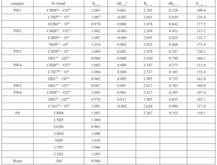
Table 1. Structural parameters (bond lengths in Å, angles in degrees) of H–bonds in PA–H2O complexes calculated at the MP2/6–311++G(d,p) level.
complex H–bonda R
X–H ΔRX–Hb RH···Y δRH···Y θX–H···Y
PW1 C8H8PA···O2PA 1.083 0.001 2.392 0.328 109.4
C5H5PA···OW 1.087 -0.001 2.681 0.039 128.4
O1H6PA···OW 0.970 0.008 1.878 0.842 177.5
PW2 C8H8PA···O2PA 1.082 -0.001 2.269 0.451 115.2
C4H4PA···OW 1.087 -0.001 2.695 0.025 135.7
NH9PA···OW 1.014 0.005 2.032 0.688 173.9
PW3 C8H8PA···OW 1.084 0.002 2.479 0.241 120.3
OH1W···O2PA 0.968 0.008 1.930 0.790 166.1
PW4 C8H8PA···O2PA 1.082 0.000 2.347 0.373 111.0
C7H7PA···OW 1.086 0.000 2.537 0.183 135.4
OH1W···O1PA 0.965 0.005 1.985 0.735 162.0
PW5 OH1W···O2PA 0.967 0.007 2.017 0.703 168.8
PW6 C8H8PA···O2PA 1.083 0.001 2.417 0.303 107.0
OH1W···O2PA 0.970 0.011 1.885 0.835 165.1
C1H1PA···OW 1.091 -0.002 2.624 0.096 117.0
PA C8H8 1.082 2.367 0.353 110.1
C5H5 1.088
O1H6 0.962
C4H4 1.088
NH9 1.010
C7H7 1.086
C1H1 1.093
Water OH 0.960
a Superscript “PA” denotes paracetamol and superscript “W” denotes H 2O b ΔR
X–H=RX–H (complexes) − RX–H(free monomer)
H–bonds are formed in PA–H2O complexes. According to QTAIM, the
H–bond, including inter– or intramolecular H–bonds, is characterized by the BCPs between H–donor (X–H) and H–acceptor (Y), and ring structure formed by multiple H–bonds is characterized by a ring critical point (RCP). The shorter distance between the RCP and corresponding BCP means less stability of the H–bond 47-50. As a note, the RCP at the center of the ring of benzene has
nothing to do with H–bond. As shown in Figs. 1 and 2, one intramolecular C8H8PA···O2PA H–bond formed between the methylene and the oxygen atom
can be found in PA monomer, which is retained in all PA–H2O complexes
except PW3 and PW5.
As shown in Fig. 2, PW3 have two H–bonds, but PW5 have one H– bond. The C8H8PA···O2PA intramolecular H–bond in PW3 is replaced by
two intermolecular H–bonds, in which water monomer acts as H–donor and H–acceptor simultaneously. Similarly, the C8H8PA···O2PA intramolecular H–
bond in PW5 is also replaced by one intermolecular H–bond formed between the hydroxyl of water moiety donating one proton to oxygen atom of the carbonyl groups in PA moiety. Therefore, it can learn that the serious structural deformations occurred in PW3 and PW5. In addition, it worth noting that there seems to be one p H–bond formed between the hydroxyl of water and the benzene ring of PA monomer, and the distance between the hydrogen atom and the center of the benzene ring is 2.738 Å. Unfortunately, such p H–bond cannot be characterized by QTAIM directly. Except for the C8H8PA···O2PA
intramolecular H–bond, other complexes have two intermolecular H–bonds. The oxygen atom of water moiety accepts two protons from the hydroxyl and methylene of PA simultaneously to form one bifurcated H–bond in PW1. Another bifurcated H–bond can be found in PW2, which is formed by the oxygen atom of water moiety accepts two protons from the hydroxyl and imino of PA simultaneously. For the PW4 and PW6 complexes, two intermolecular H–bonds are formed, in which water monomer acts as H–donor and H– acceptor, respectively.
The change (ΔRX–H) of the X–H bond with respect to the corresponding
X–H bond in free monomers (PA or water) reflects the nature of H–bond, the elongation of the X–H bond corresponds to red–shifting H–bond, while the shortening of the X–H bond represents blue–shifting one. In addition, the distance of the H···Y bond reflects the strength of the hydrogen bonding interaction as well. As shown in Table 1, for most of the complexes, the ΔRX–H
of the H–bonds taking methylene as H–donors are negative or remain little changes, which indicates that they are very weak H–bonds. All other H–bonds have positive ΔRX–H values and are red–shifting ones. The largest ΔRX–H
(0.011 Å) is found in the OH1W···O2PA H–bond of PW6, which indicates
that it is the strongest intermolecular H–bond. It is worth noting that another intermolecular H–bond (O1H6PA···OW) in PW1 is also strong, considering its
short RH···Y (1.878 Å). However, its ΔRX–H (0.008 Å) is smaller than that of
the OH1W···O2PA H–bond in PW6. Therefore, for such case, ΔR
X–H is not the
unique technical means to estimate the strength of the H–bond, while RH···Y
is an alternative choice. As shown in Table 1, the shortest of RH···Y is 1.878 Å of the intermolecular O1H6PA···OW H–bond in PW1, which seems to be
the strongest H–bond. Of course, another intermolecular H–bond in PW6, OH1W···O2PA, is also strong H–bonds due to its shorter R
H···Y (1.885 Å). For the
H–bonds in which methylene acts as H–donor in some PA complexes (PW1, PW2, PW4 and PW6), the RH···Y values are small and close to the sum of the van
der Waals radii of the H and Y atoms. Therefore, from a structural viewpoint, the interaction between the methylene and Y atom is very weak and has partial van der Waals character.
3.2 Vibrational Frequencies
The harmonic vibrational frequencies of H–bonds in PA–H2O complexes
and monomers as well as their shifts calculated at the MP2/6–311++G(d,p) level are listed in Table 2. The shift (ΔνX–H) of the X–H stretching vibrational
frequency is one of the main fingerprints of H–bonds. It is generally accepted that the X–H bond is weakened due to the formation of an H–bond, which lead to the red shift of νX–H. The larger the ΔνX–H is, the stronger the H–bond
Table 2. The X–H stretching vibrational frequencies (strength) of H–bonds in both PA–H2O complexes and monomers.
complex H–bond νX–Ha ΔνX–H
PW1 C8H8PA···O2PA 3263.2(0,s)b -4.2
C5H5PA···OW 3218.5(2,s)c 13.4
O1H6PA···OW 3724.8(684)d -151.2
PW2 C8H8PA···O2PA 3273.7(3,s)b 6.3
C4H4PA···OW 3215.2(2,s)e 10.1
NH9PA···OW 3588.2(237)d -71
PW3 C8H8PA···OW 3254.6(2,s)b -12.8
OH1W···O2PA 3962.6(107,as),3780.2(272,s) -40, -104.1
PW4 C8H8PA···O2PA 3269.7(2,s)b 2.3
C7H7PA···OW 3229.8(4,as)f 2.3
OH1W···O1PA 3969.4(138,as),3816.4(194,s)g -33.2, -67.9
PW5 OH1W···O2PA 3942.2(46,as),3799.0(169,s) -60.4, -85.3
PW6 C8H8PA···O2PA 3262.7(0,s)b -4.7
OH1W···O2PA 3961.7(108,as),3710.5(580,s)h -40.9, -173.8
C1H1PA···OW 3196.6(3,as)i,3086.6(7,s)j 18.3, 0.4
PA C8H8 3267.4(1,s)b
C5H5 3205.1(8,s)c
O1H6 3876.0(77)
C4H4 3205.1(8,s)b
NH9 3659.2(30)
C7H7 3227.5(2,as)f
C1H1 3178.3(9,as)i,3086.2(8,s)j
Water OH 4002.6(63,as),3884.3(13,s)
a All frequencies are in cm−1 and the strengths are in km·mol−1. “as” denotes the asymmetric stretching vibration mode, and “s” denotes the symmetric
stretching vibration mode.
b Mixing occurs among the C7H7 and C5H5 stretching vibration modes c Mixing occurs among the C8H8 and C4H4 stretching vibration modes
d Mixed with symmetric H–O–H stretching vibration mode of free water molecule slightly. e Mixing occurs among the C5H5 ,C7H7 and C8H8 stretching vibration modes
f Mixing occurs among the C8H8 and C4H4 stretching vibration modes g Slight mixing with O1H6 stretching vibration modes
h Slight mixing with NH9 stretching vibration modes
i Strong mixing with asymmetric H3–C2–H2 stretching vibration modes j Strong mixing with symmetric H3–C2–H2 stretching vibration modes
hard to calculate the ΔνX–H when the X–H stretching vibrational mode
mixes with other vibrational modes. For example, the mixture between C1H1 and asymmetric/symmetric H3–C2–H2 stretching vibration modes in free PA molecule are calculated to be 3178.3 and 3086.2 cm-1, respectively, so two Δν
X–H
values may be given for such H–bonds involving C1H1 as H–donor. Similar things are also seen for PA–H2O complexes. Taking PW4 as an example, the
symmetric stretching vibrational mode of OH1W···O1PA mixes with O1H6, and
the values of ΔνX–H with respect to the corresponding stretching vibration modes
in free H2O molecule are calculated to be -33.2 and -67.9 cm-1, respectively.
As shown in Table 2, the largest red–shift value of -173.8 cm-1 is found for the
OH1W···O2PA H–bond in PW6. The O1H6PA···OW (PW1) and OH1W···O2PA
(PW3) H–bonds have large red–shifts of more than -100 cm-1, so the strengths
of these H–bonds are regarded as weaker than the OH1W···O2PA H–bonds in
PW6 and stronger than other red–shifted H–bonds. Other intermolecular H– bonds in the PA–H2O complexes are weaker since their absolute values of
ΔνX–H are less than 100 cm-1. There are seven blue–shifted H–bonds which
have positive shift values of ΔνX–H, moreover, they are usually weaker than
the red–shifted ones and a partial dispersion character is attributed to them. However, the small ΔνX–H of the intramolecular C8H8PA···O2PA H–bond in PA–
H2O complexes does not mean that it is also very weak, as it originally existed
in the free PA molecule.
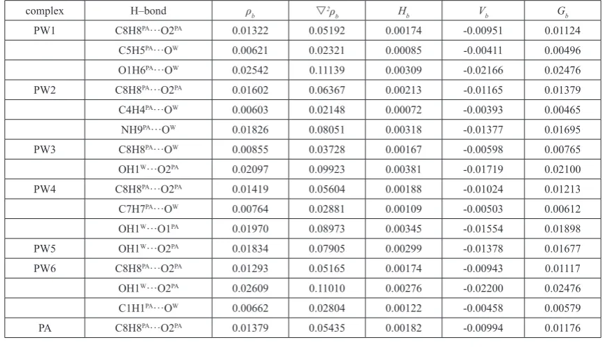
3.3 Bonding analyses
The electronic topological properties at the H···Y BCPs of H–bonds, including the electron density (ρb), the Laplacian of the electron density
(s2ρ
b), the kinetic energy density (Gb), the potential energy density (Vb), and
the total electron energy density (Hb), for all of the complexes and monomer
are listed in Table 3. As shown in Table 3, among all PA–H2O complexes and
PA monomer, both the Hb and s2ρb of all H–bonds are positive and fall in the
ranges proposed by Popelier, thus they are considered as weak or medium H– bonds. Especially, for the H–bonds taking methylene as H–donor, both ρb and
s2ρ
b are close to the lower limit of criteria proposed by Popelier, which shows
that they are very weak and partial dispersion character is attributed to them. Moreover, the H–bonds involving the hydroxyl as H-donors are stronger than other ones due to larger ρb and s2ρb. Especially, for the OH1W···O2PA (PW6)
and O1H6PA···OW (PW1) H–bonds, both ρ
b and s2ρb of them are the largest
Table 3. The electron density (ρb) and its Laplacian (s2ρb), total electron energy density (Hb), potential energy density (Vb) and Lagrangian form of kinetic
energy density (Gb) in a.u. at H···Y BCPs of H–bonds in PA–H2O complexes obtained by QTAIM analysis.
complex H–bond ρb s2ρb Hb Vb Gb
PW1 C8H8PA···O2PA 0.01322 0.05192 0.00174 -0.00951 0.01124
C5H5PA···OW 0.00621 0.02321 0.00085 -0.00411 0.00496
O1H6PA···OW 0.02542 0.11139 0.00309 -0.02166 0.02476
PW2 C8H8PA···O2PA 0.01602 0.06367 0.00213 -0.01165 0.01379
C4H4PA···OW 0.00603 0.02148 0.00072 -0.00393 0.00465
NH9PA···OW 0.01826 0.08051 0.00318 -0.01377 0.01695
PW3 C8H8PA···OW 0.00855 0.03728 0.00167 -0.00598 0.00765
OH1W···O2PA 0.02097 0.09923 0.00381 -0.01719 0.02100
PW4 C8H8PA···O2PA 0.01419 0.05604 0.00188 -0.01024 0.01213
C7H7PA···OW 0.00764 0.02881 0.00109 -0.00503 0.00612
OH1W···O1PA 0.01970 0.08973 0.00345 -0.01554 0.01898
PW5 OH1W···O2PA 0.01834 0.07905 0.00299 -0.01378 0.01677
PW6 C8H8PA···O2PA 0.01293 0.05165 0.00174 -0.00943 0.01117
OH1W···O2PA 0.02609 0.11010 0.00276 -0.02200 0.02476
C1H1PA···OW 0.00662 0.02804 0.00122 -0.00458 0.00579
PA C8H8PA···O2PA 0.01379 0.05435 0.00182 -0.00994 0.01176
Table 4. The second–order perturbation energies E(2) (in kcal·mol−1) of the H–bonds in PA–H
2O complexes obtained by NBO analysis.
complex H–bond E(2)a
PW1 C5H5PA···OW 0.38(0.11)
O1H6PA···OW 0.07(11.30)
PW2 C8H8PA···O2PA 0.51
C4H4PA···OW 0.44(0.14)
NH9PA···OW 0.05(7.17)
PW3 C8H8PA···OW 0.06(0.39)
OH1W···O2PA 3.60(1.66)
PW4 C7H7PA···OW 0.09(0.75)
OH1W···O1PA 4.05(0.48)
PW5 OH1W···O2PA 1.39(1.93)
PW6 OH1W···O2PA 2.58(7.59)
C1H1PA···OW 0.25
a The values not in parentheses refer to H–bond formation via the O sp
hybrid; those in parentheses refer to H–bond formation via the O p hybrid. See discussion in the text.
The result of NBO analysis is listed in Table 4. The O atom involved as H– acceptor in PW2 (C8H8PA···O2PA) and PW6 (C1H1PA···OW) has one sp branch,
respectively, while the O atom in other H–bonds has two branches: one has sp hybrid characteristics, and the other one has p hybrid characteristics; they corresponds to two E(2) values, respectively. Due to the largest E(2) value of 11.37 kcal·mol−1, the strongest CT effect happened in the O1H6PA···OW H–
bond of PW1 and made contribution to the hydrogen bonding interaction to a great extent. Moreover, the intermolecular OH1W···O2PA (PW6) have larger
E(2) values (10.17 kcal·mol−1), so CT effect plays an important role in it. The
E(2) values of H–bonds involving the methylene as H–donor are less than 1.0 kcal·mol−1 and are much smaller than those of the other H–bonds, which
indicates that these H–bonds are very weak and is consistent with discussion above. It is pity that no direct NBO evidence for the C8H8PA···O2PA H–bond in
some PA–H2O complexes (PW1, PW4 and PW6) was found, one reasonable
explanation is that it is too weak in these complexes, and another possible
reason is that the natural bond orbital is basically localization so that NBO cannot treat with such delocalization H–bond, which have been discussed in our previous works 47.
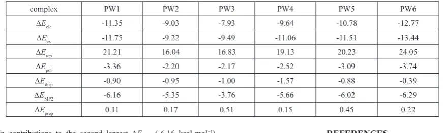
The results of LMO-EDA are listed in Table 5. As shown in Table 5, the total interaction energy (DEMP2) between PA and H2O moieties is whithin
the range of about -3.8 ~ -6.3 kcal×mol-1, and the strongest DE
MP2 of -6.29
kcal·mol−1 indicates that PW6 is the most stable PA–H
2O complex. In PW6,
the largest stabilizing force is the exchange energy (DEex) of -13.44 kcal·mol−1,
which origins from the overlap between the spin orbital of each monomer and the like–spin orbitals of the other monomer, but coming with a strong repulsion energy (DErep) of 24.05 kcal×mol-1 simultaneously. The second largest
stabilizing force is the electrostatic interaction (DEele) of -12.77 kcal·mol−1.
Moreover, the formation of the H–bond changes their orbital shapes of fragments and results in a polarization energy (DEpol) of -3.74 kcal·mol−1,
Table 5. The LMO–EDA results of PA–H2O complexes obtained at the MP2 level. Energy values are given in kcal.mol-1.
complex PW1 PW2 PW3 PW4 PW5 PW6
DEele -11.35 -9.03 -7.93 -9.64 -10.78 -12.77
DEex -11.75 -9.22 -9.49 -11.06 -11.51 -13.44
DErep 21.21 16.04 16.83 19.13 20.23 24.05
DEpol -3.36 -2.20 -2.17 -2.52 -3.09 -3.74
DEdisp -0.90 -0.95 -1.00 -1.57 -0.88 -0.39
DEMP2 -6.16 -5.35 -3.76 -5.66 -6.02 -6.29
ΔEprep 0.11 0.17 0.51 0.15 0.45 0.22
the main contributions to the second largest DEMP2 (-6.16 kcal·mol−1)
mainly come from the larger DEele (-11.35 kcal·mol−1) and DEex (-11.75
kcal·mol−1), while DE
pol (-3.36 kcal·mol−1) and DEdisp (-0.90 kcal·mol−1) make
less contribution to the DEMP2 of PW1. Similar things also happened in PW2,
PW4 and PW5 since they have almost same stabilities with each other. PW3 is the complex with less stabilities due to the smaller DEMP2 (-3.76 kcal·mol−1),
which is attributed to the weaker hydrogen bonding interactions in it. Our previous studies showed that hydrogen bonding interaction is not the unique factor for the stability of complexes involving hydrogen bonding interactions 51-55. Therefore, the influence of the deformation of the monomers
on the stability of PW complex were taken into account. On the basis of NBO theory, the preparation energy (DEprep) is the amount of energy required to
deform the separate bases from their free monomer structure to the geometry that they acquire in the pair complex,
DEprep = EPW – EPA(W) – EW(PA) (1)
here EPA(W) (or EW(PA)) is the energy of the PA (or water) monomer when
all the nucleus structure units of water (or PA) are considered as puppet atoms of carrying empty orbital. ΔEprep is positive because the structural
deformation causes the molecular energy to jump to a higher energy level, while DEMP2 is negative unless the complex is less stable than the monomers.
The preparation energies of all PA–H2O complexes are also listed in Table 5.
All complexes have small DEprep values of less than about 0.7 kcal·mol−1. The
two largest ΔEprep values are 0.51 (PW3) and 0.45 (PW5) kcal×mol-1, which
indicates that the cleavages of the intramolecular C8H8PA···O2PA H–bond in
PW3 and PW5 result in the serious structural deformation and counteracts such strong hydrogen bonding interactions to a great extent. On the contrary, the intramolecular C8H8PA···O2PA H–bond was retained in other PA–H
2O
complexes (PW1, PW2, PW4 and PW6), and the structural deformation of them are slight, which can be learned from their smaller ΔEprep values in Table 5. In one word, both hydrogen bonding interaction and structural deformation are the two important aspects of the stability of PA–H2O complexes, which is
consistent with our previous works 51-55.
4. CONCLUSIONS
The geometries, energies and IR characteristics of the H–bonds of PA–H2O
complexes were studied at the MP2/6–311++G(d,p) level. The intramolecular C8H8PA···O2PA H–bond is retained in all complexes except PW3 and PW5. The
intermolecular O1H6PA···OW (PW1) and OH1W···O2PA (PW6) H–bonds are the
two strongest ones. The H–bonds involving the methylene of PA as H–donors are very weak. Both hydrogen bonding interaction and structural deformation play important roles in the relative stabilities of the complexes. Except PW3, all PA–H2O complexes have similar stabilities, which indicates that PA inclines to
form various complexes when it meets with water solvent. These results further reinforce the concept that PA is considered as a good electron acceptor (or donor) in forming complexes with various small organic molecules. Therefore, we think that the studies on PA-H2O complexes maybe bear significance to the
understanding the hydrogen bonding interactions between PA and other small organic molecules.
ACKNOWLEDGEMENT
This work is supported by the Natural Science Foundation of Tianjin (No. 12JCYBJC13400) and the Program for Innovative Research Team in University of Tianjin (TD13-5074).
REFERENCES
1. M. Sciskalska, M. Sliwinska-Mosson, M. Podawacz, W. Sajewicz and H. Milnerowicz, Drug. Chem. Toxicol. 38, 121 (2015).
2. W.S. Waring, H. Jamie and G.E. Leggett, Hum. Exp. Toxicol. 29, 63 (2010).
3. M. Naggayi, N. Mukiibi and E. Iliya, Afr. Health. Sci. 15, 598 (2015). 4. S.B.K. Mahadevan, P.J. McKiernan, P. Davies and D.A. Kelly, Arch. Dis.
Child. 91, 598 (2006).
5. M. Cekmen, Y.O. Ilbey, E. Ozbek, A. Simsek, A. Somay and C. Ersoz, Food Chem. Toxicol. 47, 1480 (2009).
6. P. Abraham, Nephrology. 10, 623 (2005).
7. N.V. Nayyer, J. Byers and C. Marney, Brit. Dent. J. 216, 229 (2014). 8. P. Marzuillo, S. Guarino and E. Barbi, Eur. J. Pediatr. 173, 415 (2014). 9. A.-R. Marzilawati, Y.-Y. Ngau and S. Mahadeva, BMC Pharmacol.
Toxico. 13, 8 (2012).
10. H. Jaeschke, Digest. Dis. 33, 464 (2015).
11. E.W. Holt, S. DeMartini and T.J. Davern, J. Clin. Gastroenterol. 49, 790 (2015).
12. G.G. Graham, M.J. Davies, R.O. Day, A. Mohamudally and K.F. Scott, Inflammopharmacology. 21, 201 (2013).
13. M.L. Ramos, J.F. Tyson and D.J. Curran, Anal. Proc. incl. Anal. Comm.
32, 175 (1995).
14. P.A. Mosier-Boss, S.H. Lieberman and R. Newbery, Appl. Spectrosc. 49, 630 (1995).
15. B.B. Ivanova, J. Mol. Struct. 738, 233 (2005).
16. I.G. Binev, P. Vassileva-Boyadjieva and Y.I. Binev, J. Mol. Struct. 447, 235 (1998).
17. E. Dreassi, G. Ceramelli, P. Corti, M. Massacesi and P.L. Perruccio, Analyst. 120, 2361 (1995).
18. J.M. Beames and A.J. Hudson, Phys. Chem. Chem. Phys. 12, 4157 (2010). 19. Y. Danten, T. Tassaing and M. Besnard, J. Phys. Chem. A. 110, 8986
(2006).
20. S.J. Lee, A. Min, Y. Kim, A. Ahn, J. Chang, S.H. Lee, M.Y. Choi and S.K. Kim, Phys. Chem. Chem. Phys. 13, 16537 (2011).
21. M. Yoosefian and N. Etminan, RSC Adv. 5, 31172 (2015). 22. S. Rai, H. Singh and U.D. Priyakumar, RSC Adv. 5, 49408 (2015). 23. K.H. Moller, A.S. Hansen and H.G. Kjaergaard, J. Phys. Chem. A. 119,
10988 (2015).
24. M. Izadyar, M. Khavani and M.R. Housaindokht, Phys. Chem. Chem. Phys. 17, 11382 (2015).
25. Z.G. Huang, Y.M. Dai and L. Yu, Struct. Chem. 21, 863 (2010).
26. L.F. Guo, Z.G. Huang, T.T. Shen, L.L. Ma and X.Q. Niu, Chin. J. Chem.
31, 1079 (2013).
27. P.L.A. Popelier: Atoms in Molecules: An Introduction (Prentice Hall, City, 2000).
28. C.F. Matta and R.J. Boyd: The Quantum Theory of Atoms in Molecules: From Solid State to DNA and Drug Design (WILEY-VCH Verlag GmbH & Co. KGaA, City, 2007).
29. A.E. Reed, L.A. Curtiss and F. Weinhold, Chem. Rev. 88, 899 (1988). 30. P.F. Su and H. Li, J. Chem. Phys. 131, 014102 (2009).
31. J. Xi and X. Xu, Phys. Chem. Chem. Phys. 18, 6913 (2016).
32. S.K. Singh, S. Kumar and A. Das, Phys. Chem. Chem. Phys. 16, 8819 (2014).
33. J.J. Panek, A. Filarowski and A. Jezierska-Mazzarello, J. Chem. Phys.
139, 154312 (10 pp.) (2013).
35. M. Jablonski and M. Palusiak, J. Phys. Chem. A. 116, 2322 (2012). 36. L.L. Ma, Z.G. Huang, X.Q. Niu, T.T. Shen and L.F. Guo, Comput. &
Theor. Chem. 1017, 14 (2013).
37. E. Espinosa, I. Alkorta, J. Elguero and E. Molins, J. Chem. Phys. 117, 5529 (2002).
38. U. Koch and P.L.A. Popelier, J. Phys. Chem. 99, 9747 (1995). 39. W.D. Arnold and E. Oldfield, J. Am. Chem. Soc. 122, 12835 (2000). 40. S. Jenkins and I. Morrison, Chem. Phys. Lett. 317, 97 (2000).
41. S.J. Grabowski, W.A. Sokalski and J. Leszczynski, J. Phys. Chem. A. 110, 4772 (2006).
42. L.F. Pacios, J. Phys. Chem. A. 108, 1177 (2004).
43. A.E. Reed, F. Weinhold, L.A. Curtiss and D.J. Pochatko, J. Chem. Phys.
84, 5687 (1986).
44. M.J. Frisch, G.W. Trucks, H.B. Schlegel, G.E. Scuseria, M.A. Robb, J.R. Cheeseman, G. Scalmani, V. Barone, B. Mennucci, G.A. Petersson, H. Nakatsuji, M. Caricato, X. Li, H.P. Hratchian, A.F. Izmaylov, J. Bloino, G. Zheng, J.L. Sonnenberg, M. Hada, M. Ehara, K. Toyota, R. Fukuda, J. Hasegawa, M. Ishida, T. Nakajima, Y. Honda, O. Kitao, H. Nakai, T. Vreven, J.A. Montgomery Jr., J.E. Peralta, F. Ogliaro, M. Bearpark, J.J. Heyd, E. Brothers, K.N. Kudin, V.N. Staroverov, R. Kobayashi, J. Normand, K. Raghavachari, A. Rendell, J.C. Burant, S.S. Iyengar, J. Tomasi, M. Cossi, N. Rega, J.M. Millam, M. Klene, J.E. Knox, J.B. Cross, V. Bakken, C. Adamo, J. Jaramillo, R. Gomperts, R.E. Stratmann, O. Yazyev, A.J. Austin, R. Cammi, C. Pomelli, J.W. Ochterski, R.L. Martin,
K. Morokuma, V.G. Zakrzewski, G.A. Voth, P. Salvador, J.J. Dannenberg, S. Dapprich, A.D. Daniels, Ö. Farkas, J.B. Foresman, J.V. Ortiz, J. Cioslowski and D.J. Fox: Gaussian09, (Gaussian, Inc., City, 2009). 45. F. Biegler-König and J. Schönbohm: AIM2000, (University of Applied
Sciences, City, 2000).
46. M.W. Schmidt, K.K. Baldridge, J.A. Boatz, S.T. Elbert, M.S. Gordon, J.H. Jensen, S. Koseki, N. Matsunaga, K.A. Nguyen, S. Su, T.L. Windus, M. Dupuis and J.A. Montgomery, J. Comput. Chem. 14, 1347 (1993). 47. H.K. Wang, Z.G. Huang, T.T. Shen and L.F. Guo, Struct. Chem. 23, 1163
(2012).
48. H.K. Wang, Z.G. Huang, T.T. Shen and L.F. Guo, J. Mol. Model. 18, 3113 (2012).
49. L. Yu, Y.H. Wang, Z.G. Huang, H.K. Wang and Y.M. Dai, Int. J. Quantum Chem. 112, 1514 (2012).
50. X.Q. Niu, Z.G. Huang, L.L. Ma, T.T. Shen and L.F. Guo, J. Chem. Sci.
125, 949 (2013).
51. Z.G. Huang, L. Yu, Y.M. Dai and H.K. Wang, Struct. Chem. 22, 57 (2011). 52. Z.G. Huang, L. Yu and Y.M. Dai, Int. J. Quantum Chem. 111, 3915
(2011).
53. Z.G. Huang, Y.M. Dai, L. Yu and H.K. Wang, J. Mol. Model. 17, 2609 (2011).
54. Z.G. Huang, L. Yu, Y.M. Dai and H.K. Wang, J. Mol. Struct. (Theochem).
960, 98 (2010).