Journal of Education and Training Studies Vol. 6, No. 5; May 2018 ISSN 2324-805X E-ISSN 2324-8068 Published by Redfame Publishing URL: http://jets.redfame.com
The Effects of Two Different Stretching Programs on Balance Control and
Motor Neuron Excitability
Fatih Kaya1, Bilal Biçer2, Bekir Yüktaşır3, Mark E. T. Willems4, Nebil Yıldız5
1Department of Physical Education and Sport, Faculty of Education, Erzincan University, Erzincan, Turkey 2School of Physical Education and Sport, Mustafa Kemal University, Hatay, Turkey
3Sinop University, Faculty of Sport Sciences, Sinop, Turkey
4University of Chichester, Department of Sport and Exercise Sciences, Chichester, United Kingdom 5Abant Izzet Baysal University, Faculty of Medicine, Department of Neurology, Bolu, Turkey
Correspondence: Fatih Kaya, Department of Physical Education and Sport, Faculty of Education, Erzincan University, Erzincan, 24100, Turkey.
Received: February 16, 2018 Accepted: March 26, 2018 Online Published: March 31, 2018 doi:10.11114/jets.v6i5.3033 URL: https://doi.org/10.11114/jets.v6i5.3033
Abstract
We examined the effects of training (4d/wk for 6 wks) with static stretching (SS) or contract-relax proprioceptive neuromuscular facilitation (PNF) on static balance time and motor neuron excitability. Static balance time, Hmax/Mmax ratios and H-reflex recovery curves (HRRC) were measured in 28 healthy subjects (SS: n=10, PNF: n=9, control: n=9) before and after training. SS improved static balance time with a trend observed for PNF. Post training, during 150-200-250 msec interstimulus intervals, we observed a reduction in facilitation, but during 500-700-900 msec interstimulus interval; there was an increase in H2/H1 ratio in the PNF group only. Both stretching techniques improved static balance. The Ia afferent inhibitions during the acute exercises were not found after the SS and PNF training programmes. It was concluded that training with contract-relax proprioceptive neuromuscular facilitation may cause some augmentation in supraspinal and postsynaptic inhibition on the motoneuron pool.
Keywords: static stretching, contract-relax PNF stretching, static balance, Hoffmann-reflex
1. Introduction
Flexibility and balance are considered to be important for health and physical fitness. Flexibility affects muscular performance (e.g. Ferreira, Teixeira-Salmela, & Guimarães, 2007). It was shown that subjects with bilaterally shortened hamstrings improved muscle performance and range of motion (ROM) with a static stretching intervention (Ferreira et al., 2007). Effects of static stretching on ROM have been well established in other studies as well (e.g. Marek et al., 2005). However, other stretching techniques, such as proprioceptive neuromuscular facilitation (PNF) stretching which inhibits tonic reflex activity as a limiting factor during stretches, increases the ROM more markedly (Etnyre & Abraham, 1986; Guissard, Duchateau, & Hainaut, 1988). Both mechanical and neural adaptation mechanisms have been proposed to be responsible for ROM changes from stretching (Guissard & Duchateau, 2004; Youdas et al., 2010). In addition, PNF induces a larger reduction of motor neuron excitation by pre and post-synaptic inhibitory mechanisms (Guissard et al., 1988; Guissard, Duchateau, & Hainaut, 2001). This was shown by analysis of the H-waves from α-motoneuron excitability which was reduced during but not after stretch of the soleus muscle (Etnyre & Abraham, 1986; Guissard et al., 1988).
The Hoffmann reflex (H-reflex), and associated H-waves, is an established measure of motoneuron excitability. The H-reflex serves as a summative measure of excitability in the motoneuron pool in terms of reflecting influences of postsynaptic inhibition (Trimble & Koceja, 2001). The H-reflex has been instrumental in research investigating the production of purposeful movement. Interpretation of the H-reflex is complex as it is influenced by supraspinal, homonymous, and heteronymous modulation, as well as intrinsic motoneuronal properties. In addition, the H-reflex is influenced by the state of the physiologic system, with inhibition induced by factors such as joint position, muscle activity, and phase of movement (Trimble, Brunt, & Thompson, 2000).
has been related to a larger reduction of motoneuron excitation, which results from pre- and postsynaptic inhibitory mechanisms (Guissard et al., 1988; Guissard et al., 2001), assuming that greater motor pool inhibition reduces muscle contractibility and therefore allows more muscle compliance. Thus, PNF methods which involve reciprocal activation may provide the greatest potential for muscle lengthening (Etnyre & Abraham, 1986; Sharman, Cresswell, & Riek, 2006; McArdle, Katch, & Katch, 2010).
Recently, many studies focus on falls and poor balance control of elderly subjects and on the effectiveness of exercise programs to reduce the risks for falls and to improve balance (for a review see Gardner, Robertson, & Camphell, 2000).It is known that some exercises, which include stretching, are beneficial for postural balance control of the elderly (Carlson et al., 1999; Bird, Hill, Ball, & Williams, 2009). Balance control necessitates optimal muscle strength and flexibility as well as accurate visual, vestibular and proprioceptive inputs. Improvement in at least one of these factors may result in better balance control not only for the elderly but also for younger subjects. However, despite studies demonstrating the acute effects of stretching exercise on balance (Costa, Graves, Whitehurst, Jacobs, 2009; Behm, Bambury, Cahill, & Power, 2004; Lim, Nam, & Jung, 2014), the chronic effects of a consistent stretching exercise programmes on balance have not been addressed.
The aim of the present study was to investigate the chronic effects of 24 sessions of static stretching and contract-relax PNF stretching on balance control and motor neuron excitability to the hamstring and triceps surae muscles. To this end, the changes in the soleus muscle H-reflex responses, Hmax/Mmax ratio and H-reflex recovery curves were recorded. We also analyzed the changes in static balance control by measurement of single leg time. Knowledge on the chronic effects of stretching exercises on balance control, and motor neuron excitability may have implications for targeted interventions on individuals with impaired balance.
2. Method
Twenty-eight healthy male volunteers participated in the study. Subjects were all accustomed to the experimental procedures and had no signs of any neurological or orthopedic disorder. All subjects were informed about the experimental procedures, the risks involved in this study and provided informed consent (Declaration of Helsinki). Approval for the study was obtained from the Abant Izzet Baysal University’s Faculty of Medicine Ethics Committee. Subjects were randomized into three groups; a static stretching group (n =10, age 22±2 yrs, height 174±5 cm, mass 75.5±4.5 kg), a contract-relax PNF stretch group (n = 9, age 22±2 yrs, height 180±7 cm, mass 72.0±7.0 kg) and a control group (n = 9, age 22±2 yrs, height 175±6 cm, mass 70.1±8.2 kg).
All measurements were performed on the dominant leg before and after stretching interventions. Leg dominance was determined by asking to take a step forward, the preferred leg was taken as dominant leg. Investigators were blind to the treatment group of the subjects. All groups were tested before and after a 6-wk period.
Static balance time was measured with a manual chronometer (Casio) to the nearest 0.1 seconds. All subjects were instructed to focus on a target approximately three meters away at eye level. For all trials, subjects placed hands on their hips and the non-dominant foot on the knee of the dominant leg. Subsequently, subjects raised themselves upon their toes and balance time started with the elevation of the heel from the floor and stopped at loss of balance. Loss of balance included removal of one hand from the hip, removal of the foot from the knee, touching the floor with the heel, or movement of the weight-bearing foot from its original position on the floor. The trial was repeated twice and the longest time was taken as balance time.
2.1 Electrophysiological Measures and Soleus Muscle H-Reflex Studies
Initially, tibial nerve motor conduction, tibial nerve F-waves and sural nerve sensory conduction were measured and observed to be normal in all subjects. A Nicolet Viking 4 channel EMG-EP machine was used for electrophysiological assessments. Subjects were familiarized for all procedures. For H-reflex recordings, subjects were lying prone with the head in a neutral midline position and their feet resting freely over the edge of the table in a quiet room. Monopolar stimulation electrodes made of silver-silver chloride were used; the cathode was attached over the popliteal fossa superposing the tibial nerve and the anode was placed over the patella. The soleus H-reflexes were recorded with surface electrodes applied on the skin approximately 4 cm distal to the musculotendinous junction of the gastrocnemius muscle. A metal ground electrode with a diameter of 2 cm was placed between the stimulating and the recording electrodes. All electrodes were fixed tightly with a surrounding bandage. The responses were recorded using a gain of 1000-5000 µV and a filter of 10 Hz to 10 kHz. The duration of the stimulus was one ms. Peak-to-peak H-waves were measured and recorded. Electrode locations were recorded for each subject to provide similar stimulation and recording conditions for pre- and post training testing sessions.
Journal of Education and Training Studies Vol. 6, No. 5; May 2018
of equal intensity applied with varying interstimulus intervals ranging from 50 to 1000 msec (50-70-100-150-200-250-300-400-500-700-900-1000 msec). Four separate recordings were obtained at each interstimulus interval and the averages were calculated from the ratios (H2/H1) for each subject at each interstimulus interval. Ratios were expressed as percentages.
2.2 Training Program
All subject received before the start of the training program instructions on the performance of the stretching techniques. Static stretching or contract-relax PNF stretching exercises were performed four times per week for six weeks, a total of 24 sessions. The control group did not participate in any exercise program. The volume of the training programmes of the two stretching groups was similar.
2.2.1 Static Stretching Training
During the static stretching exercises, subjects lay in a supine position on the floor. For stretching of the hamstrings, the knee joint was extended with the hip held 900 of flexion while simultaneously flexing the ankle joint to 900 (neutral ankle dorsiflexion) to stretch the triceps surae for 30 seconds. Stretching was performed at the maximum range tolerated by the subjects. For each leg, the stretching was repeated four times with a rest period of 10 sec between stretches. 2.2.2 Contract-Relax PNF Stretching Training
The contract- relax PNF procedure consisted of three stages. In the first stage, with the subject lying in a supine position, the knee joint was extended with the hip held at 900 of flexion while simultaneously flexing the ankle joint to 900 (neutral ankle dorsiflexion) for 10 sec. In the second stage, hip extension and ankle plantarflexion against a force executed by the investigator was performed for five seconds. Following the subject’s five seconds voluntary contraction, the subject relaxed for five seconds followed by the investigator applying hip- and dorsi-flexion stretching forces for an additional 15 sec. After a 5 sec relaxation period, passive hip flexion and ankle dorsiflexion were performed again for 15 sec. For each leg, the stretching was repeated four times with a rest period of 10 sec between each procedure.
2.3 Statistical Analysis
For each parameter, mean, standard deviation and effect sizes were calculated. Differences in change scores in pre and post training period at each group were assessed using paired t-tests. Further, in H-reflex recovery curve evaluation, repeated measures of analysis of variance procedures were performed with checks for sphericity. The sphericity assumption was not met so the Greenhouse-Geisser correction was applied. If there was a significant F ratio, post hoc
comparisons were performed using paired t-tests with a Bonferroni adjustment of the alpha level (.05). The level of significance was determined as p<0.05 at the beginning of the survey and analyses were done by the program SPSS 20.0 for Windows.
3. Results
While the static balance time was significantly increased in SS group from pretest to posttest, a trend was observed in the PNF group with no change for in the control group (SS: t(9)=-3.064, p=.013, ߟଶ=.51, PNF: t(8)=-2.020, p=.078, ߟଶ=.34, Control: t(
8)=.175, p=.865, ߟଶ=.00) (Table 1).
In the groups, pre- and post training values of Hmax/Mmax ratios indicated a trend for the SS group but no differences for the PNF and control groups (SS: t(9)=-1.930, p=.086, ߟଶ=.29, PNF: t(8)=-.048, p=.963, ߟଶ=.00, Control: t(8)=.495, p=.634, ߟଶ=.03) (Table 1).
Pre-training, H2/H1 ratios were not different in the groups [F(2,25)=.233, p=.794, ߟଶ=.02]. In addition, post training, H2/H1 ratios were not significantly different among groups [F(2,25)=.471, p=.630, ߟଶ=.04) (Figure 1).
Table1. Static balance time and Hmax/Mmax for all three groups. Data are mean (SD)
Balance (sec) Hmax/Mmax
pretest posttest pretest posttest
Static 15.72 (9.47) 24.48 (17.51)* 0.39 (0.24) 0.50 (0.18)
PNF 18.10 (14.02) 26.14 (11.97)** 0.43 (0.23) 0.44 (0.18)
Control 15.2 9(13.16) 14.98 (11.50) 0.46 (0.15) 0.43 (0.18)
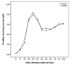
Figure 1. H-re H-reflex re In the groups ߟଶ=.00, for PN
However, the p=.018, ߟଶ=.1
reduction in f interstimulus i
Figure 2. T means (SD
Figure 3 This graph
Figure 4. C graph sh
eflex recovery ecovery curves , no differenc NF F(1,16)=.004 ere was a sign
12], in the “p facilitation but intervals in HR
This graph show D). There was
500-700 a
3. Static group h shows the sta
Control group’s hows the static
curves pre and s of control and
es in HRRC c 4, p=.951, ߟଶ=
nificant interac post training”
t then during RRC curves (F
ws the PNF gr a reduction in and 900 ms int
p’s H-reflex re atic group’s
H-s H-reflex reco c group’s H-re
d post training f d two stretchin curves were o =.00, for Contr ction between period, during 500-700 and Figure 2). roup’s H-reflex facilitation du terstimulus int covery curves -reflex recover
overy curves o flex recovery c
for all three gr ng groups in pr observed in pr rol F(1,16)=.001 n pre and post g 150-200 and 900 ms inters
x recovery curv uring 150-200 a tervals, there w
obtained at tw ry curves in pr
obtained at two curves in pre a
roups. Values a re and post trai re- and post tr 1, p=.973, ߟଶ=
t training peri d 250 ms inte stimulus interv
ves obtained a and 250 ms int was an increase
wo different pe re and post-trai
o different peri and post-trainin
are means (SD) ining periods a raining (for SS =.00) (Figure 2 iods in PNF g erstimulus inte vals, there wa
at two different terstimulus int e in HRRC cur
eriods. Values ining period ar
iods. Values ar ng period are s
). This graph sh are similar (p> S F(1,18)=.036, 2, 3, 4). group [F(11,176 ervals, we ob s an increase
t periods Valu tervals, then du rves.
are means (SD re similar (p>0
re means (SD) similar (p>0.05
hows the >0.05)
p=.852,
Journal of Education and Training Studies Vol. 6, No. 5; May 2018
4. Discussion
The current study was designed to assess the prolonged effects of two different stretching programs, performed for four times per week for six weeks, on static balance and the H-reflex activity. Despite the lack of a significant effect of PNF stretching, which may have been due to a lack of power, our results seem to indicate that that both the SS and PNF stretching exercises improved static balance, and PNF also induced some changes in HRRC curves.
The functions of the intrafusal (includes stretch receptors) muscle fibers, Golgi tendon organs and other proprioceptors is to aid in the maintenance of balance and detection of the position of the body in space (proprioception). Also, balance involves the interaction of automatic postural and voluntary motor commands of both the trunk and limb musculature (Behm et al., 2004). The results in studies regarding the effects of acute static stretching sessions on balance are conflicting. While some studies observed a worsening balance after stretch (Behm et al., 2004; La Torre et al., 2010), another study reported that longer-duration stretching protocols may not adversely affect balance (Costa et al., 2009). Behm et al. (2004) have suggested that a more compliant musculotendinous unit has more slack on the connective tissue, hence affecting muscle activation, consequently affecting balance and stability, or the proprioception of a limb.
Additionally, repeated and prolonged passive stretching has been shown to decrease reflex activity resulting from reduced sensitivity of the muscle spindles to repeated stretch (Avela, Kyrolainen, & Komi, 1999), which may partly explain the effects of stretching on balance. Again, a moderate stretching protocol may avoid possibly unfavorable reflex activity decrements. Moreover, static stretching has been shown to improve joint position sense, which investigators believe could be an increased proprioceptive feedback (Costa et al., 2009). This improvement in proprioception could be a mechanism that might, consequently, improve balance.
In the literature, Gehlsen and Whaley (1990) reported that balance, strength, and flexibility might be factors contributing to falls for the elderly. A well-designed stretching program will lessen the decline of flexibility of the elderly adults (Swank, Funk, Durham, & Roberts, 2003). It is known that some exercises, which include stretching, are beneficial for postural balance control of the elderly (Celebi, Zergeroglu, & Ergen, 2003; Marigold et al., 2005; Means, Rodell, & O’ Sullivan, 2005). In a report by Celebi et al. (2003), it was shown that loss of balance control was small in a group of the elderly subjects who performed stretching exercises. They suggested that the muscle spindles become more sensitive to muscle lengthening, thus the strength and the velocity of voluntary and reflexive muscle contractions improves leading to an amelioration in balance control (Celebi et al., 2003). Another explanation is likely to increase in muscle strength as a result of the stretching exercises. It has been suggested that muscle hypertrophy might occur as a result of chronic stretching (Stone et al., 2006). Increase in muscle strength will improve the balance control (Garrett & Kirkendall, 2000). Furthermore, it was emphasized that the stretching duration and the frequency should be sufficient to see the effects of the stretching techniques on the balance (Lim et al., 2014).
In the current study, the subjects were all young and active and had no ocular or vestibular dysfunction. Although statistically significant differences were not detected from the PNF technique, may be due to lack of power as stated above, our results indicate an improvement from pre-test to post-test in static balance by both stretching programmes (Table 1). In a report by Nielsen, Crone and Hultborn (1993), H-reflexes were of proportionally lesser amplitude in ballet dancers than in athletes. In another report by Guissard and Duchateau (2004), a training program involved 30 sessions consisting of a total of 10 minutes passive static stretching performed five times a week for 6 weeks in 12 subjects. After the 10th session, they found that muscle viscoelastic properties have progressed and after the 30th session, there have been also some neuronal changes leading to a decrease in H max and T max amplitudes. They also showed that these neuronal changes had returned to the control values more rapidly than the mechanical changes. They suggested that the decrease in H-reflex amplitudes have resulted from the reduction of the synaptic transmission from the Ia afferents to the motoneuron pool and also the more pronounced decrease in T reflex amplitude than H-reflex could be related to reduced sensitivity of the muscle spindle (Guissard & Duchateau, 2004). Guissard et al. (1988) also showed that for small amplitude passive muscle stretching, the H-reflex was reduced without any change in the T reflex amplitude but for a larger stretching amplitude both the H and the T reflexes and also the MEP response amplitudes have decreased. They proposed that decreased reflex loop activity during small-amplitude stretching should be related primarily to presynaptic inhibition on Ia afferents, and the other changes observed during large amplitude stretching were related to postsynaptic inhibition (Guissard et al., 1988). In our study, although balance improved, we did not find any changes in H-reflex amplitudes; this finding may indicate that the flexibility exercises must be performed for longer periods and more intensively than our program.
influences (Crayton & Reud, 1981; Panizza, Lelli, Nilsson, Hallet, 1990; Spaulding, Hayes, & Harburn, 1987). In the current study, there seemed to be a reduction in the secondary facilitation phase of HRRC in the PNF group. It is possible that this reduction was due to the increase of supraspinal and postsynaptic inhibition, and that the presynaptic inhibition was excluded by the unchanged Hmax/Mmax ratios. Furthermore, the HRRC seemed to be greater in the post training period than in the pre training period during the 500-700-900 msec intervals in the PNF group. This may be explained by a different adaptation mechanism resulting from supraspinal influences and interneuronal mechanisms of spinal cord or may be a rebound phenomenon with a rapid recovery reaction following the secondary inhibition phase.
5. Conclusion
In conclusion, the Ia afferent inhibition occurring during the acute exercises has not been found after our long-term training programs with either static stretching or PNF. Finally, PNF may cause some augmentation in supraspinal and postsynaptic inhibition on the human motoneuron pool.
Acknowledgments
We would like to thank Abant İzzet Baysal University, Faculty of Medicine, Department of Neurology for electrophysiological measurements. All authors declare no conflicts of interest. The authors received no financial support for the research and/or authorship of this article.
References
Avela, J., Kyrolainen, H., & Komi, P. V. (1999). Altered reflex sensitivity after repeated and prolonged passive muscle stretching. Journal of Applied Physiology, 86, 1283–1291.
http://www.physiology.org/doi/10.1152/jappl.1999.86.4.1283
Behm, D. G., Bambury, A., Cahill, F., & Power, K. (2004). Effect of acute static stretching on force, balance, reaction
time, and movement time. Medicine & Science in Sports & Exercise, 36(8), 1397-1402.
https://doi.org/10.1249/01.MSS.0000135788.23012.5F
Bird, M. L., Hill, K., Ball, M., & Williams, A. D. (2009). Effects of resistance-and flexibility-exercise interventions on balance and related measures in older adults. Journal of Aging and Physical Activity, 17(4), 444-454. https://doi.org/10.1123/japa.17.4.444
Carlson, J. E., Ostir, G. V., Black, S. A., Markides, K. S., Rudkin, L., & Goodwin, J. S. (1999). Disability in older adults 2: Physical activity as prevention. Behavioral Medicine, 24(4), 157-168
https://doi.org/10.1080/08964289.1999.11879272
Celebi, M. M., Zergeroglu, M. A., & Ergen, E. (2003). The effects of the warm up and stretching exercises on the proprioception, Medicine & Science in Sports & Exercise, 35(5), 264-264.
https://doi.org/10.1097/00005768-200305001-01461
Costa, P. B., Graves, B. S., Whitehurst, M., & Jacobs, P. L. (2009). The acute effects of different durations of static stretching on dynamic balance performance. Journal of Strength & Conditioning Research, 23(1), 141-147. https://doi.org/10.1519/JSC.0b013e31818eb052
Crayton, J. W., & Reud, R. R. (1981). An oscillatory component of the H-reflex, Journal of Neurology, Neurosurgery & Psychiatry,44(3), 239-242. Retrieved from https://www.ncbi.nlm.nih.gov/pmc/articles/PMC490899/
Etnyre, B. R., & Abraham, L. D. (1986). H-reflex changes during static stretching and two variations of proprioceptive neuromuscular facilitation techniques, Electroencephalography and Clinical Neurophysiology, 63(2), 174-179. Retrieved from https://www.ncbi.nlm.nih.gov/pubmed/2417817
Ferreira, G. N., Teixeira-Salmela, L. F., & Guimarães, C. Q. (2007). Gains in flexibility related to measures of muscular performance: impact of flexibility on muscular performance. Clinical Journal of Sports Medicine, 17(4), 276-281. https://doi.org/10.1097/JSM.0b013e3180f60b26
Gardner, M. M., Robertson, M. C., & Camphell, A. J. (2000). Exercise in preventing falls and fall related injuries in older people: A review of randomized controlled trials. British Journal of Sports Medicine, 34(1), 7-17. https://doi.org/10.1136/bjsm.34.1.7
Garrett, W. E., & Kirkendall, D. T. (Eds.). (2000). Exercise and sport science. Lippincott Williams & Wilkins
Gehlsen, G. M., & Whaley, M. H. (1990). Falls in the elderly: Part II, balance, strength, and flexibility, Archives of Physical Medicine and Rehabilitation, 71(10), 739-41. Retrieved from
https://www.ncbi.nlm.nih.gov/pubmed/2403279
Journal of Education and Training Studies Vol. 6, No. 5; May 2018
Guissard, N., Duchateau, J., & Hainaut, K. (2001). Mechanisms of decreased motoneurone excitation during passive muscle stretching. Experimental Brain Research,137(2), 163-169. https://doi.org/10.1007/s002210000648
Kagamihara, Y., Hayashi, A., Okuma, Y., Nagaoka, M., Nakajima, Y., & Tanaka, R. (1998). Reassessment of H-reflex recovery curve using the double stimulation procedure. Muscle Nerve,21(3), 352-360.
https://doi.org/10.1002/(SICI)1097-4598(199803)21:3<352::AID-MUS9>3.0.CO;2-9
La Torre, A., Castagna, C., Gervasoni, E., Cè E., Rampichini, S., Ferrarin, M., & Merati, G. (2010). Acute effects of static stretching on squat jump performance at different knee starting angles, Journal of Strength and Conditioning Research, 24(3), 687-94. https://doi.org/10.1519/JSC.0b013e3181c7b443
Lim, K. I., Nam, H. C., & Jung, K. S. (2014). Effects on hamstring muscle extensibility, muscle activity, and balance of different stretching techniques. Journal of Physical Therapy Science, 26(2), 209-213.
https://doi.org/10.1589/jpts.26.209
Marek, S. M., Cramer, J. T., Fincher, A. L., Massey, L. L., Dangelmaier, S. M., Purkayastha, S., & Fitz, K. A. (2005). Acute effects of static and proprioceptive neuromuscular facilitation stretching on muscle strength and power output. Journal of Athletic Training, 40(2), 94-103. Retrieved from
https://www.ncbi.nlm.nih.gov/pmc/articles/PMC1150232/
Marigold, D. S., Eng, J. J., Dawson, A. S., Inglis, J. T., Haris, J. E., & Gylfadottir, S. (2005). Exercise leads to faster postural reflexes, improved balance and mobility, and fewer falls in older persons with chronic stroke Journal of the American Geriatrics Society,53(3), 416-23. https://doi.org/10.1111/j.1532-5415.2005.53158.x
McArdle, W. D., Katch, F. I., & Katch, V. L. (2010). Essentials of exercise physiology. New York: Lippincott Williams & Wilkins.
Means, K. M., Rodell, D. E., & O’ Sullivan, P. S. (2005). Balance, mobility, and falls among community-dwelling elderly persons: effects of a rehabilitation exercise program, American Journal of Physical Medicine & Rehabilitation,84(4), 238-250. https://doi.org/10.1097/01.PHM.0000151944.22116.5A
Nielsen, J., Crone, C., & Hultborn, H. (1993). H-reflexes are smaller in dancers from The Royal Danish Ballet than in well-trained athletes. European Journal of Applied Physiology,66(2), 116-121.
https://doi.org/10.1007/BF01427051
Panizza, M., Lelli, S., Nilsson, I., & Hallet, M. (1990). H- reflex recovery curve and reciprocal inhibition of H- reflex in different kinds of dystonia. Neurology,40(5), 824-828. https://doi.org/10.1212/WNL.40.5.824
Sharman, M. J., Cresswell, A. G., & Riek, S. (2006). Proprioceptive neuromuscular facilitation stretching. Sports Medicine, 36(11), 929-939. https://doi.org/10.2165/00007256-200636110-00002
Spaulding, S. J., Hayes, K. C., & Harburn, K. L. (1987). Periodicity in the Hoffmann reflex recovery curve.
Experimental Neurology,98(1), 13-25. https://doi.org/10.1016/0014-4886(87)90067-7
Stone, M., Ramsey, M. W., Kinser, A. M., O'Bryant, H. S., Ayers, C., & Sands, W. A. (2006). Stretching: acute and chronic? The potential consequences. Strength & Conditioning Journal,28(6), 66-74.
https://doi.org/10.1519/1533-4295(2006)28[66:SAACTP]2.0.CO;2
Swank, A. M., Funk, D. C., Durham, M. P., & Roberts, S. (2003). Adding weights to stretching exercise increases passive range of motion for healthy elderly. Journal of Strength Conditioning Research, 17(2), 374-378. https://doi.org/10.1519/1533-4287(2003)017<0374:AWTSEI>2.0.CO;2
Trimble, M. H., & Koceja, D. M. (2001). Effect of a reduced base of support in standing and balance training on the soleus H-reflex, International Journal of Neuroscience,106(1-2), 1–20.
https://doi.org/10.3109/00207450109149734
Trimble, M. H., Du, P., Brunt, D., & Thompson, F. J. (2000). Modulation of triceps surae H-reflexes as a function of the
reflex activation history during standing and stepping, Brain Research, 858(2), 274–283.
https://doi.org/10.1016/S0006-8993(00)01956-9
Youdas, J. W., Haeflinger, K. M., Kreun, M. K., Holloway, A. M., Kramer, C. M., & Hollman, J. H. (2010). The efficacy of two modified proprioceptive neuromuscular facilitation stretching techniques in subjects with reduced hamstring muscle length. Physiotherapy Theory and Practice, 26(4), 240-250.
https://doi.org/10.3109/09593980903015292
Copyrights
Copyright for this article is retained by the author(s), with first publication rights granted to the journal.