www.wjpls.org 108 S. Swaminathan et al. World Journal of Pharmaceutical and Life Sciences
ROLE OF RED CELL CALCIUM AND MAGNESIUM IN THYROID
FUNCTION
Rajeswari S.1,Revathy K.1 andSwaminathan S.*2
1
Junior Technical Officer, Department of Biochemistry, Apollo Speciality Hospitals,
Ayanambakkam, Chennai 600 095 and Research Scholar, Department of Biochemistry, Vels
University, Pallavaram Chennai 600117.
2
Senior Consultant and Head, Department of Biochemistry, Apollo Speciality Hospitals,
Ayanambakkam, Chennai 600 095.
Article Received on 12/12/2015 Article Revised on 04/01/2016 Article Accepted on 26/01/2016
ABSTRACT
Many previous studies have established the role of metal ions like
calcium (Ca) and magnesium (Mg) in controlling the thyroid hormones
production as well as metabolism and hence an association may exist
between thyroid hormones and the above two metals. Although many
studies have been done to evaluate the role of the two principle
macrometals calcium and magnesium, but all the studies were based on
plasma levels of these metals which are variable. Very few studies
have been done to find out the red cell content of these two metals in a
variety of clinical conditions associated with altered thyroid function. The main aim of this
study is an attempt to find out the association of red cell Ca and Mg to the principle thyroid
hormones TSH, FT4 and FT3. Very Good correlations were observed between red cell Ca
and Mg to TSH and FT4 indicating that both TSH and FT4 are indeed Ca and Mg dependent
suggesting the role of both red cell Ca & Mg in regulating thyroid function. More studies are
required to establish the role of red cell Ca & Mg to evaluate all types of thyroid diseases.
KEYWORDS: Ca, Mg, FT4, FT3, TSH, Cell Ca, Cell Mg.
*Correspondence for
Author
Dr. S. Swaminathan
Senior Consultant and
Head, Department of
Biochemistry, Apollo
Speciality Hospitals,
Ayanambakkam, Chennai
600 095.
World Journal of Pharmaceutical and Life Sciences
WJPLS
www.wjpls.org 109 INTRODUCTION
Ca and Mg belong to a group of parasympathetic elements that exhibit anti inflammatory or
degenerative properties at higher amounts, Just as the human body needs adequate amounts
of water and air for survival it also needs minerals or it will die. Ca and Mg are extremely
important minerals that are often out of balance in persons with thyroid diseases. Thyroid
function is controlled by these macro metals. The objective of this study is find out the role
of red cell macro metals Ca and Mg in thyroid function.
Calcium carbonate, the most recommended form of calcium supplement can interfere with
thyroid medication, since Ca can prevent the absorption of thyroxin. Taking thyroid
supplements for hypothyrodisim in the morning and Ca supplements at night may prevent
problem like osteoporosis.[1] Thyroid diseases have widespread systemic manifestations including their effect on bone metabolism. On one hand, the effects of thyrotoxicosis
including subclinical disease have received wide attention from researchers over the last
century as an important cause of secondary osteoporosis. On the other hand, hypothyroidism
has received lesser attention as its effect on bone mineral metabolism is minimal.[2]
Basal Erythrocyte Ca2+-ATPase enzyme activity was significantly increased in the hyperthyroid group and decreased in the hypothyroid group. In vitro responsiveness of the
enzyme to calmodulin, the activator protein for Ca2+- TPase, was decreased in both hyper- and hypothyroid.[3] Thyroid hormone augments transsarcolemmal Ca influx, at least in part via slow Ca channels associated with increased numbers of these channels. T3-treated cells
appear to be more responsive to the effects of BAY k 8644 or isoproterenol on [Ca]i.[4] In L-thyroxine-induced hyperthyroidism condition, it shows a significant decrease in erythrocyte
Ca, Mg, and Zn concentrations, and a significant decrease in plasma Mg concentration.
Significant positive correlations were found for Mg and Zn both in plasma and in
erythrocytes, suggesting that the homeostasis of Ca, Mg, and Zn is altered during
experimental hyperthyroidism.[5]
Plasma and red cell Mg concentrations were low in half of the hyperthyroid subjects, but
mean values were not significantly different from controls. Urinary excretion and clearance
of Mg were lower in hypothyroid subjects, but differences were removed when expressed
relative to Chromium (Cr) excretion and clearance.[6] Peripheral thyroid hormone metabolism is altered in Mg deficiency, but this effect is dependent on the age at which the deficiency
www.wjpls.org 110
The mean Triiodothyronine (T3) and serum Thyroid Stimulating Hormone (TSH) levels in
cases were found to be significantly different from the control group. Patients in both groups
had serum Mg level more than 2 mg/dL with no significant difference. There was a
significant association between type I diabetes and serum TSH and T3 and hence assessment
of thyroid hormones in diabetic children is recommended.[8] Plasma Magnesium (P-Mg) was significantly lower in hyperthyroid patients than in euthyroid or hypothyroid individuals.
Erythrocyte Magnesium (E-Mg) was significantly higher in hypothyroid patients than in
hyperthyroid or euthyroid individuals. In the entire series of 84 subjects, both P-Mg and
E-Mg showed significant negative correlations with thyroidhormone levels, but the correlations
were greater in P-Mg than E-Mg. In hyperthyroid patients, both P-Mg and E-Mg were
negatively correlated with the duration of illness, but this correlation was greater in E-Mg
than P-Mg. Also, both P-Mg and E-Mg were significantly higher in patients with destructive
thyroiditis with a short duration of half a month such as subacute or painless thyroiditis than
in patients with Graves' disease (5 months). These results suggest that Mg metabolism
in thyroid dysfunction is affected not only by thyroid hormone levels but also by the duration
of illness.[9]
Mg increases Free Triiodothyronine (FT3) values and reduces Total Triiodo Thyronine (TT3)
values in all groups. After supplementation, sedentars receiving 10 mg/Kg/day had higher
Thyroxine (T4) values than sedentars and control subjects practicing sports but receiving Mg
supplement and the sportsperson receiving Mg supplement for 90-120 min/day had higher T4
values than the third group. Results of this research show that training until exhaustion causes
reduction in thyroid hormone activity in sedentars and sportsperson. It has been established
that Mg supplementation however, prevents reduction in thyroid hormone activity in
sedentars and sportsperson.[10] There was no significant difference in ionized or total serum Mg concentration between the 2 groups, but there was a significant difference in the ratio of
ionized to total serum Mg concentrations between the healthy cats and the hyperthyroid cats
with T4 concentrations at or above the median. There was a significant correlation between
the ionized and total Mg concentrations in the hyperthyroid cats. The hyperthyroid cats had a
significantly lower total serum protein concentration than the healthy cats. A significant
negative correlation was detected between Mg and logarithmically transformed T4
concentrations in the hyperthyroid cats, which suggests that the severity of hyperthyroidism
www.wjpls.org 111
Parameters of bone turnover showed a decrease when hyperthyroid patients became
euthyroid: serum Ca, acid phosphatase, alkaline phosphatase, the Ca/creatinine ratio and the
hydroxyproline/creatinine ratio in the urine. These parameters showed an increase when
hypothyroid patients became euthyroid: serum Ca, alkaline phosphatase and the
hydroxyproline/creatinine ratio in the urine. Changes in the Ca regulating hormones,
parathyroid hormone, calcitonin and vitamin D metabolites were not observed when
hyperthyroid patients became euthyroid. When hypothyroid patients were treated a decrease
in serum levels of 1.25-dihydroxyvitamin D was observed. Serum growth hormone levels
decreased when hypothyroid patients became euthyroid. An additional factor could be
somatomedin, that might also be involved in changes in bone turnover in hyper- and
hypothyroidism.[12]
Increased thyroid activity causes more Mg to be consumed by the tissue, thus favoring
hypomagnesaemia. T4 diminishes Mg content in serum, but Mg content of the ratio
containing the high dosage of T4 is increased and its effect is cancelled and Mg content of the
serum is more or less to normal.[13] Serum T4 response to TRH challenge was reduced in Mg deficient due to an impaired T4 synthesis or release in Mg deficient rats.[14]
MATERIALS AND METHODS
50 patients in the age group of 17-70 years consisting of males and female were selected for
this study. All 50 patients who attended the Master Health Checkup and who were
investigated for Thyroid function (FT3, FT4 and TSH) were included for the study. The main
aim of this study was to find out the role of red cell Ca and Mg in thyroid function.
Diuri CS 1300 B fully automatic analyser and Dialab reagents were used to measure Ca and
Mg and Siemens Advia Centaur CP fully automatic analyser and their reagents were used to
analyse FT4, FT3 & TSH. The accuracy of all analytes were validated by the use of Bio-Rad
accuracy controls at two levels for Ca & Mg and trilevel for thyroid hormones.
Inclusion Criteria
Patients who attended Endocrinology Clinic and who were investigated for Thyroid function
tests and routine Master Health Checkup patients who were investigated for the analytes used
www.wjpls.org 112 Exclusion Criteria
Patients who attended Endocrinology Clinic and Master Health Checkup and who were not
investigated for thyroid function tests were excluded.
Preparation and Analysis of Haemolysate for red cell Ca and Mg measurements
1) Centrifuge the EDTA blood sample at 2500 rpm at 5 minutes.
2) Separate the plasma and buffy coat completely with the use of a teat pipette.
3) Wash the blood cells with 0.9% ice cold sodium chloride. Mix well and centrifuge at
2500 rpm.
4) Repeat the above washing procedures two times. Finally remove the supernatant
completely and to the washed cells add equal volume of deionized water to lyse the cells.
5) Store it at -50C for overnight.
6) Bring the lysed cells to room temperature.
7) Add 0.2 mL of haemolysate to 0.8 mL of 3% Tri-Chloroacetic Acid (TCA).
8) Mix well and leave for 5 minutes.
9) Centrifuge at 2500 rpm and collect the filtrate to analyse red cell Ca and Mg.
10)Ca was measured using Arsenazo III dye binding and Mg by Xylidyl blue dye binding.
For Statistical analysis of data, a software downloaded from the website http://
www.graphpadqucikcalcs.com was used to calculate Student ‘t’ distribution (t) and
probability (p) between analytes.
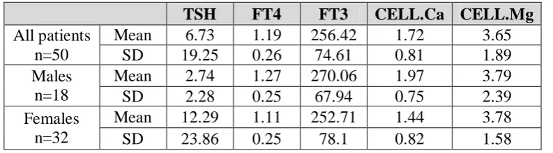
Table I. Mean & SD for all study groups n=50.
Table I shows the Mean & SD for all the patients studied. An observation of this Table gives
a rough idea about the correlation that may be predicted from the difference in mean values
of the metals and hormones.
TSH FT4 FT3 CELL.Ca CELL.Mg
All patients n=50
Mean 6.73 1.19 256.42 1.72 3.65 SD 19.25 0.26 74.61 0.81 1.89 Males
n=18
Mean 2.74 1.27 270.06 1.97 3.79 SD 2.28 0.25 67.94 0.75 2.39 Females
n=32
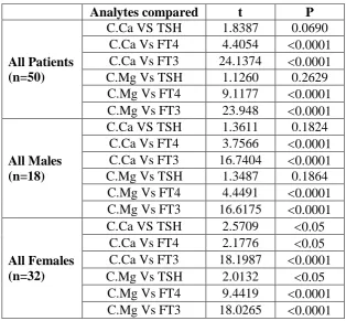
www.wjpls.org 113 Table II. Statistical Parameters (t & P): for the study group.
Ca = Cell Calcium, C.Mg = Cell Magnesium
Table II presents the statistical parameters viz t and p between individual cell content of the
metals and the hormones. In the case of all patients a highly significant correlations were
observed between cell content of Ca & Mg to both FT4and FT3, and only a moderate
significance was see between cell Ca and TSH.
In Males, both C.Ca & C.Mg shows highly significant correlations to both FT4 & FT3, while
TSH does not show any correlation. In Females, C.Ca shows a moderate association to both
TSH and FT4and a highly significant association to FT3. C.Mg shows a moderate association
to TSH, but highly significant association to both FT4 & FT3.
DISCUSSION
As already stated in the abstract, studies linking red cell macrometals to thyroid hormones are
scares. Only few studies have been done. A study done in 1989 has found an association of
red cell Mg in patients with hyperthyroid induced by thyroxin supplements and our study has
linked both red cell Ca & Mg to the three principle thyroid hormones TSH, FT4 & FT3.[5] Many studies carried out previously have linked only plasma levels and predicted low values
of these two metals in hyperthyroidism but no association was found between plasma levels
Analytes compared t P
All Patients (n=50)
C.Ca VS TSH 1.8387 0.0690 C.Ca Vs FT4 4.4054 0.0001 C.Ca Vs FT3 24.1374 0.0001 C.Mg Vs TSH 1.1260 0.2629
C.Mg Vs FT4 9.1177 0.0001 C.Mg Vs FT3 23.948 0.0001
All Males (n=18)
C.Ca VS TSH 1.3611 0.1824 C.Ca Vs FT4 3.7566 0.0001 C.Ca Vs FT3 16.7404 0.0001 C.Mg Vs TSH 1.3487 0.1864
C.Mg Vs FT4 4.4491 0.0001 C.Mg Vs FT3 16.6175 0.0001
All Females (n=32)
C.Ca VS TSH 2.5709 0.05 C.Ca Vs FT4 2.1776 0.05 C.Ca Vs FT3 18.1987 0.0001 C.Mg Vs TSH 2.0132 0.05
www.wjpls.org 114
of Ca & Mg to thyroid hormones.[6,7] Our study has established that red cell content of both Ca & Mg may be very useful compared to plasma level to evaluate the role of these two
metals to study the functions of thyroid gland and its hormones.
CONCLUSION
The outcome of this study strongly suggests that both the macro metals Ca & Mg may play a
significant role in the functioning of thyroid gland and that there exists an inverse correlation
between the thyroid hormones TSH, FT4 & FT3 to both cell Ca & Mg. Further studies with a
large number of population are to be done to recommend the measurement of red cell Ca &
Mg for augmenting the diagnosis of thyroid disorders and to suggests their supplementation.
Further, routine measurement of these two macro metals in erythrocyte may be included
along with thyroid profile tests.
CONFLICT OF INTEREST: None
ACKNOWLEDGEMENT
The authors would like to thank Dr. Mitra Ghosh, Chief of Lab Services at Apollo Speciality
Hospital, Ayanambakkam, Chennai, Tamil Nadu for giving up permission to undertake this
study.
REFERENCE
1. Don’t take Calcium and thyroid mestogether. JAMA, 2000; 21(283): 2822-2825.
2. Dinesh Kumar Dhanwal. Thyroid disorders and bone mineral metabolism. Indian J
Endocrinol Metab, 2011; 15(2): S107–S112.
3. Marjory P. Dube, Faith B. Davis, Paul J. Davis, Marion Schoenl, and Susan D. Blas.
Effects of Hyperthyroidism and Hypothyroidism on Human Red Blood Cell Ca2+-ATPase Activity. The Journal of Clinical Endocrinology & Metabolism, February 1986; 62(2):
253.
4. D Kim, T W Smith, and J D Marsh. Effect of thyroid hormone on slow calcium channel
function in cultured chick ventricular cells. J Clin Invest, 1987; 80(1): 88–94.
5. Gönül Simsek, Gülnur Andican, Ethem Ünal, Hüsrev Hatemi, Günnur Yigit, Gülden
Candan. Calcium, magnesium, and zinc status in experimental hyperthyroidism.
www.wjpls.org 115
6. Dolev E, Deuster PA, Solomon B, Trostmann UH, Wartofsky L, Burman KD.
Alterations in magnesium and zinc metabolism in thyroid disease. Metabolism, 1988;
37(1): 61-7.
7. Mahoney CP, Alster FA, Carew LB Jr. Growth, thyroid function, and serum
macromineral levels in magnesium-deficient chicks. Poult Sci., 1992; 71(10): 1669-79.
8. Derakhshan R, Balaee P, Darakhshan S, Masoodpoor N, Banihosseini SS. Lipid
profile, thyroid function, and serum magnesium level in type I diabetic children. Minerva
Pediatr, 2011; 63(1): 27-33.
9. Shibutani Y, Yokota T, Iijima S, Fujioka A, Katsuno S, Sakamoto K.Plasma and
erythrocyte magnesium concentrations in thyroid disease: relation to thyroid function and
the duration of illness. Jpn J Med., 1989; 28(4): 496-502.
10.Cinar V.The effects of magnesium supplementation on thyroid hormones of sedentars and
Tae-Kwon-Do sportsperson at resting and exhaustion. Neuro Endocrinol Lett., 2007;
28(5): 708-12.
11.Cornelia V. Gilroy, Barbara S. Horney, Shelley A. Burton, and Allan L. MacKenzie.
Evaluation of ionized and total serum magnesium concentrations in hyperthyroid cats.
Can J Vet Res. 2006; 70(2): 137–142.
12.Bijlsma JW, Duursma SA, Roelofs JM, der Kinderen PJ.Thyroid function and bone
turnover. Acta Endocrinol (Copenh), 1983; 104(1): 42-9.
13. S. Swaminathan1, G. Priya and Mitra Ghosh. Association Between Thyroid Hormones
And Macro Metals Calcium And Magnesium. JPBMS, 2010; 4: 17.
14.Mecafffrey C, Quamme,G A : Effects of thyroid status on renal Calcium and Magnesium