R E S E A R C H A R T I C L E
Open Access
Loss of Dgcr8-mediated microRNA expression in
the kidney results in hydronephrosis and renal
malformation
Malte P Bartram
1, Claudia Dafinger
1, Sandra Habbig
1,2, Thomas Benzing
1,3,4, Bernhard Schermer
1,3,4and Roman-Ulrich Müller
1,3,4*Abstract
Background:Small non-coding RNA molecules (miRNAs) play a pivotal role in regulating gene expression in development. miRNAs regulate key processes at the cellular level and thereby influence organismal and tissue development including kidney morphogenesis. A miRNA molecule is initially synthesized as a longer hairneedle-shaped RNA transcript and then processed through an enzymatic complex that contains the RNA-processing enzyme Drosha and its essential interactor Dgcr8. Resulting pre-miRNAs are then cleaved by Dicer. Recent data showed that loss ofDicerresulted in severe developmental kidney phenotypes. However, as Dicer has multiple miRNA-independent functions, it was not entirely clear whether the observed renal phenotypes could be exclusively attributed to a lack of miRNA expression.
Methods:We analyzed the role of miRNAs in kidney development by conditional gene deletion ofDgcr8in the developing kidney using a transgenic mouse line that expresses Cre recombinase in the distal nephron and derivatives of the ureteric bud in kidney development.
Results:Animals with a gene deletion ofDgcr8in these tissues developed severe hydronephrosis, kidney cysts, progressive renal failure and premature death within the first two months after birth, a phenotype strongly resembling
Dicerdeletion.
Conclusions:Here we show that conditional gene deletion of the essential miRNA-processing enzymeDgcr8in the developing renal tubular system results in severe developmental defects and kidney failure. These data confirm earlier findings obtained inDicerknock-out animals and clearly illustrate the essential role of miRNAs in kidney development. The data suggests that miRNA dysregulation may play an important, yet ill-defined role in the pathogenesis of inborn defects of the genitourinary system and indicate that miRNA defects may be causative in the development of human disease.
Keywords:CAKUT, Dgcr8, Dicer, Hydronephrosis, Kidney, miRNA
Background
MicroRNAs are important regulators of gene expression and have been shown to be crucial to developmental processes in many different tissues [1]. To study the role of miRNAs in the kidney several publications have ad-dressed this question using a conditional knockout of
Dicer, the RNAse III enzyme catalyzing the maturation from pre-miRNA to mature microRNAs [2-9]. This strategy revealed major defects in both tubular and glomerular development and maintenance. The loss of Dicer in derivatives of the ureteric bud and in the tubu-lar system lead to a severe hydronephrosis coupled with cystic kidneys and loss of functional parenchyme [2-4,6]. These phenotypes strongly resemble congenital anomal-ies of the kidney and urinary tract (CAKUT) in the clin-ical setting. However, whether this is truly due to loss of microRNAs has remained elusive since Dicer fulfills * Correspondence:roman-ulrich.mueller@uk-koeln.de
1
Department II of Internal Medicine and Center for Molecular Medicine, University of Cologne, Kerpener Str. 62, Cologne 50937, Germany 3
Cologne Excellence Cluster on Cellular Stress Responses in Aging-Associated Diseases, University of Cologne, Cologne, Germany
Full list of author information is available at the end of the article
several other important functions which may well be in-volved in renal development [10]. Among these are its role as a DNAse in genomic DNA fragmentation during apoptosis, the processing of endogenous siRNA and the detoxification of repeat elements [11-13]. In an elegant study elucidating the role of microRNAs in skin devel-opment this issue has been addressed using the condi-tional knockout of genes involved in different steps of miRNA processing displaying a phenotypic overlap [14,15]. As to the kidney this has been done successfully regarding the effects of podocyte-specific loss of micro-RNAs using a conditional knockout allele of Drosha [16], which confirmed previous studies based on Dicer knockout in podocytes [7-9].
Consequently, we set out to confirm the role of micro-RNAs in renal development using a conditional allele of
Dgcr8. Dgcr8 interacts with Drosha and is essential for its role in processing pri-microRNAs in the nucleus [17].
Methods Mice
Dgcr8 fl/flanimals were described before [18] and gener-ously provided by Elaine Fuchs (Rockefeller University, NYC, USA). To generate a kidney tubulus specificDgcr8 knockout these mice were crossed to aKspCretransgenic line (contributed by Peter Igarashi, UT Southwestern Medical Center, Dallas, USA) that expresses the Cre re-combinase under the control of a ksp-cadherin promotor resulting in Cre expression in the developing genitouri-nary tract and kidney tubulus system was performed as described before [19]. Animals were housed in standard-ized specific pathogen-free conditions in the animal facil-ity of the CMMC (Universfacil-ity of Cologne).
All animal procedures were performed according to European (EU directive 86/609/EEC), national (TierSchG), and institutional guidelines and were approved by local governmental authorities (LANUV NRW).
Histology
The kidneys were fixed in formalin, embedded in paraffin and stained with PAS according to standard protocols. To analyse the expression of Ki-67, slides of fixed and paraffin-embedded mouse kidneys were de-paraffinized using Xylol and descending concentrations of ethanol. Antigen retrieval was carried out by warming kidney slides in citrate buffer (10 mM, pH6) for 10 min using a micro-wave. After blocking with 3% H2O2and Avidin and Biotin (Vector Laboratories, Inc.) for 15 min each, slides were se-quentially incubated with the Ki-67 antibody (rabbit Ki-67 ab16667, abcam, 1:500 dilution, over night at 4°C) and after washing with PBS with biotinylated anti-rabbit IgG (Jackson ImmunoResearch, West Grove, PA, USA; 1 h at room temperature). Kidney slides were labelled with ABC kit (Vector Laboratories, Inc.), and development was
carried out using diaminobenzidine solution (Sigma Aldrich). Slides were counterstained with hematoxylin (Sigma-Aldrich), dehydrated and afterwards mounted with Histomount (National Diagnostics). Stained slides were scanned using a Slidescanner (Leica) and analyzed using the ImageScope software (version 12.0.1.5030, Aperio).
Laboratory medicine
Heparinized blood was obtained by cardiac puncture. Plasma was prepared by centrifugation at 3000 rpm for 10 min. Urea was measured in the central laboratory medicine unit of the University Hospital of Cologne using the kinetic UV test (Roche Diagnostics). Signifi-cance was calculated using a two-tailed Student’s t test for all measurements (urea, body weight of mice).
qPCR
RNA was extracted from whole mouse kidneys using acid guanidinium thiocyanate-phenol-chloroform extraction [20]. RT reactions were performed using the Taqman microRNA Reverse Transcription Kit (ABI). Expression of mir-192 (assay ID 000491), and miR-200b (assay ID 4426961) was analyzed using Taqman assays (ABI), and snoRNA135 (assay ID 001230) served as endogenous con-trol. All qPCR experiments were performed on the ABI 7900HT System. All data points were generated using the number of biological replicates indicated in the figure. Data analysis and statistics were performed using the Ex-pression Suite v1.3 software package applying the com-parative Ct method and using one standard deviation for error bar calculation (LifeTechnologies).
TUNEL assay
To analyse apoptosis in the kidneys of Dgcr8 knockout and littermate control mice we utilized the Promega DeadEnd Fluorometric Kit according to the manufac-turers protocol. Pictures were taken with an inverted microscope (Axiovert200, equipped with an ApoTome system and an AxioCam MRm camera. Objective used: Plan Apochromat 20×/0.8 NA. Carl Zeiss) using Axiovi-sion 4.8 (Carl Zeiss).
Results
To analyze the role of Dgcr8 and thereby miRNAs in the developing tubular system independent of a Dicer mouse model, we crossed a Dgcr8 fl/fl mouse line [18] with
KspCremice [19], resulting in a conditional knockout of
Dgcr8 in the developing urogenital tract and tubulus system.
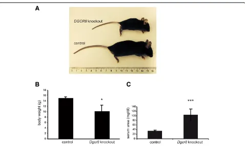
Dgcr8 fl/fl; KspCre positive mice (afterwards named
Dgcr8 knockout) showed an obvious delay in growth (Figure 1A) in comparison to the control littermates and a significantly reduced body weight (Figure 1B). This
phenotype was most likely caused by the developing renal failure in these mice, since they showed a marked elevation in serum urea (Figure 1C). The phenotype be-came apparent in the first weeks of life, several mice died during the weaning period leading to a significantly reduced number of Dgcr8knockout animals after wean-ing in comparison to the control genotypes (Additional file 1: Figure S1A). None of the Dgcr8 knockout mice analyzed so far survived longer than 8 weeks, most likely due to development of end stage renal disease. In order to confirm that loss ofDgcr8 abrogates miRNA biogen-esis we quantified two miRNAs that had been shown be-fore to be primarily expressed in renal tubular cells and to be depleted by KspCre driven loss of Dicer [3,6,21]. Both expression of miR-192 and miR-200b were greatly reduced in the conditional Dgcr8 knockout mouse line (Figure 2).
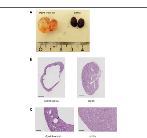
Further examination of the kidneys of the Dgcr8 knockout mice showed the macroscopic picture of se-vere hydronephrosis with a dilated ureter and kidney pelvis (Figure 3A). There were no signs for a complete obstruction of the ureter, since the bladder of the knock-out animals was filled with urine (data not shown).
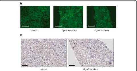
Histopathological analyses of the kidneys confirmed the diagnosis of hydronephrosis and obstructive nephropathy. In most affected animals nearly the entire medulla was missing, and the cortex was very thin (Figure 3B). In addition, some kidneys displayed dilated tubuli and cysts were observed (Figure 3C). As described forDicer knock-out mice before [3] theDgcr8knockout kidneys showed a reduced glomerular density pointing towards a branching defect (Additional file 1: Figure 1B). To further analyse the cellular basis to this phenotype we performed TUNEL as-says and Ki-67 stainings. These revealed a strong induc-tion of apoptosis and cellular proliferainduc-tion in tubular cells ofDgcr8knockout animals (Figure 4).
Discussion
Figure 2KspCre-mediated loss ofDgcr8induces depletion of tubulus-specific miRNAs. Both expression of miR-192 and miR-200b is strongly reduced inDgcr8knockout kidneys when compared to WT littermates (4-7 week old animals; ** = p < 0.01; error bars represent SEM).
Figure 3Hydronephrosis and cystic kidneys ofDgcr8knockout animals.AConditional knockout ofDgcr8in the renal tubulus system leads to hydronephrosis.B + CHistological analysis confirms hydronephrosis with severe loss of kidney parenchyma especially in the medulla region, a thinned cortex and kidney cysts (bar = 1000μm (B) and 200μm (C)).
phenotype in both mouse models including hydronephro-sis and renal failure is caused by the loss of microRNA processing. This finding is of great importance and en-couraging to plan and perform follow-up studies now ad-dressing the role of specific microRNAs in development and maintenance of renal architecture.
Interestingly, a small number of microRNAs is either independent from Dicer or from the Drosha/Dgcr8 com-plex [24,25]. As an example the so-called mirtrons are processed by the spliceosome in the nucleus instead of Drosha/Dgcr8 [26]. As for Dicer miR-451 is not proc-essed by this enzyme but depends on Ago2 in its matur-ation [27-29]. Consequently, our study does not only confirm the crucial role of microRNAs in renal develop-ment but also narrows down the list by excluding any small RNAs processed by only one of the two enzymes.
In contrast to our previous study on Dicer knockout animals with a penetrance of the phenotype of about 66% [3] the Dgcr8 knockout described in this study has a complete penetrance with no Dgcr8 knockout animal surviving longer than 8 weeks. Whether this may be due to a partial rescue of theDicerknockout animals by an-other enzyme – e.g. for microRNAs that are generally processed by Dicer but may be processed by Ago2 as well – remains elusive and will be subject to future studies.
In summary, our results underline the relevance of microRNAs during kidney development and will encour-age further functional studies examining single microRNAs
and their target mRNA interactions–such as miR-20 and its targets PKD1 and PKD2 [3,6,30,31] - as regulators of renal organogenesis. This will be fundamental for gaining a better understanding of developmental defects in human kidney formation as observed in CAKUT–the predomin-ant cause of end-stage renal disease in children.
Conclusions
MiRNAs are key regulators of intracellular signaling and development. In this study we show that loss of Dgcr8 dependent miRNAs in the kidney epithelium leads to se-vere hydronephrosis, kidney cysts and rapid kidney fail-ure. This confirms an essential role for miRNAs in renal development and disease.
Additional file
Additional file 1: Figure S1.A Genotyping after weaning at 3–4 weeks of age reveals that the knockout mice did not reach weaning at a Mendelian ratio suggesting death before the timepoint of weaning and genotyping. In line with this finding several mice of unknown genotype had died before weaning. B Glomerular density is reduced inDgcr8 knockout kidneys (n = 3 per group; error bars represent SEM; ** = p <0.01 using an unpaired Student’st-test; 5 high power fields of the kidney cortex were counted per animal) C Table showing the number of mice revealing either kidney cysts or hydronephrosis at weaning.
Abbreviations
CAKUT:Congenital anomalies of the kidney and urinary tract;
Dgcr8: DiGeorge syndrome critical region 8; PKD: Polycystic kidney disease.
Competing interests
The authors declare that they have no competing interests. The results presented in this paper have not been published previously in whole or part.
Authors’contributions
MPB and RUM were involved in all experimental steps and drafting of the manuscript. CD and SH were involved in histological analysis and image aquisition. TB and BS coordinated the study and drafted the manuscript. All authors read and approved the final manuscript.
Acknowledgments
This work was funded by the Deutsche Nierenstiftung and the Deutsche Forschungsgemeinschaft MU3629/2-1 to R.-U.M., SFB829 to T.B. and SCHE1562-2 to B.S. We thank Elaine Fuchs and Peter Igarashi for providing theDgcr8 fl/fland
KspCremouse lines, respectively and members of the laboratory for helpful discussion. We would like to thank Sonja Kunath and Nadine Urban for their support with the animal experiments and Martyna Brütting for excellent technical help. We thank the CECAD Imaging Facility for their technical support with the Slidescanner.
Author details 1
Department II of Internal Medicine and Center for Molecular Medicine, University of Cologne, Kerpener Str. 62, Cologne 50937, Germany. 2
Department of Pediatrics, University of Cologne, Cologne, Germany. 3Cologne Excellence Cluster on Cellular Stress Responses in Aging-Associated
Diseases, University of Cologne, Cologne, Germany.4Systems Biology of Ageing Cologne, University of Cologne, Cologne, Germany.
Received: 21 October 2014 Accepted: 7 April 2015
References
1. Tüfekci KU, Meuwissen RLJ, Genç S. The role of microRNAs in biological processes. Methods Mol Biol Clifton NJ. 2014;1107:15–31.
2. Pastorelli L, Wells S, Fray M, Smith A, Hough T, Harfe B, et al. Genetic analyses reveal a requirement for Dicer1 in the mouse urogenital tract. Mamm Genome. 2009;20:140–51.
3. Bartram MP, Höhne M, Dafinger C, Völker LA, Albersmeyer M, Heiss J, et al. Conditional loss of kidney microRNAs results in congenital anomalies of the kidney and urinary tract (CAKUT). J Mol Med Berl Ger. 2013;91:739–48. 4. Nagalakshmi VK, Ren Q, Pugh MM, Valerius MT, McMahon AP, Yu J. Dicer
regulates the development of nephrogenic and ureteric compartments in the mammalian kidney. Kidney Int. 2011;79:317–30.
5. Chu JYS, Sims-Lucas S, Bushnell DS, Bodnar AJ, Kreidberg JA, Ho J. Dicer function is required in the metanephric mesenchyme for early kidney development. Am J Physiol Renal Physiol. 2014;306:F764–72.
6. Patel V, Hajarnis S, Williams D, Hunter R, Huynh D, Igarashi P. MicroRNAs regulate renal tubule maturation through modulation of Pkd1. J Am Soc Nephrol JASN. 2012;23:1941–8.
7. Harvey SJ, Jarad G, Cunningham J, Goldberg S, Schermer B, Harfe BD, et al. Podocyte-specific deletion of dicer alters cytoskeletal dynamics and causes glomerular disease. J Am Soc Nephrol JASN. 2008;19:2150–8.
8. Ho J, Ng KH, Rosen S, Dostal A, Gregory RI, Kreidberg JA. Podocyte-specific loss of functional micrornas leads to rapid glomerular and tubular injury. J Am Soc Nephrol JASN. 2008;19:2069–75.
9. Shi S, Yu L, Chiu C, Sun Y, Chen J, Khitrov G, et al. Podocyte-selective deletion of dicer induces proteinuria and glomerulosclerosis. J Am Soc Nephrol JASN. 2008;19:2159–69.
10. Johanson TM, Lew AM, Chong MMW. MicroRNA-independent roles of the RNase III enzymes drosha and dicer. Open Biol. 2013;3:130144.
11. Calabrese JM, Seila AC, Yeo GW, Sharp PA. RNA sequence analysis defines Dicer’s role in mouse embryonic stem cells. Proc Natl Acad Sci U S A. 2007;104:18097–102.
12. Tam OH, Aravin AA, Stein P, Girard A, Murchison EP, Cheloufi S, et al. Pseudogene-derived small interfering RNAs regulate gene expression in mouse oocytes. Nature. 2008;453:534–8.
13. Babiarz JE, Ruby JG, Wang Y, Bartel DP, Blelloch R. Mouse ES cells express endogenous shRNAs, siRNAs, and other Microprocessor-independent, Dicer-dependent small RNAs. Genes Dev. 2008;22:2773–85.
14. Yi R, Pasolli HA, Landthaler M, Hafner M, Ojo T, Sheridan R, et al. DGCR8-dependent microRNA biogenesis is essential for skin development. Proc Natl Acad Sci U S A. 2009;106:498–502.
15. Teta M, Choi YS, Okegbe T, Wong G, Tam OH, Chong MMW, et al. Inducible deletion of epidermal Dicer and Drosha reveals multiple functions for miRNAs in postnatal skin. Dev Camb Engl. 2012;139:1405–16. 16. Zhdanova O, Srivastava S, Di L, Li Z, Tchelebi L, Dworkin S, et al. The
inducible deletion of Drosha and microRNAs in mature podocytes results in a collapsing glomerulopathy. Kidney Int. 2011;80:719–30.
17. Landthaler M, Yalcin A, Tuschl T. The human DiGeorge syndrome critical region gene 8 and Its D. melanogaster homolog are required for miRNA biogenesis. Curr Biol CB. 2004;14:2162–7.
18. Wang Y, Medvid R, Melton C, Jaenisch R, Blelloch R. DGCR8 is essential for microRNA biogenesis and silencing of embryonic stem cell self-renewal. Nat Genet. 2007;39:380–5.
19. Shao X, Somlo S, Igarashi P. Epithelial-specific Cre/lox recombination in the developing kidney and genitourinary tract. J Am Soc Nephrol JASN. 2002;13:1837–46.
20. Chomczynski P, Sacchi N. Single-step method of RNA isolation by acid guanidinium thiocyanate-phenol-chloroform extraction. Anal Biochem. 1987;162:156–9.
21. Jenkins RH, Martin J, Phillips AO, Bowen T, Fraser DJ. Pleiotropy of microRNA-192 in the kidney. Biochem Soc Trans. 2012;40:762–7. 22. Luhur A, Chawla G, Wu Y-C, Li J, Sokol NS. Drosha-independent DGCR8/
Pasha pathway regulates neuronal morphogenesis. Proc Natl Acad Sci U S A. 2014;111:1421–6.
23. Macias S, Plass M, Stajuda A, Michlewski G, Eyras E, Cáceres JF. DGCR8 HITS-CLIP reveals novel functions for the microprocessor. Nat Struct Mol Biol. 2012;19:760–6.
24. Xie M, Steitz JA. Versatile microRNA biogenesis in animals and their viruses. RNA Biol. 2014;11(6):673–81.
25. Chong MMW, Zhang G, Cheloufi S, Neubert TA, Hannon GJ, Littman DR. Canonical and alternate functions of the microRNA biogenesis machinery. Genes Dev. 2010;24:1951–60.
26. Berezikov E, Chung W-J, Willis J, Cuppen E, Lai EC. Mammalian mirtron genes. Mol Cell. 2007;28:328–36.
27. Dueck A, Meister G. MicroRNA processing without Dicer. Genome Biol. 2010;11:123.
28. Cheloufi S, Dos Santos CO, Chong MMW, Hannon GJ. A dicer-independent miRNA biogenesis pathway that requires Ago catalysis. Nature.
2010;465:584–9.
29. Cifuentes D, Xue H, Taylor DW, Patnode H, Mishima Y, Cheloufi S, et al. A novel miRNA processing pathway independent of Dicer requires Argonaute2 catalytic activity. Science. 2010;328:1694–8.
30. Patel V, Williams D, Hajarnis S, Hunter R, Pontoglio M, Somlo S, et al. miR-17∼92 miRNA cluster promotes kidney cyst growth in polycystic kidney disease. Proc Natl Acad Sci. 2013;110(26):10765–70.
31. Marrone AK, Stolz DB, Bastacky SI, Kostka D, Bodnar AJ, Ho J. MicroRNA-17 ~ 92 is required for nephrogenesis and renal function. J Am Soc Nephrol JASN. 2014;25:1440–52.
Submit your next manuscript to BioMed Central and take full advantage of:
• Convenient online submission
• Thorough peer review
• No space constraints or color figure charges
• Immediate publication on acceptance
• Inclusion in PubMed, CAS, Scopus and Google Scholar
• Research which is freely available for redistribution
Submit your manuscript at www.biomedcentral.com/submit