ARTICLE OPEN ACCESS
Chorea-acanthocytosis
Homozygous 1-kb deletion in
VPS13A
detected by whole-genome sequencing
Susan Walker, PhD, Rubina Dad, MPhil, Bhooma Thiruvahindrapuram, MSc, Muhammed Ikram Ullah, PhD, Arsalan Ahmad, MD, Muhammad Jawad Hassan, PhD, Stephen W. Scherer, PhD, and Berge A. Minassian, MD
Neurol Genet2018;4:e242. doi:10.1212/NXG.0000000000000242
Correspondence
Dr. Minassian Berge.Minassian@ UTSouthwestern.edu
Abstract
Objective
To determine a molecular diagnosis for a large multigenerational family of South Asian ancestry with seizures, hyperactivity, and episodes of tongue biting.
Methods
Two affected individuals from the family were analyzed by whole-genome sequencing on the Illumina HiSeq X platform, and rare variants were prioritized for interpretation with respect to the phenotype.
Results
A previously undescribed, 1-kb homozygous deletion was identified in both individuals se-quenced, which spanned 2 exons of theVPS13Agene, and was found to segregate in other family members.
Conclusions
VPS13Ais associated with autosomal recessive chorea-acanthocytosis, a diagnosis consistent with the phenotype observed in this family. Whole-genome sequencing presents a compre-hensive and agnostic approach for detecting diagnostic mutations in families with rare neu-rologic disorders.
From the Centre for Applied Genomics (S.W., B.T., S.W.S.), The Hospital for Sick Children; Program in Genetics and Genome Biology (S.W., R.D., B.T., S.W.S., B.A.M.), The Hospital for Sick Children, Toronto, Ontario, Canada; Atta-ur Rahman School of Applied Biosciences (R.D., M.J.H.), National University of Sciences and Technology (NUST), Islamabad; Department of Biochemistry (I.M.U.), University of Health Sciences, Lahore; Division of Neurology (A.A.), Shifa International Hospital, Shifa Tameer e Millat University, Islamabad, Pakistan; Department of Molecular Genetics (S.W.S.), University of Toronto; McLaughlin Centre (S.W.S.), University of Toronto, Canada; and Department of Pediatrics (B.A.M.), University of Texas Southwestern, Dallas.
Funding information and disclosures are provided at the end of the article. Full disclosure form information provided by the authors is available with the full text of this article at Neurology.org/NG.
The Article Processing Charge was funded by the authors.
Chorea-acanthocytosis (CHAC; MIM number 200150) is an autosomal recessive neurodegenerative disorder characterized by chorea and blood cells with abnormal morphology (acanthocytosis). Additional common features include dys-tonia, seizures, tics, and uncontrollable tongue biting. CHAC is caused by homozygous or compound heterozygous muta-tions inVPS13A,1which encodes chorein, a protein of un-known function. Here, we report a family with previously unexplained epilepsy found by whole-genome sequencing (WGS) to carry a novel mutation in VPS13A, leading to a diagnosis of CHAC.
Clinical report
A large multigeneration Pakistani family was recruited from the province of Khyber Pakhtunkhwa, Pakistan, presenting with a history of seizures, episodes of hyperactive and psy-chiatric problems and headache, nausea, sleep problems, and persistent tics with onset beginning late in the third decade of life. Within the family, there are 4 affected individuals across 3 generations and an additional younger participant suspected to be in the early stages of developing the same disorder.
Clinical details of index case IV:16
The index patient, a 37-year-old woman, first visited the neurologist at Shifa International Hospital, Islamabad, pre-senting with hyperactive behavior beginning 1 year prior, with episodes of tongue biting commencing 6 months later and 3 generalized tonic-clonic seizures during the month before the consultation. She had been diagnosed with epilepsy and mi-graine 3 years earlier but since developed hyperactivity and difficulty in concentrating in daily activities. She also suffered frequent tongue bites while speaking. On examination, she was found to have bilateral ptosis, but other cranial nerves were normal. Tone and power in all 4 limbs were normal, as were plantar responses. Higher mental function and speech were also typical. She was diagnosed as having a congenital syndrome with epilepsy, Tourette syndrome, and bilateral ptosis and prescribed escitalopram, procyclidine, carbamaze-pine, and later, haloperidol. One genetic test was sent for, namely Huntington disease, for which CAG repeats at theHD
locus were normal (27 repeats). Red blood cell morphology was studied and reported normal.
Her parents were reportedly nonrelated, and the symptoms were also present in 3 other family members: participants III: 1, IV:13, and V:8 in thefigure. Individual V:6 was also sus-pected of being in the early stages of developing the same disorder. The family history and clinical features of affected individuals are described in table 1. Six additional family members (individuals III:16, III:21, V:2, V:4, V:9, and V:13
[figure]) were suspected to have a clinically separate neuro-logic disorder with symptoms including intellectual disability, seizures, migraine, depression, and osteoarthritis.
Methods
DNA from 22 family members, including the 5 affected individuals, was extracted from whole blood using a standard phenol-chloroform method. Participants III-1 and IV-16 (index case) were selected for analysis by WGS. WGS was performed at The Centre for Applied Genomics (Toronto, Canada) with total genomic DNA following standard proto-cols. Library preparation was performed from 100 ng of DNA using the Illumina TruSeq Nano DNA Library Prep Kit fol-lowing the manufacturer’s recommended protocol. Libraries were pooled in equimolar quantities and sequenced on an Illumina HiSeq X platform following Illumina’s recom-mended protocol to generate paired-end reads of 150 bases in length. Base calling and data analysis were performed using Illumina HiSeq Analysis Software version 2-2.5.55.1311. Reads were mapped to the hg19 reference sequence using Isaac alignment software (Isaac alignment software: SAAC00776.15.01.27), and SNV and small indel variants were called using the Isaac variant caller (Isaac Variant Caller [Starling]: 2.1.4.2). Rare variants were defined as those with less than 1% frequency in population databases.2–4 CNVs were detected using the read depth methods as previously described.5Rare CNVs were defined as those less than 1% frequency among unrelated, unaffected individuals sequenced using the same technology.6Participants were screened for regions of homozygosity using PLINK (v1.90) “Runs of homozygosity”implementation.
Standard protocol approvals, registrations and patient consents
Informed consent was obtained from all participants, and this study was approved by The Hospital for Sick Children Research Ethics Board, Toronto, Canada.
Results
Through analysis of rare CNVs detected from WGS data, a 1168-bp deletion was detected, spanning exons 8 and 9 of VPS13A (chr9:79827422-79828590); c.556_696del; p.(Thr186_Leu232del). The deletion was homozygous in both individuals sequenced and would likely result in an in-frame deletion of 47 amino acids. PCR amplification and Sanger sequencing confirmed the deletion and were used to test the pattern of segregation among all family members, where DNA was available (figure). Homozygosity mapping
Glossary
from the WGS data for participants III-1 and IV-16 showed that the deletion resides in an extended region of homozy-gosity in both individuals (chr9:78,311,259-98,376,367 in III-1 and chr9:79,45III-1,734-97,0III-16,027 in IV-III-16). Individuals IV: 13, V:8, and V:6 were found to be homozygous for the de-letion. Family relationships were confirmed by the analysis of short tandem repeats. Analysis of single nucleotide variants and small insertion/deletions did not yield any pathogenic or likely pathogenic variants (all homozygous variants in coding regions identified in either individual are shown in table e-1, links.lww.com/NXG/A50). On peripheral blood smear, acanthocytes were detected in 3 of the 4 individuals with the primary condition (III:1, IV:13, and V:8) and in V:6, but not in the index case IV:16.
Discussion
The phenotype of the affected individuals is in keeping with the known CHAC phenotype. All 4 experienced choreoathetosis, seizures, and cognitive/psychiatric symptoms. Four members of the family were found to have acanthocytes on peripheral blood smear, supporting the diagnosis of CHAC. The molec-ular findings and hematology testing also confirmed the di-agnosis of the youngest individual, V:6, presenting with the early stages of the condition. None of the 6 individuals with the second phenotype was homozygous for the deletion in VPS13A (3 did not carry the variant, and 3 were heterozygous), confirming that they likely have a second, distinct disorder.
There are 4 neuroacanthocytosis syndromes (CHAC, McLeod syndrome, Huntington disease-like 2, and pantothenate
kinase–associated neurodegeneration) with similar but distinct phenotypic features, and the clinical presentation of CHAC also overlaps with other disorders, presenting a diagnostic challenge.7,8The presence of acanthocytes can be helpful to suggest a diagnosis of CHAC, but hematology results are inconsistent,7,8 as is the case in this family. With WGS, a precise diagnosis could be rapidly achieved without the requirement of any prior awareness of the disorder by the referring clinician. Achieving a correct diagnosis now allows accurate anticipation of the course of the disorder (particu-larly in the case of the youngest, mildly affected individual) and facilitates appropriate treatment.9 The diagnosis also prevents further unnecessary testing for possible nutritional or infectious etiologies and helps to remove any stigma as-sociated with behaviors in the family.
The nature of the mutation in this family further demonstrates the benefit of using WGS compared with other nontargeted technologies. Such a small deletion would be too small to detect by microarray analysis and challenging to identify using exome sequencing. It may also not have been detected using a targeted gene or gene panel test, depending on the specific methodologies used. Small CNVs affectingVPS13A, such as the one identified in this family, have been found previously in cases with CHAC, although the deletion we describe had not been reported to date.10
Here, we present a large multigenerational family from Pakistan with chorea-acanthocytosis attributable to a 1168-bp, 2-exon deletion inVPS13A. This family showcases the potential for WGS as afirst-tier diagnostic test in neurologic disorders.
FigurePedigree of the family
Author contributions
S. Walker: acquisition of data and analysis and in-terpretation of data. R. Dad and B. Thiruvahindrapuram: analysis and interpretation of data. I.M. Ullah: sample col-lection. A. Ahmad: acquisition of data and analysis and interpretation of data. M.J. Hassan, S.W. Scherer, and B.A. Minassian: critical revision of the manuscript and study supervision.
Acknowledgment
The authors thank the family for their participation in the study and The Centre for Applied Genomics for their analytical and technical support. This work was supported by The Centre for Applied Genomics, the University of Toronto McLaughlin Centre, Genome Canada/Ontario Genomics Institute, the Canadian Institutes of Health Research (CIHR), the Canadian Institute for Advanced Research,
and the Canada Foundation for Innovation. R.D. gratefully acknowledges her funding by the Higher Education Commission of Pakistan under the International Research Support Initiative Program (HEC-IRSIP). S.W.S. is funded by the GlaxoSmithKline-CIHR Chair in Genome Sciences at the University of Toronto and The Hospital for Sick Children. B.A.M holds the University of Toronto Michael Bahen Chair in Epilepsy Research and the University of Texas Southwestern Jimmy Elizabeth Westcott Distin-guished Chair in Pediatric Neurology.
Study funding
This work was supported by The Centre for Applied Genomics, the University of Toronto McLaughlin Centre, Genome Canada/Ontario Genomics Institute, the Canadian Institutes of Health Research (CIHR), the Canadian Institute for Advanced Research, and the Canada Foundation for Innovation.
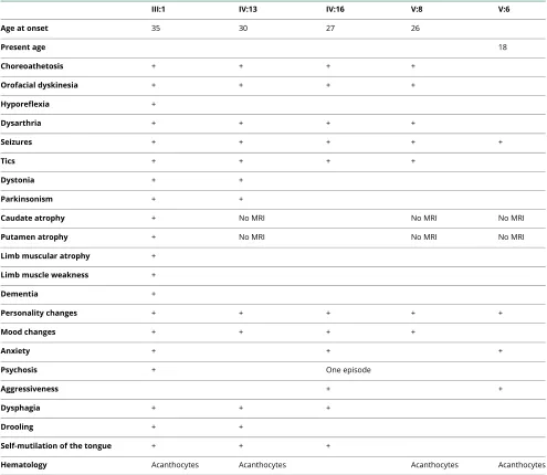
Table 1Clinical features of affected individuals
III:1 IV:13 IV:16 V:8 V:6
Age at onset 35 30 27 26
Present age 18
Choreoathetosis + + + +
Orofacial dyskinesia + + + +
Hyporeflexia +
Dysarthria + + + +
Seizures + + + + +
Tics + + + +
Dystonia + +
Parkinsonism + +
Caudate atrophy + No MRI No MRI No MRI
Putamen atrophy + No MRI No MRI No MRI
Limb muscular atrophy +
Limb muscle weakness +
Dementia +
Personality changes + + + + +
Mood changes + + + +
Anxiety + + +
Psychosis + One episode
Aggressiveness + +
Dysphagia + + +
Drooling + +
Self-mutilation of the tongue + + +
Disclosure
S. Walker, R. Dad, B. Thiruvahindrapuram, M.I. Ullah, A. Ahmad, and M.J. Hassan report no disclosures. S.W. Scherer holds the GlaxoSmithKline-CIHR Chair in Genome Scien-ces at the University of Toronto and The Hospital for Sick Children; is on the scientific advisory board of Deep Genomics; has served on the scientific advisory board of Population Diagnostics; has served on the editorial boards ofGenomic Medicine,Genes,Genomes,Genetics,the Journal of Personalized Medicine,the Open Genomics Journal,the Hugo Journal,Genome Medicine,the Journal of Neurodevelopmental Disorders,Autism Research,PathoGenetics,Comparative and Functional Genomics, BMC Medical Genomics, and Cytoge-netics and Genome Research; and has received research sup-port from the Genome Canada/Ontario Genomics Institute, Canadian Institutes of Health Research, Canadian Institute for Advanced Research, McLaughlin Centre, Canada Foun-dation for Innovation, Government of Ontario, and NIH. B.A. Minassian holds patents for diagnostic testing of the following genes:EPM2A, EPM2B, MECP2, andVMA21; has received research support from the NIH; and has received license fee payments/royalty payments from patents for di-agnostic testing of the following genes: EPM2A, EPM2B,
MECP2, andVMA21. Full disclosure form information pro-vided by the authors is available with the full text of this article at Neurology.org/NG.
Received January 10, 2018. Accepted infinal form April 5, 2018.
References
1. Rampoldi L, Dobson-Stone C, Rubio JP, et al. A conserved sorting-associated protein is mutant in chorea-acanthocytosis. Nat Genet 2001;28:119–120.
2. Lek M, Karczewski KJ, Minikel EV, et al. Analysis of protein-coding genetic variation in 60,706 humans. Nature 2016;536:285–291.
3. 1000 Genomes Project Consortium. A global reference for human genetic variation. Nature 2015;526:68–74.
4. Fu W, O’Connor TD, Jun G, et al. Analysis of 6,515 exomes reveals the recent origin of most human protein-coding variants. Nature 2013;493:216–220.
5. Trost B, Walker S, Wang Z, et al. A comprehensive workflow for read depth-based identification of copy-number variation from whole-genome sequence data. Am J Hum Genet 2018;102:142–155.
6. Yuen RKC, Merico D, Bookman M, et al. Whole genome sequencing resource identifies 18 new candidate genes for autism spectrum disorder. Nat Neurosci 2017; 20:602–611.
7. Walterfang M, Evans A, Looi JCL, et al. The neuropsychiatry of neuroacanthocytosis syndromes. Neurosci Biobehav Rev 2011;35:1275–1283.
8. Walker RH. Untangling the thorns: advances in the neuroacanthocytosis syndromes. J Mov Disord 2015;8:41–54.
9. Benninger F, Afawi Z, Korczyn AD, et al. Seizures as presenting and prominent symptom in choreaacanthocytosis with c.2343del VPS13A gene mutation. Epilepsia 2016;57:549–556.
DOI 10.1212/NXG.0000000000000242
2018;4;
Neurol Genet
Susan Walker, Rubina Dad, Bhooma Thiruvahindrapuram, et al.
whole-genome sequencing
Chorea-acanthocytosis: Homozygous 1-kb deletion in VPS13A detected by
This information is current as of May 18, 2018
Services
Updated Information &
http://ng.neurology.org/content/4/3/e242.full.html including high resolution figures, can be found at:
References
http://ng.neurology.org/content/4/3/e242.full.html##ref-list-1 This article cites 10 articles, 0 of which you can access for free at:
Subspecialty Collections
http://ng.neurology.org//cgi/collection/chorea Chorea
following collection(s):
This article, along with others on similar topics, appears in the
Permissions & Licensing
http://ng.neurology.org/misc/about.xhtml#permissions its entirety can be found online at:
Information about reproducing this article in parts (figures,tables) or in
Reprints
http://ng.neurology.org/misc/addir.xhtml#reprintsus Information about ordering reprints can be found online:
reserved. Online ISSN: 2376-7839.
Published by Wolters Kluwer Health, Inc. on behalf of the American Academy of Neurology.. All rights an open-access, online-only, continuous publication journal. Copyright Copyright © 2018 The Author(s).
is an official journal of the American Academy of Neurology. Published since April 2015, it is