*Corresponding author:Priyanka
Department of Radio-diagnosis, JJM Medical College, Davangere, India ISSN: 0976-3031
Research Article
MRI PATTERNS OF HYPOXIC ISCHEMIC ENCEPHALOPATHY IN TERM INFANTS AND
CORRELATION WITH NEUROSONOGRAPHIC APPEARANCE & CLINICAL OUTCOME
Priyanka*., Jeevika M.U., Joish Upendra Kumar., Pallavi Sinha., Ramya Konareddy.,
Dipti Kumari and Shivanand H.G
Department of Radio-diagnosis, JJM Medical College, Davangere, India
ARTICLE INFO ABSTRACT
MRI is the imaging modality of choice for the diagnosis and follow-up of infants with moderate to severe hypoxic-ischemic encephalopathy. This study was undertaken to evaluate the various MRI patterns of term hypoxic ischemic encephalopathy and to correlate the MRI appearances with clinical outcome. It was also undertaken to compare wherever possible, the neurosonogrpahic appearances with the MRI findings. Differences in the patterns of hypoxic ischemic encephalopathy are likely to result from severity of birth asphyxia. MRI is non-invasive and has no radiation hazards associated with it .The excellent gray–white matter resolution and multiplanar imaging capabilities provide an advantage to MRI over other modalities. The addition of DWI improves the diagnostic capacities of MRI .There is a strong relation between the MRI appearances of birth asphyxia and the clinical outcome. Therefore MRI has a strong role in prognosticating lesions.
INTRODUCTION
Perinatal asphyxia, more appropriately known as hypoxic ischemic encephalopathy (HIE) is characterized by clinical and laboratory evidence of acute or subacute brain injury due to asphyxia. The primary causes of this condition are systemic hypoxia and / or reduced cerebral blood flow (CBF). Birth asphyxia causes an estimated 840,000 deaths or 23% of all neonatal deaths worldwide annually.(1,2,3) Despite major advances in monitoring technology and knowledge of fetal and neonatal pathologies, perinatal asphyxia or more appropriately hypoxic ischemic encephalopathy remains a significant condition that cause mortality and long term morbidity.
MRI is the imaging modality of choice for the diagnosis and follow-up of infants with moderate to severe hypoxic-ischemic encephalopathy.(4,5,6,7) Conventional MRI sequences (T1W and T2W) provide information on the status of myelination and pre-existing developmental defects of the brain. When performed after the first day (and particularly after day 4), conventional images may accurately demonstrate the injury pattern as areas of hyperintensity. Conventional images are most helpful at 7-10 days age, when the diffusion – weighted imaging
(DWI) findings have pseudonormalized .DWI allows earlier identification of injury patterns in the first 24-48 hours. MRI is also a useful tool in the determination of prognosis. Studies indicate that infants with predominant injuries to the basal ganglia or thalamus have an unfavourable neurological outcome when compared with infants with a white matter predominant pattern of injury. Both conventional images (T1 and T2 weighted) and diffusion techniques (DWI and ADC maps) have a good specificity (>90%) and positive predictive value (>85%) in predicting death or major disability at age 2 years.
MATERIALS AND METHODS
This is a prospective study. A total of 45 hemodynamically stable term neonates with a clinical staging of HIE, referred to the department of Radio-diagnosis for imaging studies, from pediatric department at tertiary care hospital. The study requires that the patients be adequately sedated to tolerate the prolonged examination time inside the MRI machine.
All patients who fulfill the criteria for the study, referred to the department of Radio diagnosis with clinically suspected Hypoxic Ischemic Encephalopathy in a period of 1 year from July 2015 to June 2016 will be subjected for
Recent Scientific
Research
International Journal of Recent Scientific Research
Vol. 8, Issue, 3, pp. 15994-15998, March, 2017
Copyright © Priyanka et al, 2017, this is an open-access article distributed under the terms of the Creative Commons Attribution License, which permits unrestricted use, distribution and reproduction in any medium, provided the original work is properly cited.
Article History:
Received 05thDecember, 2016 Received in revised form 08th January, 2017
Accepted 10thJanuary, 2017 Published online 28stMarch, 2017
DOI: 10.24327/IJRSR
Key Words:
Birth asphyxia, Perinatal asphyxia, Brain injury.
the study. Study sample was calculated by power based sample size calculation formula.
Following MRI patients were clinically followed up for a period of five months to assess for neurological outcome. The study exclude preterm neonates with birth asphyxia, haemodynamically unstable patients who may not tolerate prolonged examination times in the isolated setting of MRI,
Mri Protocol
The MR imaging protocol is part of our routine protocol and comprise T2W (axial and coronal), T1W (axial), FLAIR (axial), DWI (axial), GRE (axial), SWI (axial) and MRA (TOF-circle of willis and neck). The total imaging time with 1.5T whole body MR imager was approximately 20 minutes 53 seconds and head coil was used.
RESULTS
A total of 45 children underwent for MRI brain. Most of the neonates included in our study belonged to the age group of 6 to 10 days. This study recognizes the limitations imposed because of this selection bias as diffusion findings have been classically known to pseudonormalize after a period of 7 days. 75 % of the patients included in our study were male. The rest were female. 90% of the patients included in our study were normal weight. Only10 % patients were classed as low birth weight.
Out of 45 babies who were part of the study, 35 babies (77.8%) were clinically staged as Stage II, 6 patients were graded Stage I (13.3%) and 4 out of 45 were categorized as Stage III, corresponding to 8.9% of the total study population. The number of subjects with Grade III injuries that who could be included in the study was less due to the unstable clinical picture associated with the same and the difficulties in obtaining MRI in patients on respiratory support.
Clinical outcome
Out of the 45 patients included in the study, 21 (46.67%) had abnormal outcomes at 5 months follow up, 18 (40%) had normal outcomes at 5 months and 6 (13.33%) were lost to follow up. This study recognizes the limitations of a five month follow up as many of the neurological sequelae of birth asphyxia are known to manifest in a delayed fashion.
MRI findings
Out of 45 patients, 36 patients demonstrated findings positive for HIE. The remaining 9 MRI scans were either normal or demonstrated pathology different from HIE.
Out of these 9 patients, 4 showed presence of subdural haemorrhage, 3 showed evidence of venous sinus thrombosis, 2 patients showed intra - axial hemorrhages.
MRI findings that correlated with HIE in our patients fit into the four categories, i.e. central, periventricular, watershed territory infarcts and mixed findings.
The largest group (17 patients – 37.78%) demonstrated changes in the central structures only. These structures
included the thalami, basal ganglia, internal capsules and the corpus callosum.
The next significant group by numbers was the patient population with mixed findings. 14 patients out of 45 (31.1%) patients had findings that categorized them in the mixed category.
The next group in our study was three study belonged to the watershed infarction corresponding to 6.67% of the study population and two with periventricular changes alone. Two patient in our study belonged to this category, corresponding to 4.44% of the study population.
In a review article titled Prognostic tests in term neonates with Hypoxic Ischemic ncephalopathy: A systematic Review(8), the following results were published. In imaging, diffusion weighted MRI performed best on specificity (0.89 [0.62-0.98]) and T 1 /T2- weighted MRI performed best on sensitivity (0.98 [0.80-1.00 ]). Magnetic resonance spectroscopy demonstrated a sensitivity of 0.75 (0.26-0.96) with poor specificity (0.58 [0.23-0.87]). The results obtained in our study are in close correlation with the statistical results for MRI published in the review article.
It was also found that neurosonography had a poorer sensitivity compared to MRI as it detected abnormalities in only 44% cases where it was performed as compared to MRI which was abnormal in a much larger percentage of patients. However, the positive predictable value and the specificity are comparable to that of MRI.
Table - 1 Distribution of Neurosonograpic Findings
Neurosonogram Number Percent
Abnormal 11 24.44 Normal 14 31.11 Not performed 20 44.45 Total 45 100
Table - 2 Mri Pattern of Hie
MRI pattern Number Percentage
Central 17 37.78 Mixed 14 31.11 Watershed infarction 3 6.67
Periventricular 2 4.44
Other 9 20
Table - 3 Distribution of Clinical Outcome
Outcome Number Percent
Abnormal 21 46.67
Normal 18 40
Lost of Followup 6 13.33
Total 45 100
Table - 4 Correlation of Restricted Diffusion In Posterior Limb of Internal Capsule With Clinical
Outcome Diffusion PLIC Abnormal outcome Normal outcome Lost to follow up Total
DISCUSSION
James Barkovich et al (9) undertook a study to detect changes of neonatal Hypoxic Ischemic Encephalopathy. They evaluated the results for MRI patterns and divided into four groups based on the tissue structures affected.
1) Primarily deep grey matter involvement. 2) Primarily cortical injury.
3) Periventricular pattern of injury. 4) Mixed pattern of injury.
In our study a larger number of patients (20 patients) had restricted diffusion in the posterior limb of the internal capsule as compared to the number of patients (11 patients) who had loss of the normal signal intensity on T1 in the posterior limb of the internal capsule. 55 % of patients with loss of the normal signal in the posterior limb of the internal capsule had abnormal outcomes at 5 months compared to 55.55 % of PLIC diffusion restriction cases. This is in partial concordance with the observations provided by Rutherford et al (10) in their studies who found that all patients with abnormal signal intensity in the Posterior limb of the Internal capsule had abnormal neuro developmental outcome.
In our study three out of the 45 patients were found to have restricted diffusion in the region of the thalamus and out of these 3 patients, 1 patient also had T1 hyperintensity in the thalamus. All these patients had abnormal neuro developmental outcomes at 5 months follow up.
Seven patients had restricted diffusion within the basal ganglia, of which five (71.4%) had abnormal outcomes and 2 patient ( 28.5% ) had a normal outcome.
10 patients had T1 hyperintensity in the basal ganglia of which 7 (70%) had an abnormal outcome, 2 had a normal outcome (20%) and 1 patient (10%) was lost to follow up. Rutherford et al (11) conducted studies on infants with history of birth asphyxia. They found that outcome was severe in patients with bilateral basal ganglia abnormalities and our study matches with the data obtained in this study.
Out of 45 neonates, 25 (55.56%) neonates showed presence of restricted diffusion within the splenium of the corpus callosum, 10 neonates (22.22%) showed restriction in the genu and 8 neonates (17.78%) showed restriction in the body. All these three groups had comparable outcomes with outcomes from diffusion restriction in the genu and that in the splenium matching more closely.
poorly with clinical outcome as patients in both groups (those with abnormal and normal neurosonograms) had similar outcomes.
Table - 5 Comparison of Thalamic Diffusion Restriction with Outcome
Diffusion Thalamus
Abnormal Outcome Normal Outcome Lost t o follow-up FrequencyPercentageFrequencyPercentageFrequencyPercentage
Positive 3 100.0 0 0.0 0 0.0
Table - 6 Comparison of Restricted Diffusion in the Basal Anglia with Outcome
Diffusion BG
Abnormal Outcome Normal Outcome Lost to follow-up FrequencyPercentageFrequencyPercentageFrequencyPercentage
Yes 5 71.43 2 28.57 0 0.0
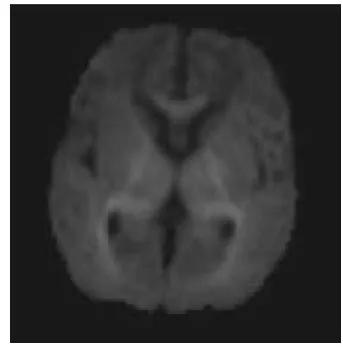
Case 1 Mri Findings In Case of Central Hypoxicischemic Encephalopathy
Restricted diffusion in the genu of the corpus callosum and both internal capsules
Diffusion restriction in the corpus callosal splenium
ADC image showing reduced ADC values in the corpus callosul splenium
Diffusion restriction in the corpus callosal splenium
Loss of normal signal intensity in the posterior limb of internal capsule bilaterally on T1W images
Case 2 Mixed Pattern of Hypoxic Ischemic Encephalopathy in a Neonate
T1 hyperintensity noted involving bilateral basal ganglia
Restriction of diffusion in the splenium of the Corpus Callosum and the posterior limbs of bilateral internal capsules
Restriction of water diffusion in thalami bilateral basal ganglia
Corresponding ADC images showing reduced ADC values
Limitation
The limitation of this study is the lack of systematic follow up neuro-imaging. Also patients could be followed up for a maximum of 5 months time at follow up clinics and it has been recognized that subtle neuro-developmental abnormalities can occur years after birth asphyxia.
CONCLUSION
This study attempted to evaluate the various magnetic resonance imaging changes in hypoxic ischemic encephalopathy in term infants. It set out to categorize and suitably classify the various MRI patterns of disease in these patients. Additionally, an attempt to correlate the MRI findings with neurosonographic appearances was undertaken wherever feasible.
It was found that MRI is superior to other imaging modalities in the evaluation of neonatal hypoxic ischemic encephalopathy. There is a strong and consistent correlation between the various MRI findings and the final clinical outcome.
MRI has a high sensitivity and specificity in the evaluation of the above condition. It is non-invasive and has no radiation hazards. It offers excellent gray - white matter resolution which Computed Tomography is incapable of especially in the neonate with a large proportion of unmyelinated brain parenchyma.
sensitivity and provides information not seen on the other conventional sequences. Many of the signs of hypoxic ischemic encephalopathy are based on diffusion weighted imaging.
References
1. Ferriero DM. Neonatal brain injury. N Engl J Med Nov.42004; 351(19):1985-95.
2. Perlman JM. Brain injury in the term infant. Semin
Perinatol Dec.2004; 28(6):415-24.
3. Grow J, Barks JD. Pathogenesis of hypoxic - ischemic cerebral injury in the term infant: Current concepts.
Clinical Perinatology Dec. 2002; 29(4):585-602.
4. Huang BY, Castillo M. Hypoxic - ischemic brain injury: imaging findings from birth to adulthood. Radiographics Mar - Apr 2008; 28(2):417-39; Quiz 617.
5. Latchaw RE, Truwit Cue. Imaging of perinatal hypoxic -ischemic brain injury. Semin Pediatr Neurol Mar 1995; 2(1):72-89.
6. Rutherford M, Pennock J, Schwieso J, Cowan F, Dubowitz L. Hypoxic ischaemic encephalopathy: early and late magnetic resonance imaging findings in relation to outcome. Arch Dis Child Fetal Neonatal Ed.1996; 75:F145-F15.
7. Rutherford M, Biarge MM, Allsop J, Counsell S, Cowan F. MRI of perinatal brain injury. Pediatr Radiol 2010; 40(6):819-33.
8. Prognostic tests in Term Neonates with Hypoxic Ischemic Encephalopathy; A systematic Review, Henriette Van Laerhoven, Timo R. De Haan, Martin Offringa, Bart Post and Johanna H. Van der Lee Pediatrics 2013; 131:88.
9. A James Barkovich, Kaye Westmark, Colin Patridge, Augusto Sola and Donna M. Ferreiro, AJNAR Am J
Neuroradiol 16; 427-438, March-95.
10. Rutherford MA, Pennock J, Counsell S et al. Abnormal magnetic resonance signal in the internal capsule predicts poor developmental outcome in infants with hypoxic–ischaemic encephalopathy. Pediatrics1998;
102:323–328.
11. Rutherford MA, Pennock JM, Murdoch - Eaton DM et al. Athetoid cerebral palsy and cysts in the putamen after hypoxic–ischaemic encephalopathy. Arch Dis Child
1992; 67:846–850.
Transfontanelle neurosonogram in the patient reveals increased echogenicity in the region of the basal ganglia and the deep white
matter with compression of the ventricles.
*******
How to cite this article: