donor-reactive memory CD8
+
T cells
Hidetoshi Tsuda, … , Anna Valujskikh, Robert L. Fairchild
JCI Insight.
2018;
3(4)
:e96940.
https://doi.org/10.1172/jci.insight.96940
.
Recipient endogenous memory T cells with donor reactivity pose an important barrier to
successful transplantation and costimulatory blockade–induced graft tolerance. Longer
ischemic storage times prior to organ transplantation increase early posttransplant
inflammation and negatively impact early graft function and long-term graft outcome. Little is
known about the mechanisms enhancing endogenous memory T cell activation to mediate
tissue injury within the increased inflammatory environment of allografts subjected to
prolonged cold ischemic storage (CIS). Endogenous memory CD4
+and CD8
+T cell
activation is markedly increased within complete MHC-mismatched cardiac allografts
subjected to prolonged versus minimal CIS, and the memory CD8
+T cells directly mediate
CTLA-4Ig–resistant allograft rejection. Memory CD8
+T cell activation within allografts
subjected to prolonged CIS requires memory CD4
+T cell stimulation of graft DCs to
produce p40 homodimers, but not IL-12 p40/p35 heterodimers. Targeting p40 abrogates
memory CD8
+T cell proliferation within the allografts and their ability to mediate CTLA-4Ig–
resistant allograft rejection. These findings indicate a critical role for memory CD4
+T cell–
graft DC interactions to increase the intensity of endogenous memory CD8
+T cell activation
needed to mediate rejection of higher-risk allografts subjected to increased CIS.
Research Article
Immunology
Transplantation
Find the latest version:
R E S E A R C H A R T I C L E
Authorship note: HT and CAS contributed equally to this work and are co–first authors.
Conflict of interest: The authors have declared no conflict of interest exists.
Submitted: August 17, 2017 Accepted: December 28, 2017 Published: February 22, 2018 Reference information:
JCI Insight. 2018;3(4):e96940. https://doi.org/10.1172/jci. insight.96940.
Allograft dendritic cell p40 homodimers
activate donor-reactive memory
CD8
+
T cells
Hidetoshi Tsuda,1,2 Charles A. Su,1,3 Toshiaki Tanaka,1,2 Katayoun Ayasoufi,1 Booki Min,1
Anna Valujskikh,1 and Robert L. Fairchild1,2,3
1Lerner Research Institute and 2Transplant Center, Cleveland Clinic, and 3Department of Pathology, Case Western Reserve
University School of Medicine, Cleveland, Ohio, USA.
Introduction
The generation of T cell memory provides effective protection to recurrent infectious agents. When compared with antigen-reactive naive T cells, memory T cells have many distinct features enhanc-ing their function to rapidly respond to and eliminate viral and bacterial infections, includenhanc-ing rapid trafficking into peripheral tissue inflammatory sites and activation that occurs independently of the many costimulatory signals required for naive T cell activation (1–3). Moreover, memory CD8+ T cell
responses within peripheral tissue inflammatory sites often occurs without the CD4+ T cell generated
helper signals required for optimal naive antigen–reactive CD8+ T cell responses (4–6). Memory T cells
with reactivity to allogeneic MHC molecules are an important barrier to the function and survival of organs transplanted to treat end-stage organ disease (7–9). Seminal studies by Heeger and colleagues indicated that the presence of high numbers of memory T cells with donor reactivity in the peripheral blood of kidney transplant patients prior to the transplant resulted in higher incidence of delayed graft function, acute graft rejection, and poorer long-term graft outcomes (10, 11). Memory CD4+ and CD8+
T cell repertoires generated during exposure to viral and bacterial infections often contain high frequen-cies of T cells reactive to allogeneic class II or class I MHC molecules, providing donor-reactive mem-ory T cells that can undermine successful transplantation (12–15). Mice also contain readily detectable populations of CD4+ and CD8+ T cells with memory phenotypes, and a proportion of this memory T
cell repertoire in naive mice infiltrate cardiac allografts within hours of graft reperfusion and respond to donor class I and class II MHC alloantigens (16–18). Endogenous memory CD8+ T cells infiltrating
allografts subjected to minimal periods of cold ischemic storage (CIS) are activated to produce IFN-γ
and other mediators that increase graft inflammation. This endogenous memory CD8+ T cell response
occurs independently of CD4+ T cells and CD28/CD80/86- and CD40/CD154-mediated
costimula-tion, but it is insufficient to directly mediate graft rejection.
Recipient endogenous memory T cells with donor reactivity pose an important barrier to successful transplantation and costimulatory blockade–induced graft tolerance. Longer ischemic storage times prior to organ transplantation increase early posttransplant inflammation and negatively impact early graft function and long-term graft outcome. Little is known about the mechanisms enhancing endogenous memory T cell activation to mediate tissue injury within the increased inflammatory environment of allografts subjected to prolonged cold ischemic storage (CIS). Endogenous memory
CD4+ and CD8+ T cell activation is markedly increased within complete MHC-mismatched cardiac
allografts subjected to prolonged versus minimal CIS, and the memory CD8+ T cells directly mediate
CTLA-4Ig–resistant allograft rejection. Memory CD8+ T cell activation within allografts subjected to
prolonged CIS requires memory CD4+ T cell stimulation of graft DCs to produce p40 homodimers,
but not IL-12 p40/p35 heterodimers. Targeting p40 abrogates memory CD8+ T cell proliferation
within the allografts and their ability to mediate CTLA-4Ig–resistant allograft rejection. These findings indicate a critical role for memory CD4+ T cell–graft DC interactions to increase the intensity
of endogenous memory CD8+ T cell activation needed to mediate rejection of higher-risk allografts
The initial inflammation induced in transplanted organs arises from their removal from a source of oxygen following graft harvest and the subsequent reexposure to oxygen during graft revascularization that generates ROS and danger-associated molecules, potent activators of innate immunity (19–21). Imposition of longer ischemic storage times on clinical transplants generates marked increases in graft inflammation during reperfusion and negatively impacts early graft function and long-term survival (22–26). We recently reported that increasing the time of CIS imposed on cardiac allografts quickly provokes a highly inflam-matory environment after revascularization that increases endogenous memory CD4+ and CD8+ T cell
numbers within the allograft and increases in endogenous memory CD8+ T cell expression of IFN-γ,
perfo-rin, and granzyme B (27). Whereas peritransplant CTLA-4Ig treatment extended the survival of allografts subjected to minimal (e.g., 30 minutes) CIS by more than 7 weeks, this treatment extended the survival of allografts subjected to prolonged (e.g., 6–8 hours) CIS by only a few days; this rejection was mediated by the endogenous memory CD8+ T cells. These results were reminiscent of the costimulatory
blockade–resis-tant endogenous memory CD8+ T cell responses that mediate acute rejection and decrease graft survival in
nonhuman primate (NHP) and human allograft recipients (28–31). How such endogenous memory CD8+
T cells are activated within allografts to mediate this early severe graft injury and failure has not been deter-mined. The goal of the current study was to identify mechanisms promoting endogenous memory CD8+ T
cell activation within allografts subjected to increased CIS. Overall, the results indicate an intricate series of activation events within higher-risk allografts during the first 1–2 days after transplant initiated by endoge-nous memory CD4+ T cell interaction with graft DCs to generate a unique cytokine required for activation
of donor-reactive memory CD8+ T cells to mediate acute allograft injury and rejection.
Results
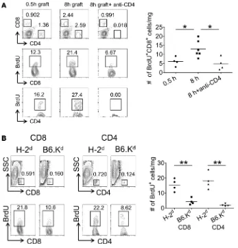
Donor-specific alloreactivity of early graft–infiltrating endogenous memory CD8+ T cells. To begin to further assess polyclonal donor-reactive memory CD8+ T cell infiltration into highly ischemic cardiac allografts,
respons-es of adoptively transferred donor-sensitized memory CD8+ T cells were studied. Briefly, memory CD8+ T
cells were enriched from the spleens and lymph nodes of CD45.2+ C57BL/6 recipients of A/J skin
trans-plants and adoptively transferred to congenic CD45.1+ recipients that were then transplanted with A/J
car-diac allografts subjected to minimal or prolonged (0.5 vs.8 hours) CIS. Similar to the endogenous memory CD8+ T cells from recipients without such adoptively transferred memory CD8+ T cells (27), transferred
donor antigen–primed memory CD45.2+CD8+ T cells were detectable in A/J heart allografts subjected to
either minimal or prolonged CIS within 12–16 hours after graft reperfusion, but transferred memory CD8+
T cell numbers were increased in allografts subjected to longer CIS (Figure 1A). The adoptively transferred CD8+ T cells from A/J skin allograft–primed mice also infiltrated third-party (DBA/1) allografts subjected
to prolonged CIS but at the decreased levels seen in A/J cardiac allografts subjected to minimal CIS. The donor reactivity of endogenous memory CD8+ T cells infiltrating A/J cardiac allografts subjected
to 8 hours of CIS was directly investigated by isolating the CD8+ T cells from the allografts on day 3 after
transplant, labeling the T cells with CFSE, and testing their ability to proliferate in response to various splenocyte stimulator cells in vitro. The purified graft-infiltrating memory CD8+ T cells exhibited little
reac-tivity to syngeneic stimulator cells but robustly proliferated to graft donor A/J stimulator cells (Figure 1B). Consistent with their infiltration into DBA/1 cardiac allografts, the A/J graft–infiltrating memory CD8+ T
cells also demonstrated a lower but significant response to third-party DBA/1 stimulator cells. Mixing A/J and DBA stimulators did not yield a synergistic proliferative response when compared with the response to the A/J stimulators alone, suggesting that the third-party alloreactive memory CD8+ T cells are contained
within the A/J donor–reactive population. Overall, these results establish the donor reactivity of endoge-nous memory CD8+ T cells infiltrating heart allografts and complement studies investigating the infiltration
of donor-reactive transgenic CD8+ T cells into allografts (32).
The substantial percentage of allograft-infiltrating CD8+ T cells that did not respond to donor cells
prompted a more comprehensive analysis of the endogenous CD4+ and CD8+ T cell populations
infiltrat-ing complete MHC-mismatched heart allografts subjected to minimal vs. prolonged CIS 48 hours after reperfusion of the allografts. The greatest percentage of CD4+ T cells infiltrating the allografts subjected to
either minimal or prolonged CIS were effector memory (CD62LlowCD44high) cells with a lower percentage
of central memory (CD62LhighCD44high) cells (Supplemental Figure 1A; supplemental material available
online with this article; https://doi.org/10.1172/jci.insight.96940DS1). Naive (CD62LlowCD44high and
the largest percentage of CD8+ T cells in allografts subjected to minimal vs. prolonged CIS, but infiltrating
effector memory (CD62LlowCD44high) CD8+ T cells were prominent with a smaller percentage of central
memory (CD62LhighCD44high) CD8+ T cells (Supplemental Figure 1B).
Consistent with previous studies (27), macrophages (F4/80+) and neutrophils (Ly6G+) also
infiltrat-ed allografts subjectinfiltrat-ed to minimal and prolonginfiltrat-ed CIS, with greater numbers in the allografts subjectinfiltrat-ed to prolonged CIS (Supplemental Figure 2, A and B). Smaller but equivalent numbers of B lymphocytes (CD19+B220+) and barely detectable numbers of NK (CD3ε–NK1.1+CD49b+) cells were observed in
allografts subjected to minimal and prolonged CIS (Supplemental Figure 2C and data not shown).
Increased proliferation of endogenous memory CD8+ T cells within allografts subjected to prolonged CIS. The greater numbers of endogenous memory CD8+ T cells rapidly accumulating in allografts subjected to
pro-longed vs. minimal CIS led us to hypothesize that reperfusion of allografts subjected to propro-longed CIS generated an inflammatory environment promoting the proliferation of early graft–infiltrating memory T cells. To test this, recipients of A/J cardiac allografts subjected to 0.5 or 8 hours of CIS were pulsed with BrdU, and its incorporation into DNA of graft-infiltrating memory CD8+ and CD4+ T cells was assessed
on day 2 after transplant. As previously reported (27), increasing CIS time prior to transplant markedly increased endogenous CD4+ and CD8+ T cell numbers in the allografts (Figure 1C). Compared with T
cells infiltrating allografts subjected to minimal CIS, CD8+ and CD4+ T cells within allografts subjected
to prolonged CIS exhibited a roughly 2-fold increase in proliferation at this time point. Interestingly, even in allografts subjected to prolonged CIS prior to transplant, a relative minority (< 30%) of total graft-infil-trating T cells was proliferating on day 2 after transplant. Endogenous CD4+ and CD8+ T cell proliferation
was not observed 24 hours after reperfusion of the highly ischemic allografts, but it was clearly evident and increased with time after transplant thereafter (Figure 1D).
The populations of allograft-infiltrating endogenous CD4+ and CD8+ T cells proliferating in allografts
subjected to minimal and prolonged CIS were examined (Supplemental Figure 3, A and B). Effector mem-ory CD4+ and CD8+ T cell proliferation was stronger in allografts that had been subjected to prolonged
vs. minimal CIS. In contrast to the absence of central memory CD4+ and CD8+ T cell proliferation within
allografts subjected minimal CIS, central memory CD4+ and CD8+ T cells proliferated within allografts
that had been subjected to 8 hours of CIS.
Anti-CD154 mAb prolongs survival of cardiac allografts subjected to prolonged CIS and inhibits endogenous memory T cell proliferation within allografts. Endogenous memory CD8+ T cells infiltrating allografts
sub-jected to prolonged CIS are activated to directly mediate graft rejection that is resistant to CTLA-4Ig (27). Consistent with this resistance, peritransplant CTLA-4Ig treatment had a minimal effect on the early proliferation of endogenous memory CD4+ and CD8+ T cells within A/J allografts subjected to 8
hours of CIS (Figure 2A). In contrast to CTLA-4Ig, peritransplant treatment with a different costimu-latory blockade agent, anti-CD154 mAb, markedly attenuated total numbers and numbers of proliferat-ing/BrdU+ endogenous memory CD4+ and CD8+ T cells within allografts subjected to 8 hours of CIS
when assessed 48 hours after transplant (Figure 2B). Similar to our previous results (27), peritransplant treatment with CTLA-4Ig extended the survival of heart allografts subjected to minimal CIS (Figure 2C) but had little impact on the survival of allografts subjected to 8 hours of CIS (median time of sur-vival [MTS], 7 vs. 11 days; control Ig vs. CTLA-4Ig treatment) (Figure 2C). In contrast, peritransplant
R E S E A R C H A R T I C L E
treatment with anti-CD154 mAb resulted in a substantial prolongation in survival of the highly isch-emic cardiac allografts (MTS, 36 days).
Endogenous memory CD8+ T cell proliferation within highly ischemic cardiac allografts is dependent on CD4+
T cells. The anti-CD154 mAb–mediated inhibition of endogenous memory CD8+ T cell proliferation
within highly ischemic cardiac allografts suggested a role for CD4+ T cells in this response. Groups
of C57BL/6 recipients were treated with control Ig or with depleting anti-CD4 mAb on 3 consecutive days prior to transplant, with A/J heart allografts subjected to 0.5 or 8 hours of CIS, and endogenous memory CD8+ T cell proliferation within the allografts was examined 48 hours after transplant. In
recipients depleted of CD4+ T cells, the increases in endogenous memory CD8+ T cell proliferation in
highly ischemic allografts were reduced to the proliferation observed in allografts subjected to minimal CIS (Figure 3A) and were accompanied by decreases in total memory CD8+ T cell numbers in the
allografts (not shown).
The role of memory CD4+ T cells in promoting the increased endogenous memory CD8+ T cell
pro-liferation within allografts subjected to prolonged CIS was further tested by comparing C57BL/6 recipient endogenous memory CD4+ and CD8+ T cell responses within cardiac allografts from complete
MHC-mis-matched BALB/c donors (H-2d) vs. single class I MHC–mismatched B6.Kd donors (transgenic Kd on
H-2b background) that had been subjected to 8 hours of CIS. Total numbers and proliferation of
endoge-nous memory CD4+ and CD8+ T cells were significantly reduced in single class I MHC–disparate B6.Kd
allografts when compared with complete MHC-mismatched allografts (Figure 3B). These results are con-sistent with the need for graft class II MHC alloantigen expression to activate the recipient memory CD4+
T cells required for infiltrating memory CD8+ T cell activation to the donor class I MHC alloantigen.
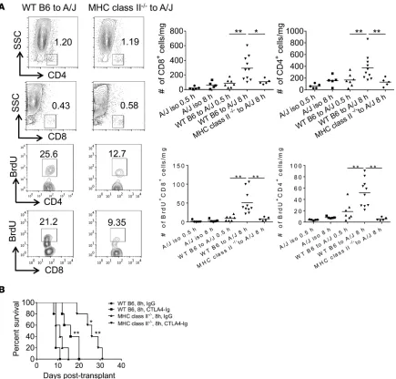
The requirement for allograft class II MHC expression was directly tested by comparing endoge-nous memory CD4+ and CD8+ T cell proliferation within WT C57BL/6 vs. B6.class II MHC–deficient
cardiac allografts subjected to prolonged CIS before transplantation to A/J recipients. When compared with WT allografts, there was a marked decrease of endogenous memory CD4+ and CD8+ T cell
num-bers and proliferation in the highly ischemic class II MHC–/– allografts that were similar to the numbers
and proliferation observed in WT allografts subjected to minimal CIS and in A/J isografts subjected
Figure 3. Endogenous memory CD8+ T cell proliferation in allografts subjected to prolonged CIS requires recipient CD4+ T cells. (A) Groups of C57BL/6
R E S E A R C H A R T I C L E
to 0.5 or 8 hours of CIS (Figure 4A). Consistent with the memory CD8+ T cell responses in the WT
vs. class II MHC–deficient allografts, mRNA expression of the effector CD8+ T cell mediators IFN-γ,
FasL, perforin, and granzyme B was markedly increased in WT allografts subjected to 8 vs. 0.5 hours of CIS but was significantly decreased in class II MHC–/– allografts subjected to prolonged CIS
(Sup-plemental Figure 4). In contrast, no significant difference was observed in the mRNA expression of the neutrophil chemoattractant CXCL1 in any of the iso- and allograft groups. Whereas CTLA-4Ig treatment only extended survival of WT C57BL/6 allografts subjected to prolonged CIS in A/J recip-ients from day 9 (MTS) to day 16 MTS, CTLA-4Ig treatment significantly extended survival of highly ischemic B6.class II MHC–/– allografts from day 11 MTS to day 26 MTS (Figure 4B).
Endogenous memory CD4+ and CD8+ T cell proliferation within highly ischemic heart allografts requires graft
expres-sion of CD40 and graft DCs. The inhibition of endogenous memory CD4+ and CD8+ T cell activation within
highly ischemic allografts by anti-CD154 mAb suggested a requirement for CD40 engagement for this response.
Figure 4. Endogenous memory CD8+ T cell proliferation in allografts subjected to prolonged CIS requires recipient graft class II MHC expression. (A) Cardiac
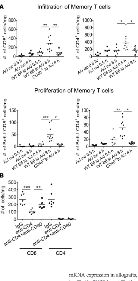
When compared with memory T cell responses within WT C57BL/6 allografts subjected to 8 hours of CIS, marked decreases in total numbers and proliferating endogenous memory CD4+ and CD8+ T
cells were observed in B6.CD40–/– allografts 48 hours after transplant to
A/J recipients (Figure 5A). The importance of CD40-mediated signal-ing for increased endogenous memory CD8+ T cell accumulation within
allografts subjected to prolonged CIS was further indicated by the abili-ty of an agonist anti-CD40 mAb to restore endogenous memory CD8+
T cell numbers in highly ischemic A/J allografts in C57BL/6 recipients depleted of CD4+ T cells (Figure 5B).
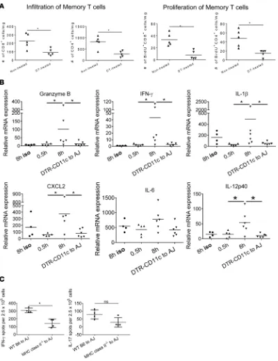
Endogenous memory T cell proliferation was compared in control and DC-depleted C57BL/6 cardiac allografts subjected to 8 hours of CIS and transplanted to A/J recipients. Diphtheria toxin (DT) receptor– CD11c transgenic (B6.DTR-CD11c) mice were treated with vehicle or with DT on 2 consecutive days prior to harvest of hearts and transplant to A/J recipients, and endogenous memory CD4+ and CD8+ T cell
prolif-eration within the allografts was determined 48 hours later. The absence of graft DCs markedly decreased both endogenous memory CD4+ and
CD8+ T cell numbers and proliferation within the heart allografts (Figure
6A). Graft DC depletion also resulted in decreased IFN-γ and granzyme B mRNA expression in allografts, as well as in decreased expression of proinflammatory cytokine genes includ-ing IL-1β, CXCL2, and IL-12 p40, but not IL-6 (Figure 6B). On day 8 after transplant, the impact of allograft DC depletion on the de novo priming of donor-reactive naive T cells was also determined by enumerating donor-reactive T cells producing IFN-γ and IL-17 in the spleens from each allograft recipient group. While IFN-γ–producing cell numbers were significantly decreased in recipients of the DC-depleted allografts, Il-17– producing T cell numbers were similar to recipients of WT allografts (Figure 6C).
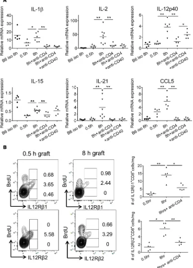
p40 homodimers mediate proliferation of endogenous memory CD8+ T cells within highly ischemic allografts. Candidate factors driving the CD4+ T cell–dependent proliferation of endogenous memory CD8+ T cells
within the highly ischemic cardiac allografts were next investigated. First, mRNA expression of different cytokines within cardiac allografts subjected to either 30 minutes or 8 hours of CIS was compared on day 2 after transplant. Of a large panel of test cytokines, IL-1β, IL-2, IL-12 p40, IL-21, and CCL5 mRNA had the largest increases in cardiac allografts subjected to prolonged vs. minimal CIS (Figure 7A). Depletion of recipient CD4+ T cells prior to the transplant reduced mRNA expression levels of these cytokines in
the highly ischemic cardiac allografts to the levels observed in isografts and in allografts subjected to min-imal CIS. In contrast, treating CD4+ T cell–depleted allograft recipients with an agonist anti-CD40 mAb
restored the expression of IL-12 p40 mRNA in the highly ischemic allografts but not IL-1β, IL-2, IL-21, or CCL5 mRNA. Similar to the observed mRNA expression levels, increases in proinflammatory cytokine protein were observed in allografts subjected to increased CIS, and this increase was decreased to back-ground by depletion of recipients CD4+ T cells (Supplemental Figure 5).
Figure 5. Endogenous memory CD8+ T cell proliferation in allografts subjected to prolonged CIS requires graft CD40 expression. (A) Groups of A/J mice (n = 5–10) received WT C57BL/6 or B6.CD40–/– cardiac allografts subjected to 0.5 or 8 hours of CIS and were injected with 100 μg BrdU i.p. on days 0 and 1 after transplant. The next day, allografts were harvested and digested, and aliquots of single cell suspensions were stained with antibody and analyzed by flow cytometry to quantitate the infiltration and proliferation of memory CD8+ and CD4+ T cells. *P < 0.05, **P < 0.01, ***P < 0.001, as determined by the Mann-Whitney nonpara-metric test. (B) Groups of C57BL/6 (n = 5–7/group) mice were treated with 200
R E S E A R C H A R T I C L E
Figure 6. Endogenous memory CD8+ T cell proliferation in allografts subjected to prolonged CIS requires graft DCs. (A)Groups of A/J mice (n = 5/
To begin to test a potential role for IL-12 in endogenous memory CD8+ T cell proliferation within
cardiac allografts subjected to prolonged CIS, infiltrating memory CD8+ T cell expression of the IL-12
receptor β1 and β2 subunits (IL-12R β1 and IL-12R β2, respectively) was examined. CD8+ T cells
within allografts subjected to either minimal or prolonged CIS expressed both IL-12R β1 and IL-12R
β2 (Figure 7B), with greater memory CD8+ T cell numbers expressing both IL-12 receptor subunits in
allografts subjected to prolonged CIS, and these numbers decreased in the absence of recipient CD4+
T cells. The expression of both IL-12 receptor subunits was restricted to those infiltrating memory CD8+ T cells with intermediate incorporation of BrdU and was absent on memory CD8+ T cells with
the highest incorporation of BrdU. Expression of IL-12R β1 was expressed on a minority of memory CD4+ T cells infiltrating allografts subjected to either minimal or prolonged CIS, whereas prolonged
CIS induced marked increases in allograft-infiltrating memory CD4+ T cells expressing IL-12R β2
(data not shown).
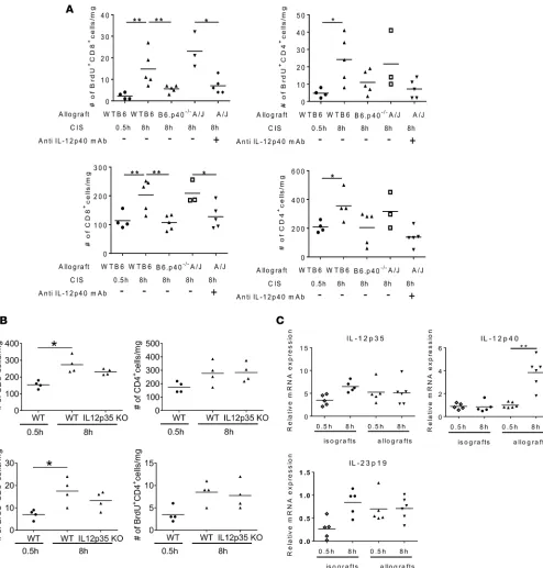
The roles of IL-12 p35/p40 heterodimers and p40 on the proliferation and accumulation of endog-enous memory CD4+ and CD8+ T cells within allografts was then investigated. Endogenous memory
CD8+ T cell, but not memory CD4+ T cell, proliferation and accumulation was significantly reduced
in B6.p40–/– cardiac allografts that had been subjected to 8 hours of CIS before transplantation to A/J
recipients when compared with WT C57BL/6 allografts (Figure 8A). Similarly, recipient C57BL/6 treatment with p40 neutralizing mAb resulted in significant reductions of endogenous memory CD8+
T cell proliferation and numbers in A/J allografts subjected to 8 hours of CIS and more modest reduc-tions of endogenous memory CD4+ T cell proliferation and accumulation (Figure 8A). However, no
dif-ferences in endogenous memory CD8+ and CD4+ T cell proliferation and accumulation were observed
in B6.p35–/– and WT C57BL/6 heart allografts that had been subjected to 8 hours of CIS before
trans-plantation to A/J recipients (Figure 8B).
To further investigate the indicated role of p40 but not p35 in endogenous memory CD8+ T cell
pro-liferation within allografts subjected to prolonged CIS, mRNA levels of IL-12 p35, IL-23 p19, and IL-12 p40 in cardiac iso- and allografts were compared 2 days after transplantation. Although p35 mRNA was expressed at 6- to 8-fold higher levels than p19 mRNA, expression of IL-23 p19 and IL-12 p35 mRNA did not change in allografts subjected to either minimal or prolonged CIS or in isografts sub-jected to prolonged CIS time before transplant (Figure 8C). Only expression of p40 mRNA increased in allografts subjected to the prolonged vs. minimal CIS time before transplantation.
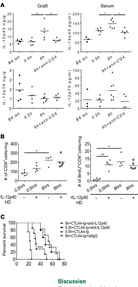
When assessed 48 hours after transplant, p40 protein was increased in allografts subjected to prolonged vs. minimal CIS, as well as in the recipient serum, and these increases were dependent on recipient CD4+
T cells (Figure 9A). In contrast, IL-12 p70 heterodimer protein levels were equivalent in heart allografts subjected to either minimal or prolonged CIS, and the absence of recipient CD4+ T cells did not alter p70
levels in the allografts subjected to prolonged CIS.
Macrophages and DC often produce p40 homodimers during inflammatory stimulation and antag-onize the delivery of IL-12 p35/p40 heterodimer signaling to T cells during antigen priming (33, 34). A role for p40 homodimers in the proliferation of the endogenous memory CD8+ T cells within the
highly ischemic cardiac allografts was directly addressed by testing the ability of p40 homodimers to induce endogenous memory CD8+ T cell proliferation within cardiac allografts subjected to minimal
or prolonged CIS. Injection of p40 homodimers into mice on the day of and 1 day after transplant provoked a marked increase in memory CD8+ T cell proliferation in allografts subjected to 0.5 hours
of CIS when assessed on day 2 after transplant but had no effect on memory CD8+ T cell proliferation
within allografts subjected to prolonged CIS (Figure 9B). Injection of the p40 homodimers had little to no effect on proliferation and numbers of endogenous memory CD4+ T cells in allografts subjected to
either minimal or prolonged CIS (not shown).
R E S E A R C H A R T I C L E
Figure 8. Endogenous memory CD8+ T cell proliferation within allografts subjected to prolonged CIS requires graft p40 but not p35. (A) Groups of
R E S E A R C H A R T I C L E
Discussion
This study investigated the early posttransplant function of polyclonal donor-reactive memory CD4+ and
CD8+ T cells that comprise the endogenous memory T cell repertoire in unprimed heart allograft
recipi-ents. Our previous studies had indicated the CD4+ T cell–independent activation of endogenous memory
CD8+ T cells to produce IFN-γ within allografts transplanted as quickly as possible from donor harvest to
revascularization in the recipient, but this activation was insufficient to mediate acute rejection (16, 18). In contrast, prolonging CIS time prior to transplant increased endogenous memory CD4+ and CD8+ T
cell numbers within the allografts and activation of the memory CD8+, but not CD4+, T cells to directly
reject the allograft (27). Such direct memory CD8+ T cell activation within tissue sites is often dependent
on inflammatory signals that compensate for the absence of CD4+ T cell–generated helper signals (35–
37). A key finding of the current study is that the increased activation of endogenous memory CD8+ T
cells to mediate acute injury and rejection of allografts subjected to prolonged CIS requires endogenous memory CD4+ T cell activation, suggesting that a critical difference within allografts subjected to
min-imal vs. prolonged CIS is that the increased inflammation in the latter grafts generates an environment promoting memory CD4+ T cell interaction with graft DC to produce helper signals required for optimal
donor-reactive memory CD8+ T cell activation.
While memory T cells cross-reactive to allogeneic MHC are able to disrupt strategies administered to recipients to induce allograft tolerance (30, 38, 39), it had remained unclear if such endogenous memory CD8+ T cells have the ability to directly mediate graft injury independently of CD4+ T cell help. Although
a requirement for CD4+ T cell help to elicit donor-reactive memory CD8+ T cell responses to complete
MHC-mismatched allografts has not been observed, previous studies investigating memory CD4+ and
CD8+ T cell activation to allografts have primarily utilized sensitized recipients or transfer of
donor-primed memory T cells to naive recipients (39–43). In addition to CD4+ T cell–independent activation,
these donor alloantigen–primed memory CD8+ T cell responses are typically resistant to both CD28- and
CD154-mediated costimulatory blockade, and we have also observed that memory T cells primed to donor skin allografts are resistant to CD40-CD154 costimulatory blockade when transferred to recipients of donor heart allografts subjected to prolonged CIS (H. Tsuda, unpublished results). Clearly, these donor antigen priming strategies bias the reactive memory CD8+ T cell repertoire to those cells with strong reactivity to
the sensitizing antigen, and this is likely to confer their CD4+ T cell–independent activation and resistance
to many different costimulatory blockade strategies. Alternatively, the endogenous memory T cell reper-toire is likely to be composed of cells with a range of avidities for allogeneic MHC, and the current study demonstrates that peritransplant treatment with anti-CD154 mAb inhibits endogenous memory CD8+ T
cell activation within allografts subjected to prolonged CIS and enhances graft survival.
A second key finding of the current study is the identification of unique memory CD4+ T cell–induced
helper factors, IL-12 p40 homodimers, required for the proliferation and accumulation of endogenous memory CD8+ T cells within the highly ischemic allografts. This increase in infiltrating memory CD8+
T cell numbers within allografts is not a trivial matter, as previous studies titrating donor antigen–primed memory T cells have demonstrated that a critical threshold is needed to mediate allograft rejection and resistance to costimulatory blockade (39). IL-12 p40/p35 (i.e., p70) heterodimers direct naive antigen–spe-cific CD4+ T cell differentiation to IFN-γ–producing cells during initiation of T cell responses to
intracel-lular bacterial infections and potentiate elicitation of primary and memory CD8+ T cell responses (44–49).
Although IL-12 p70 heterodimers are required for rejection of single minor antigen OVA-expressing heart allografts, their absence does not decrease donor-reactive effector CD4+ and CD8+ T cell priming or
rejec-tion of complete MHC-mismatched cardiac allografts (50–52). The absence of allograft p40, but not p35, or antibody neutralization of p40 abrogates endogenous memory CD8+ T cell activation within allografts
subjected to increased CIS, implicating p40 homodimers, and not IL-12 p70 heterodimers, as critical help-er factors driving endogenous memory CD8+ T cell proliferation within the allografts. The upregulation
of p40, but not p35, mRNA and the absence of this upregulation in allografts depleted of DC in cardiac allografts subjected to prolonged, but not minimal, CIS suggests that the increased inflammatory environ-ment promotes memory CD4+ T cell interaction with graft DCs to induce p40, but not p35, transcription
and translation. Indeed, p40 homodimers were initially viewed as antagonists for the IL-12R β1 and –β2 heterodimer, resulting in modulation of the potency of the IL-12 p70 heterodimer to promote antigen-spe-cific CD4+ T cell development to IFN-γ–producing cells (33, 34). Recent studies suggest that p40
homod-imer binding to dhomod-imers or oligomers of IL-12R β1 on macrophages and DCs activates NF-κB to induce chemokine production (53, 54).
A final key finding is the requirement for graft cells producing p40 and graft DCs for endogenous memory CD8+ T cell activation within the highly ischemic allografts. DCs in heart and kidney allografts subjected to
minimal CIS are rapidly replaced by recipient DCs — up to 90% within 24 hours (55, 56) — and we have observed similar replacement in the cardiac allografts subjected to prolonged CIS prior to transplant (H. Tsu-da, data not shown). The results of the current study suggest that the recipient DC infiltrating p40-deficient allografts cannot supply the IL-12 p40 homodimers required for memory CD8+ T cell activation. Recipient
R E S E A R C H A R T I C L E
Collectively, the results of this study document a unique donor-reactive memory T cell response that is a consequence of increased ischemia-reperfusion injury within the organ allograft and provide potentially novel insights into the early mobilization and expansion of donor-reactive endogenous memory CD8+ T cells within cardiac allografts subjected to prolonged CIS. This response requires the
coordinated and inter-dependent activation of donor-reactive endogenous memory CD4+ and CD8+ T
cells within the ischemic allografts that generates functional memory CD8+ T cells that directly mediate
acute tissue injury and graft rejection. The initiation and progression of this endogenous memory CD8+
T cell response is resistant to CTLA-4Ig but is sensitive to agents that block the CD4+ T cell–graft DC
interactions generating the p40 homodimers. It is worth noting that these activation events are analo-gous to those occurring in secondary lymphoid organs during de novo activation of reactive CD8+ T
cells by antigen-presenting DCs, where CD4+ T cell licensing of antigen-presenting DC is required for
optimal naive antigen–reactive CD8+ T cell development to functional effector cells. Considering the
high incidence of acute rejection episodes observed in kidney transplant patients treated with CTLA-4Ig in place of cyclosporine A (59, 60), the results suggest other targets to inhibit or attenuate this rejection. Given the detrimental impact of ischemia-reperfusion injury and memory T cells on allograft function and survival, strategies to minimize the postreperfusion inflammatory milieu of higher-risk grafts by inhibiting early memory T cell proliferation and activation to express the effector functions mediating graft tissue injury should prove beneficial to transplant outcomes.
Methods
Mice. C57BL/6 (H-2b), congenic B6.PL-Thy1a/CyJ mice (H-2b), B6.MHC class II–/–, B6.CD40–/–,
B6.p40–/–, B6.p35–/–, and B6.diphtheria-toxin receptor–CD11c (DTR-CD11c) transgenic mice and A/J
(H-2a), BALB/c (H-2d), and DBA/1 (H-2q) mice were purchased from the Jackson Laboratory. The
gen-eration of C57BL/6 mice transgenically expressing Kd (i.e., B6.Kd) has been previously detailed (61), and
colonies of B6.Kd mice were established and maintained at our facility. Male mice, 8–12 weeks of age,
were used in the studies.
Heterotopic cardiac transplantation and recovery. Murine heterotopic intraabdominal cardiac
transplan-tation and recovery were adapted from the method of Corry and colleagues as previously reported by our laboratory (18, 62). Briefly, the donor aorta and pulmonary artery were anastomosed to the recipient abdominal aorta and inferior vena cava in the peritoneal cavity. Following harvest from the donor, hearts were stored for either 0.5 or 8 hours in cold UW solution (BUW-0001, Bridge to Life) on ice prior to trans-plant. Graft survival was monitored daily by recipient abdominal palpation, and rejection was confirmed visually by laparotomy. At the time of cardiac graft recovery, 10 ml of Ringer’s solution (Ringer’s Irriga-tion, USP, Baxter Healthcare Corporation) was flushed into the recipient circulatory system. Grafts and recipient spleens were removed and were snap-frozen in liquid nitrogen or placed in media for digestion and analysis of graft-infiltrating cells.
In vivo antibody treatments. A list of all antibodies used in this study and their sources is provided
in Supplemental Table 1. CD4+ T cell depletion in graft recipients was performed using a 1:1 cocktail
of anti-CD4 mAb, (YTS191 and GK1.5, Bio X Cell), 0.2 mg given i.p. on days –3, –2, and –1 prior to transplant. CD4+ T cell depletion was ≥ 98% in peripheral blood and spleen in treated sentinel animals.
CTLA-4Ig (Bio X Cell) was given at a daily dose of 0.25 mg i.p. on days 0 and +1 after transplant. Anti-CD154 mAb (MR-1, Bio X Cell) was administered at 0.25 mg i.p. daily on days 0 and +1. Agonist anti-CD40 mAb (FGK4.5, Bio X Cell) was administered at 0.1 mg i.v. daily on days 0 and +1. Anti-p40 mAb (C17.8, catalog 505305, BioLegend) was given at 0.2 mg i.p. daily on days 0 and +1. In all cases, equal amounts of normal rat IgG (catalog I4131, MilliporeSigma) were administered to control groups. Recombinant p40 homodimers (BioLegend) were given at 0.2 μg i.v. on day +1 after transplant.
BrdU labeling and flow cytometry analysis of graft-infiltrating CD4+ and CD8+ T cells. To assess memory CD4+ and CD8+ T cell proliferation within allografts, recipient mice were injected i.p. with 100 μg
553049), and APC-anti-CD8 mAb (53-6.7, BD Biosciences, catalog 553045). Intracellular staining to detect BrdU incorporation was performed using the FITC BrdU Kit according to the manufacturer’s protocol (BD Biosciences). Total numbers of each leukocyte population were determined by the follow-ing equation: (the total number of leukocytes counted) × (% of the leukocyte population counted in the CD45+ cells)/100.
Memory T cell priming, purification, and adoptive transfer. To generate donor-reactive memory T cell
populations, WT CD45.2+ C57BL/6 recipients received full thickness A/J skin allografts. Recipient
spleen suspensions were prepared 6 weeks later, and total CD8+ T cells were enriched by negative
selection (Stemcell Technologies). Purified CD8+ T cell populations (roughly 50% were of an activated/
memory phenotype, CD44high) were transferred i.v. (2 × 10⁶/mouse) to congenic CD45.1+ B6 recipients.
After 72 hours, recipient mice remained untransplanted or received cardiac allografts subjected to min-imal or prolonged CIS.
In vitro alloreactivity assay. Graft-infiltrating CD8+ T cells were purified from allografts on day 3
after transplant by surface staining with PE-anti-CD8 mAb, followed by positive selection with anti-PE mAb microbeads (Miltenyi Biotec). Purified responder CD8+ T cells were labeled with 3 μM CFSE
(Invitrogen) in PBS for 10 minutes at room temperature. An equal volume of FCS (Atlanta Biologicals) was added to stop the staining. Stimulator spleen cells from C57BL/6, A/J, or DBA (third party) were depleted of T cells using mouse pan T Dynabeads (Invitrogen) and treated with 25 μg/ mitomycin C (MilliporeSigma). A 1:4 mixture of responder and stimulator cells was cultured for 4 days in 200 μl complete RPMI (Mediatech Inc.) in a U-bottomed 96-well plate, and CFSE dilution on the graft-puri-fied responder CD8+ T cells was detected by flow cytometry analysis.
RNA extraction and quantitative analysis of chemokines. RNA was isolated from harvested iso- and
allografts using Fibrous Tissue kits (QIAGEN) and reverse transcribed using the High-Capacity cDNA Reverse Transcription Kit (Applied Biosystems) as previously described (65). Duplicate runs of isolated RNA from 3–5 individual grafts per group were tested, and the data for each group are expressed as mean test cytokine expression level ± SEM.
IL-12 p70 and –p40 ELISA. Harvested grafts were immediately snap-frozen, and lysates were prepared
in 1.5% Triton-X in PBS with protease inhibitors as previously detailed (18). Quantification of IL-12 heterodimer and p40 homodimer protein was performed on heart iso- and allograft lysates, and serum was collected at 48 hours after transplant using Mouse IL-12 p70 and Mouse p40 Quantikine ELISA Kits, respectively, from R&D Systems.
Statistics. Data analysis was performed using GraphPad Prism Pro (GraphPad Software Inc.).
Car-diac allograft survival was plotted using Kaplan-Meier cumulative survival curves, and differences in survival between groups were determined using the Log-rank (Mantel-Cox) test. Differences between experimental and control or naive groups for cellular infiltration and proliferation were evaluated by the Mann-Whitney nonparametric test to determine significance. Statistical comparison between groups for the in vitro alloreactivity assay was determined by 1-way ANOVA with Bonferroni’s multi-ple comparison post test. P < 0.05 was considered a significant difference between groups. Error bars indicate ± SEM.
Study approval. All procedures involving animals were approved by the IACUC at the Cleveland Clinic.
Author contributions
HT, CAS, TT, KA, and BM performed experiments. AV provided advice on experimental strategies, and RLF designed the research and wrote the paper.
Acknowledgments
This work was supported by NIH RO1-AI40459 and PO1-AI087586 (RLF and AV). CAS was supported in part by NIH TL1-24991, T32-AI089474, and the Case Western University School of Medicine MSTP.
R E S E A R C H A R T I C L E
1. Cho BK, Wang C, Sugawa S, Eisen HN, Chen J. Functional differences between memory and naive CD8 T cells. Proc Natl Acad
Sci USA. 1999;96(6):2976–2981.
2. Barber DL, Wherry EJ, Ahmed R. Cutting edge: rapid in vivo killing by memory CD8 T cells. J Immunol. 2003;171(1):27–31. 3. Ahmed R, Gray D. Immunological memory and protective immunity: understanding their relation. Science. 1996;272(5258):54–60. 4. West EE, et al. Tight regulation of memory CD8(+) T cells limits their effectiveness during sustained high viral load. Immunity.
2011;35(2):285–298.
5. Khanolkar A, Badovinac VP, Harty JT. CD8 T cell memory development: CD4 T cell help is appreciated. Immunol Res. 2007;39(1-3):94–104.
6. Chowdhury FZ, Ramos HJ, Davis LS, Forman J, Farrar JD. IL-12 selectively programs effector pathways that are stably expressed in human CD8+ effector memory T cells in vivo. Blood. 2011;118(14):3890–3900.
7. Espinosa JR, Samy KP, Kirk AD. Memory T cells in organ transplantation: progress and challenges. Nat Rev Nephrol. 2016;12(6):339–347.
8. Benichou G, Gonzalez B, Marino J, Ayasoufi K, Valujskikh A. Role of Memory T Cells in Allograft Rejection and Tolerance.
Front Immunol. 2017;8:170.
9. Ford ML, Larsen CP. Overcoming the memory barrier in tolerance induction: molecular mimicry and functional heterogeneity among pathogen-specific T-cell populations. Curr Opin Organ Transplant. 2010;15(4):405–410.
10. Augustine JJ, Siu DS, Clemente MJ, Schulak JA, Heeger PS, Hricik DE. Pre-transplant IFN-gamma ELISPOTs are associated with post-transplant renal function in African American renal transplant recipients. Am J Transplant. 2005;5(8):1971–1975. 11. Heeger PS, et al. Pretransplant frequency of donor-specific, IFN-gamma-producing lymphocytes is a manifestation of
immuno-logic memory and correlates with the risk of posttransplant rejection episodes. J Immunol. 1999;163(4):2267–2275.
12. Brehm MA, Markees TG, Daniels KA, Greiner DL, Rossini AA, Welsh RM. Direct visualization of cross-reactive effector and memory allo-specific CD8 T cells generated in response to viral infections. J Immunol. 2003;170(8):4077–4086.
13. Burrows SR, Khanna R, Burrows JM, Moss DJ. An alloresponse in humans is dominated by cytotoxic T lymphocytes (CTL) cross-reactive with a single Epstein-Barr virus CTL epitope: implications for graft-versus-host disease. J Exp Med. 1994;179(4):1155–1161.
14. Selin LK, et al. Memory of mice and men: CD8+ T-cell cross-reactivity and heterologous immunity. Immunol Rev. 2006;211:164–181.
15. Amir AL, et al. Allo-HLA reactivity of virus-specific memory T cells is common. Blood. 2010;115(15):3146–3157. 16. El-Sawy T, Miura M, Fairchild R. Early T cell response to allografts occurring prior to alloantigen priming up-regulates
innate-mediated inflammation and graft necrosis. Am J Pathol. 2004;165(1):147–157.
17. Schenk AD, Gorbacheva V, Rabant M, Fairchild RL, Valujskikh A. Effector functions of donor-reactive CD8 memory T cells are dependent on ICOS induced during division in cardiac grafts. Am J Transplant. 2009;9(1):64–73.
18. Schenk AD, Nozaki T, Rabant M, Valujskikh A, Fairchild RL. Donor-reactive CD8 memory T cells infiltrate cardiac allografts within 24-h posttransplant in naive recipients. Am J Transplant. 2008;8(8):1652–1661.
19. Eltzschig HK, Eckle T. Ischemia and reperfusion--from mechanism to translation. Nat Med. 2011;17(11):1391–1401. 20. Land WG. Emerging role of innate immunity in organ transplantation: part I: evolution of innate immunity and oxidative
allograft injury. Transplant Rev (Orlando). 2012;26(2):60–72.
21. Zhai Y, Petrowsky H, Hong JC, Busuttil RW, Kupiec-Weglinski JW. Ischaemia-reperfusion injury in liver transplantation--from bench to bedside. Nat Rev Gastroenterol Hepatol. 2013;10(2):79–89.
22. Banner NR, et al. The importance of cold and warm cardiac ischemia for survival after heart transplantation. Transplantation. 2008;86(4):542–547.
23. Debout A, et al. Each additional hour of cold ischemia time significantly increases the risk of graft failure and mortality follow-ing renal transplantation. Kidney Int. 2015;87(2):343–349.
24. Kayler LK, Magliocca J, Zendejas I, Srinivas TR, Schold JD. Impact of cold ischemia time on graft survival among ECD trans-plant recipients: a paired kidney analysis. Am J Transtrans-plant. 2011;11(12):2647–2656.
25. Russo MJ, et al. The effect of ischemic time on survival after heart transplantation varies by donor age: an analysis of the Unit-ed Network for Organ Sharing database. J Thorac Cardiovasc Surg. 2007;133(2):554–559.
26. Terasaki PI, Cecka JM, Gjertson DW, Takemoto S. High survival rates of kidney transplants from spousal and living unrelated donors. N Engl J Med. 1995;333(6):333–336.
27. Su CA, Iida S, Abe T, Fairchild RL. Endogenous memory CD8 T cells directly mediate cardiac allograft rejection. Am J
Trans-plant. 2014;14(3):568–579.
28. Larsen CP, et al. Rational development of LEA29Y (belatacept), a high-affinity variant of CTLA4-Ig with potent immunosup-pressive properties. Am J Transplant. 2005;5(3):443–453.
29. Lo DJ, et al. A pilot trial targeting the ICOS-ICOS-L pathway in nonhuman primate kidney transplantation. Am J Transplant. 2015;15(4):984–992.
30. Nadazdin O, et al. Host alloreactive memory T cells influence tolerance to kidney allografts in nonhuman primates. Sci Transl
Med. 2011;3(86):86ra51.
31. Samy KP, et al. Selective Targeting of High-Affinity LFA-1 Does Not Augment Costimulation Blockade in a Nonhuman Pri-mate Renal Transplantation Model. Am J Transplant. 2017;17(5):1193–1203.
32. Walch JM, et al. Cognate antigen directs CD8+ T cell migration to vascularized transplants. J Clin Invest. 2013;123(6):2663–2671. 33. Ling P, et al. Human IL-12 p40 homodimer binds to the IL-12 receptor but does not mediate biologic activity. J Immunol.
1995;154(1):116–127.
34. Gillessen S, et al. Mouse interleukin-12 (IL-12) p40 homodimer: a potent IL-12 antagonist. Eur J Immunol. 1995;25(1):200–206. 35. Raué HP, Beadling C, Haun J, Slifka MK. Cytokine-mediated programmed proliferation of virus-specific CD8(+) memory T
cells. Immunity. 2013;38(1):131–139.
36. Richer MJ, Nolz JC, Harty JT. Pathogen-specific inflammatory milieux tune the antigen sensitivity of CD8(+) T cells by enhancing T cell receptor signaling. Immunity. 2013;38(1):140–152.
Immunol. 2011;41(11):3176–3186.
38. Adams AB, Pearson TC, Larsen CP. Heterologous immunity: an overlooked barrier to tolerance. Immunol Rev. 2003;196:147–160. 39. Adams AB, et al. Heterologous immunity provides a potent barrier to transplantation tolerance. J Clin Invest.
2003;111(12):1887–1895.
40. Chen Y, Heeger PS, Valujskikh A. In vivo helper functions of alloreactive memory CD4+ T cells remain intact despite donor-specific transfusion and anti-CD40 ligand therapy. J Immunol. 2004;172(9):5456–5466.
41. Valujskikh A, Pantenburg B, Heeger PS. Primed allospecific T cells prevent the effects of costimulatory blockade on prolonged cardiac allograft survival in mice. Am J Transplant. 2002;2(6):501–509.
42. Welsh RM, et al. Virus-induced abrogation of transplantation tolerance induced by donor-specific transfusion and anti-CD154 antibody. J Virol. 2000;74(5):2210–2218.
43. Zhai Y, Meng L, Gao F, Busuttil RW, Kupiec-Weglinski JW. Allograft rejection by primed/memory CD8+ T cells is CD154 blockade resistant: therapeutic implications for sensitized transplant recipients. J Immunol. 2002;169(8):4667–4673.
44. Flynn JL, Goldstein MM, Triebold KJ, Sypek J, Wolf S, Bloom BR. IL-12 increases resistance of BALB/c mice to Mycobacteri-um tuberculosis infection. J Immunol. 1995;155(5):2515–2524.
45. Zhou P, et al. IL-12 prevents mortality in mice infected with Histoplasma capsulatum through induction of IFN-gamma.
J Immunol. 1995;155(2):785–795.
46. Hsieh CS, Macatonia SE, Tripp CS, Wolf SF, O’Garra A, Murphy KM. Pillars article: development of TH1 CD4+ T cells through IL-12 produced by Listeria-induced macrophages. 1993. Science 260(5107): 547-549. J Immunol. 2008;181(7):4437–4439. 47. Keppler SJ, Theil K, Vucikuja S, Aichele P. Effector T-cell differentiation during viral and bacterial infections: Role of direct
IL-12 signals for cell fate decision of CD8(+) T cells. Eur J Immunol. 2009;39(7):1774–1783.
48. Gately MK, Wolitzky AG, Quinn PM, Chizzonite R. Regulation of human cytolytic lymphocyte responses by interleukin-12.
Cell Immunol. 1992;143(1):127–142.
49. Curtsinger JM, et al. Inflammatory cytokines provide a third signal for activation of naive CD4+ and CD8+ T cells. J Immunol. 1999;162(6):3256–3262.
50. Filatenkov AA, Jacovetty EL, Fischer UB, Curtsinger JM, Mescher MF, Ingulli E. CD4 T cell-dependent conditioning of dendrit-ic cells to produce IL-12 results in CD8-mediated graft rejection and avoidance of tolerance. J Immunol. 2005;174(11):6909–6917. 51. Piccotti JR, et al. Alloantigen-reactive Th1 development in IL-12-deficient mice. J Immunol. 1998;160(3):1132–1138.
52. Piccotti JR, Chan SY, Goodman RE, Magram J, Eichwald EJ, Bishop DK. IL-12 antagonism induces T helper 2 responses, yet exacerbates cardiac allograft rejection. Evidence against a dominant protective role for T helper 2 cytokines in alloimmunity.
J Immunol. 1996;157(5):1951–1957.
53. Jana M, Dasgupta S, Saha RN, Liu X, Pahan K. Induction of tumor necrosis factor-alpha (TNF-alpha) by interleukin-12 p40 monomer and homodimer in microglia and macrophages. J Neurochem. 2003;86(2):519–528.
54. Pahan K, Sheikh FG, Liu X, Hilger S, McKinney M, Petro TM. Induction of nitric-oxide synthase and activation of NF-kap-paB by interleukin-12 p40 in microglial cells. J Biol Chem. 2001;276(11):7899–7905.
55. Oberbarnscheidt MH, et al. Non-self recognition by monocytes initiates allograft rejection. J Clin Invest. 2014;124(8):3579–3589. 56. Zhuang Q, et al. Graft-infiltrating host dendritic cells play a key role in organ transplant rejection. Nat Commun. 2016;7:12623. 57. Liu Q, et al. Donor dendritic cell-derived exosomes promote allograft-targeting immune response. J Clin Invest.
2016;126(8):2805–2820.
58. Marino J, et al. Donor exosomes rather than passenger leukocytes initiate alloreactive T cell responses after transplantation. Sci
Immunol. 2016;1(1):aaf8759.
59. Durrbach A, et al. A phase III study of belatacept versus cyclosporine in kidney transplants from extended criteria donors (BENEFIT-EXT study). Am J Transplant. 2010;10(3):547–557.
60. Vincenti F, et al. A phase III study of belatacept-based immunosuppression regimens versus cyclosporine in renal transplant recipients (BENEFIT study). Am J Transplant. 2010;10(3):535–546.
61. Honjo K, Yan Xu X, Kapp JA, Bucy RP. Evidence for cooperativity in the rejection of cardiac grafts mediated by CD4 TCR Tg T cells specific for a defined allopeptide. Am J Transplant. 2004;4(11):1762–1768.
62. Corry RJ, Winn HJ, Russell PS. Primarily vascularized allografts of hearts in mice. The role of H-2D, H-2K, and non-H-2 anti-gens in rejection. Transplantation. 1973;16(4):343–350.
63. Afanasyeva M, et al. Quantitative analysis of myocardial inflammation by flow cytometry in murine autoimmune myocarditis: correlation with cardiac function. Am J Pathol. 2004;164(3):807–815.
64. Setoguchi K, et al. LFA-1 antagonism inhibits early infiltration of endogenous memory CD8 T cells into cardiac allografts and donor-reactive T cell priming. Am J Transplant. 2011;11(5):923–935.