O R I G I N A L
Referred pain from myofascial trigger points in head
and neck–shoulder muscles reproduces head pain features
in children with chronic tension type headache
Ce´sar Ferna´ndez-de-las-Pen˜as•Daniel M. Ferna´ndez-Mayoralas•
Ricardo Ortega-Santiago•Silvia Ambite-Quesada•Domingo Palacios-Cen˜a•
Juan A. Pareja
Received: 6 November 2010 / Accepted: 6 December 2010 / Published online: 27 February 2011 ÓThe Author(s) 2011. This article is published with open access at Springerlink.com
Abstract Our aim was to describe the referred pain pat-tern and areas from trigger points (TrPs) in head, neck, and shoulder muscles in children with chronic tension type headache (CTTH). Fifty children (14 boys, 36 girls, mean age: 8±2) with CTTH and 50 age- and sex- matched children participated. Bilateral temporalis, masseter, superior oblique, upper trapezius, sternocleidomastoid,
suboccipital, and levator scapula muscles were examined for TrPs by an assessor blinded to the children’s condition. TrPs were identified with palpation and considered active when local and referred pains reproduce headache pain attacks. The referred pain areas were drawn on anatomical maps, digitalized, and also measured. The total number of TrPs was significantly greater in children with CTTH as compared to healthy children (P\0.001). Active TrPs were only present in children with CTTH (P\0.001). Within children with CTTH, a significant positive associ-ation between the number of active TrPs and headache duration (rs=0.315;P=0.026) was observed: the greater the number of active TrPs, the longer the duration of headache attack. Significant differences in referred pain areas between groups (P\0.001) and muscles (P\ 0.001) were found: the referred pain areas were larger in CTTH children (P\0.001), and the referred pain area elicited by suboccipital TrPs was larger than the referred pain from the remaining TrPs (P\0.001). Significant positive correlations between some headache clinical parameters and the size of the referred pain area were found. Our results showed that the local and referred pains elicited from active TrPs in head, neck and shoulder shared similar pain pattern as spontaneous CTTH in children, supporting a relevant role of active TrPs in CTTH in children.
Keywords Chronic tension-type headache
Trigger pointsReferred painChildren
Introduction
Tension-type headache is the most common form of headache in both adults [1] and adolescents [2]. Different C. Ferna´ndez-de-las-Pen˜as (&)R. Ortega-Santiago
Department of Physical Therapy, Occupational Therapy, Rehabilitation and Physical Medicine, Facultad de Ciencias de la Salud, Universidad Rey Juan Carlos, Avenida de Atenas s/n, 28922 Alcorco´n, Madrid, Spain
e-mail: cesar.fernandez@urjc.es
C. Ferna´ndez-de-las-Pen˜asR. Ortega-Santiago
Esthesiology Laboratory of Universidad Rey Juan Carlos, Alcorco´n, Spain
C. Ferna´ndez-de-las-Pen˜as
Department of Health Science and Technology, Centre for Sensory-Motor Interaction (SMI), Aalborg University, Aalborg, Denmark
D. M. Ferna´ndez-Mayoralas
Hospital Quiro´n de Madrid, Madrid, Spain
D. M. Ferna´ndez-Mayoralas Centro ‘‘CADE’’, Madrid, Spain
S. Ambite-Quesada
Department of Anatomy, Universidad Rey Juan Carlos, Madrid, Spain
D. Palacios-Cen˜a
Department of Health Sciences II,
Universidad Rey Juan Carlos, Madrid, Spain
J. A. Pareja
Neurology Department, Hospital Fundacio´n Alcorco´n, Madrid, Spain
studies have reported an overall prevalence rate for tension type headache ranging from 5.5 to 26% in children between 6 and 12 years old [3–6]. In a recent epidemio-logical study in Germany, the 6-month prevalence of headaches was 53.2% among children from 7 to 14 years [7]. In fact, Lewis et al. [8] estimated that about 20% of the children with primary headache need medical therapy. Furthermore, as tension type headache is also problematic for children, this headache needs further study [9,10].
Although there has been an increasing interest in the pathogenic mechanisms of tension type headache, the true patho-anatomical mechanisms remain inconclusive [11]. It seems clear that hyper-excitability of peripheral and central nociceptive pain pathways plays an important role in ten-sion type headache [12], as several studies have demon-strated the presence of pressure pain hyperalgesia in children with tension type headache [13–15].
Nevertheless, it has been postulated that tension type headache-related pain may be originated, at some extent, from referred pain from muscle trigger points (TrPs) located in head, neck and shoulder muscles [16, 17]. Myofascial/muscle TrPs are usually defined as the hyper-sensitive spots in a taut band of a skeletal muscle that elicit a referred distant pain upon examination [18]. From a clinical point of view, TrPs may be active or latent. Active TrPs are those which both local and referred pain repro-duce pain symptoms and the pain is recognized as a usual or familiar pain by the subjects. In tension type headache, active TrPs are those reproducing pain symptoms similar to those the patients perceive during their headache attacks.
Different studies have demonstrated the relevance of active TrPs in adults with chronic tension type headache (CTTH) [19]. In fact, a series of studies reported that the referred pain elicited by active TrPs in suboccipital [20], upper trapezius [21], temporalis [22], superior oblique [23], and sternocleidomastoid [24] muscles reproduced the head pain pattern in CTTH. Although muscle TrPs may be also involved in the development of CTTH in children, the literature on this topic is scarce. In fact, a recent case series has suggested that myofascial TrPs may play an additional role in a subgroup of children with tension-type headache [25]. This study showed that treatment targeted at inacti-vating TrPs may be effective for reducing the intensity, duration, and frequency of headache in children with ten-sion type headache. However, this was a non-controlled study which only included nine girls [25]. To the best of the authors’ knowledge; no previous study has investigated the referred pain areas of myofascial TrPs in children with CTTH in a systematically way. Our aims were to examine the presence of myofascial TrPs in head, neck, and shoulder muscles in children with CTTH and healthy controls and to compare the referred pain patterns and size of the areas in relation to clinical features of CTTH pain.
Materials and methods
Subjects
Consecutive children diagnosed with CTTH by an experi-enced paediatric neurologist from the Pediatric Neurology Department of the General Hospital Quiro´n were screened for eligibility criteria. In all children headache features, temporal profile of the headache, and family history were assessed. To be included children had to describe all the characteristics typical of CTTH according to the ICHD-II criteria [26]: bilateral location, pressing or tightening pain, mild/moderate intensity (B6 on a numerical pain rate scale) and no aggravation of headache during physical activity. Only one, either photophobia or phonophobia, was per-mitted. No children reported vomiting or evident nausea during pain attacks. Other primary headaches, medication-overuse, and secondary headaches were excluded [26]. None of the children was taken prophylactic drugs at the time of the study.
Additionally, age- and sex- matched children without history of head or neck pain symptoms were recruited from volunteers who responded to a local announcement. Ethical Approval was granted by Local Ethics Committee (FHA 043). Informed consent was obtained from both the chil-dren and parents and all procedures were conducted according to the Declaration of Helsinki.
Self-reported measures
Children completed a headache diary for 4 weeks in order to complement the diagnosis [27]. A 11-point numerical pain rate scale [28] (NPRS; range: 0 =no pain to 10 = maxi-mum pain) was used to assess headache intensity. The diary was completed daily, irrespective of presence or absence headache with the assistance of their parents. It was rec-ommended that the children fill out the diary once per day before going to bed to record the information for the whole day. One assessor made weekly telephone calls to increase compliance with filling out the daily diary.
The headache diary was used to calculate the following variables: (1) headache intensity, calculated from the mean of the NPRS of the days with headache; (2) headache frequency, calculated by dividing the number of days with headache by the number of the analyzed weeks (days/ week) and (3) headache duration, calculated by dividing the sum of the total hours of headache by the number of days with headache (hours/day).
Muscle trigger point examination
muscles by an examiner with more than 10 years experi-ence in TrP examination, and who was blinded to the children’s condition. TrP diagnosis in these muscles was done following the criteria as described by Simons et al. [18] and by Gerwin et al. [29]: (1) presence of a palpable taut band in a skeletal muscle; (2) presence of a hyperir-ritable tender spot within the taut band; (3) local twitch response elicited by the snapping palpation of the taut band; and (4) presence of referred pain in response to TrP compression (approximately 20 N force for 5 s).
Additionally, TrPs within superior oblique and suboc-cipital muscles were also examined following previous guidelines [20,23]. Briefly, TrP diagnosis in the superior oblique muscles was made when there was local tenderness in the trochlear region, referred pain with maintained pressure for 10 s and increased referred pain with both contraction (downward-medial gaze) and stretching (upward-lateral gaze) of the superior oblique muscle [23]. TrP diagnosis within the suboccipital muscles was made when there was local tenderness in the suboccipital region, referred pain with maintained pressure for 10 s and increased referred pain with active extension of the upper cervical spine [20].
TrPs were considered active if both the local and the referred pain evoked by the compression reproduced the spontaneous pain symptoms of the children and the elicited pain was recognized by the children [18], whereas TrPs were considered latent if the local and referred pain elicited by the compression did not reproduce any pain symptom familiar to the children [18].
TrP examination was performed in a blinded fashion. After TrP assessment on each muscle, children were asked by a second assessor: ‘‘When I pressed these muscles, did you feel any pain or discomfort locally, and in other area (referred pain). Please tell me whether the pain that you felt in the other area reproduced any symptoms that you suffer from.’’ Children had to indicate whether the pain elicited by palpation reproduced a familiar or usual pain or another non-familiar type of pain. In such a way, assessment of TrPs was performed in blinded fashion. The order of TrP evaluation was randomized between participants.
Assessment of referred pain area and quality
Local pain was defined as a pain located around the com-pression site, and referred pain was defined as the pain located at least 1 cm outside the local pain area evoked by TrP palpation. Finally, children were asked to draw the distribution of the referred pain on an anatomical map after palpation of each TrP. The spontaneous pain symptoms and the referred pain areas were measured with a digitizer (ACECAD D9000, Taiwan) [30,31].
Statistical analysis
Data were analysed with the SPSS statistical package (16.0 Version). Results are expressed as mean, standard devia-tion (SD) or 95% confidence interval (95% CI). The Kol-mogorov–Smirnov test was used to analyse the normal distribution of the variables (P[0.05). Quantitative data without a normal distribution (i.e., pain history, headache intensity, headache frequency, headache duration, and number of active muscle TrPs) were analysed with non-parametric tests, and data with a normal distribution (referred pain areas) were analysed with parametric tests. Differences in the number of active TrPs between groups were assessed with the non-parametric Mann–Whitney U test. The Chi-square (v2) test was used to assess the differences in the size of distribution of TrPs for each muscle on either side within both study groups. A 3-way analysis of variance (ANOVA) was used to compare the areas of referred pain (arbitrary units) between sides (dominant/non dominant) and muscles (i.e., temporalis, masseter, superior oblique, upper trapezius, and sterno-cleidomastoid) as within-subject factors and group (patients, controls) as between-subject factor. A similar 2-way ANOVA was used for the referred pain areas from the suboccipital muscles but without side as factor. The Bonferroni test was used for post-hoc analyses. The Spearman’s rho (rs) test was used to analyse the association between the number of TrPs, the referred pain areas and clinical variables of the headache. The statistical analysis was conducted at 95% confidence level. A P value less than 0.05 was considered statistically significant.
Results
Demographic and clinical data of the sample
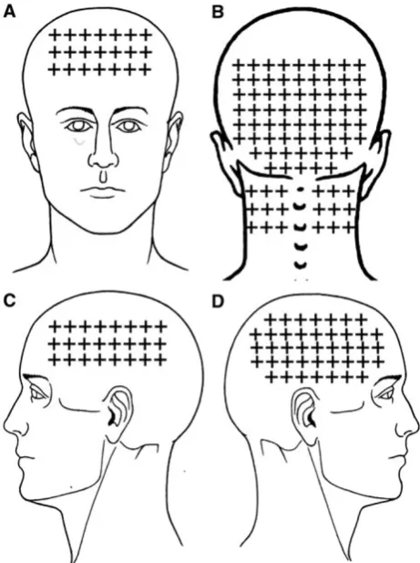
The mean spontaneous pain area reported by CTTH children was 57.7 arbitrary units (95% CI 50.0–65.6) in the frontal region (n=41, 82%), 59.8 (95% CI 44.8–74.8) in the occipital region (n =24, 58%) including the posterior part of the neck), 32.4 (95% CI 25.8–39.1) on the dominant side of the head (n=35, 70%), and 28.7 (95% CI 23.0–34.3) in the non dominant of the face (n=33, 66%). The overall spontaneous pain area of children with CTTH is illustrated in Fig.1. Significant positive correlations between headache intensity and spontaneous pain areas within dominant (rs=0.543; P\0.001) and non domi-nant (rs=0.467;P=0.005) sides were found: the higher the headache pain intensity, the larger the spontaneous pain area in both sides.
In addition, 50 age- and sex- matched children without headache, 14 boys and 36 girls, aged 6–12 years (mean: 8±1.6 years old) also participated (P=0.897).
Muscle TrPs
The mean±SD number of TrPs of each child with CTTH was 4±2, being all of them active TrPs. Healthy children showed latent, but not active, TrPs (mean±SD: 0.3±
0.7). Therefore, the total number of TrPs was significantly higher within children with CTTH (z= -8.939; P\ 0.001). In children with CTTH, a significant positive association between the number of active TrPs and head-ache duration (rs =0.315; P=0.026) was observed: the greater the number of active TrPs, the longer the duration of headache attack. No other association between the number of active TrPs and length of headache (rs=0.090; P=0.533), headache intensity (rs=0.261;P =0.067) or headache frequency (rs=0.220;P=0.125) was found.
In all the examined muscles, active TrPs were only present within children with CTTH compared to healthy children (P\0.001). The distribution of myofascial TrPs between patients and controls was significantly different for the upper trapezius (both sides: v2=14.967, P\ 0.001), sternocleidomastoid (dominant side: v2=15.674, P\0.001; non-dominant side: v2=6.383, P=0.012); temporalis (dominant: v2=61.763, P\0.001; non-dom-inant: v2=51.515, P\0.001), superficial masseter (dominant side: v2=9.538, P=0.008; non-dominant side: v2=5.263, P=0.022), suboccipital (v2=66.780, P\0.001), superior oblique (dominant: v2=9.337, P=0.009; non-dominant: v2=10.494, P=0.005), and dominant levator scapulae (v2=6.383, P=0.012) mus-cles, but not for the non-dominant levator scapulae muscle (v2=0.709,P =0.400). In fact, suboccipital muscle TrPs were the most prevalent (80%, n =40), followed by temporalis TrPs (70%,n=38 dominant side; 32%,n =16 non-dominant side) and superior oblique muscle TrPs (28%, n=14 dominant side; 30%, n=15 non-dominant side) within children with CTTH. Table1summarizes the distribution of TrPs in both children with CTTH and healthy children.
Referred pain area
The referred pain elicited by upper trapezius TrPs spread to the lateral aspect of the neck (10/10 both sides) and to the temple (8/10 both sides). Sternocleidomastoid muscle TrPs were associated with the referred pain to the temple (13/13 dominant side and 6/6 non-dominant side). Temporalis muscle TrPs referred pain to the temple perceived inside the head (38/38 dominant side, 16/16 non-dominant). The referred pain elicited by superficial masseter TrPs was perceived into the forehead (8/5 dominant side, 5/5 non-dominant side). Superior oblique muscle TrPs referred pain to the forehead (14/14 dominant side, 15/15 non-dominant side) and behind the eye (10, in both sides). Levator scapulae TrPs referred pain to the posterior part of the neck (6/6 dominant side, 4/4 non-dominant side). Finally, sub-occipital muscle TrP referred pain was perceived inside the head particularly into the forehead and behind the eyes (40/40 bilaterally). Figure2 depicts the referred pain
patterns elicited by active TrPs. In fact, the combination of the referred pain patterns from active TrPs fully reproduced the overall spontaneous clinical pain pattern in children with CTTH (Fig.1).
A 3-way ANOVA showed significant differences in referred pain areas between groups (F=18.687, P\ 0.001) and muscles (F=3.239, P=0.008), but not between sides (F =0.264; P=0.613). No significant interactions between group9muscle (F=0.382, P= 0.821), group9side (F=1.073, P=0.317), side9 muscle (F=0.209,P=0.958) or group 9muscle 9side (F=0.064,P=0.802) were found. A 2-way ANOVA for the suboccipital musculature revealed similar results (group: F=36.551, P\0.001; muscle: F=4.211, P= 0.001; group9muscle: F=1.735, P=0.406). Thus, children with CTTH had larger pain areas compared to healthy children (P\0.001). Bonferroni post-hoc analyses revealed that the referred pain area elicited by suboccipital muscle TrPs was significantly larger than the referred pain elicited from all other muscles (P\0.001), except from levator scapulae muscle TrPs (P=0.212). In addition, referred pain areas from the upper trapezius, sternocleido-mastoid, temporalis, masseter, and superior oblique muscle TrPs were not significantly different in size (P[0.312).
Table2details the size of the referred pain areas in all the examined muscles.
Finally, significant positive correlations between head-ache clinical parameters and the size of the referred pain area were found: (a) years with headache were significantly associated with the referred pain area in both upper tra-pezius muscles (rs=0.700; P=0.011); (b) headache intensity was significantly associated with the size of the referred pain area in both sternocleidomastoid muscles (rs=0.753;P=0.007); (c) the duration of headache was significantly associated with the referred pain area from both masseter (rs=0.763;P =0.017) and superior obli-que (rs=0.599; P=0.024) muscles. In summary, the greater the length of the onset of headache, the higher headache intensity, or the longer the mean duration of headache, the larger the referred pain areas elicited by TrP palpation.
Discussion
The current controlled and blinded study showed the existence of multiple active TrPs in head and neck-shoul-der muscles in children with CTTH. Both local and
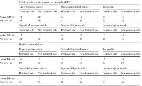
Table 1 Number of children with chronic tension type headache and healthy children (n) with muscle trigger points (trps) located in head and neck-shoulder musculature
Children with chronic tension type headache (CTTH)
Upper trapezius muscle Sternocleidomastoid muscle Temporalis
Dominant side Non dominant side Dominant side Non dominant side Dominant side Non dominant side
Active TrPs (n) 10 10 13 6 38 16
No TrPs (n) 40 40 37 44 12 34
Superficial masseter muscle Superior oblique muscle Levator scapulae muscle
Dominant side Non dominant side Dominant side Non dominant side Dominant side Non dominant side
Active TrPs (n) 8 5 14 15 6 4
No TrPs (n) 42 45 36 35 44 46
Healthy control children
Upper trapezius muscle Sternocleidomastoid muscle Temporalis
Dominant side Non dominant side Dominant side Non dominant side Dominant side Non dominant side
Latent TrPs (n) 0 0 1 0 3 0
No TrPs (n) 50 50 49 50 47 50
Superficial masseter muscle Superior oblique muscle Levator scapulae muscle
Dominant side Non dominant side Dominant side Non dominant side Dominant side Non dominant side
Latent TrPs (n) 1 0 4 4 0 2
referred pain characteristics elicited from manual palpation of active TrPs reproduced the pain pattern in all children with CTTH. Additionally, TrP referred pain areas were larger in children with CTTH than in healthy children. The
size of referred pain areas of some muscles was positively related to some headache clinical parameters.
A recent study has found that children with CTTH exhibit lesser cervical range of motion than children
Fig. 2 Referred pain from active TrPs in upper trapezius (a), sternocleidomastoid (b), temporalis (c), superficial masseter (d), suboccipital (e), combined suboccipital-upper trapezius (f), levator scapulae (g), and superior oblique (h) muscles in children with
chronic tension type headache. *Note that the posterior referred pain in figure f comes from the suboccipital muscles (?) and from the upper trapezius (*)
Table 2 Referred pain areas of myofascial trigger points in head and neck–shoulder muscles in children with chronic tension type headache and healthy control children (n=50)
Children with chronic tension type headache# Healthy controls children
Upper trapezius Dominant side (n=10) 32.5±15.8 (21.8–39.2) Dominant side (n=0) –
Non dominant side (n=10) 28.8±18.1 (18.6–39.1) Non dominant side (n=0) –
Sternocleidomastoid Dominant side (n=13) 27.1±14.6 (16.1–38.2) Dominant side (n=1) 12.2
Non dominant side (n=6) 29.1±14.9 (19.7–38.5) Non dominant side (n=0) –
Temporalis Dominant side (n=38) 32.6±16.9 (26.7–38.6) Dominant side (n=3) 14.6±8.1 (10.1–19.3)
Non dominant side (n=16) 31.1±18.1 (25.4–36.9) Non dominant side (n=0) –
Superficial masseter Dominant side (n=8) 22.9±9.3 (14.4–31.5) Dominant side (n=1) 3.4
Non dominant side (n=5) 28.7±12.9 (20.6–36.8) Non dominant side (n=0) –
Superior oblique Dominant side (n=14) 38.2±18.8 (26.9–49.4) Dominant side (n=4) 25.4±9.1 (15.2–35.6)
Non dominant side (n=15) 35.5±14.4 (24.7–46.3) Non dominant side (n=4) 26.6±8.9 (18.5–34.7)
Levator scapulae Dominant side (n=6) 52.0±11.5 (32.4–71.6) Dominant side (n=0) –
Non dominant side (n=4) 55.1±16.6 (29.7–70.4) Non dominant side (n=2) 22.8±6.1 (11.1–36.7)
Suboccipital* Bilateral (n=10) 75.4±18.3 (64.2–86.6) Bilateral (n=1) 16.2
Referred pain areas (arbitrary units) are expressed as means±standard deviation (95% confidence interval) # Significant differences between groups (P\0.001)
without headache, particularly in flexion/extension and lateral-flexion [32]. It may be possible that the presence of TrPs in the neck muscles can reduce cervical range of motion [18]. In the current study, we found active TrPs in the upper trapezius and sternocleidomastoid muscles in children with CTTH, which may reduce extension and lateral-flexion motions. Future studies should investigate the relationship between restricted cervical range of motion and the presence of active TrPs in neck and shoulder muscles in children with headache.
Referred pain from active TrPs in children with CTTH Active myofascial TrPs in head, neck and shoulder muscles elicited a referred pain that reproduced headache pain pattern in children with CTTH. When active TrPs were explored, children reported: ‘‘Yes, this is the pain that I feel during pain attacks.’’ These findings support the view that active TrPs in these muscles may be involved in the pathophysiology and manifestation of CTTH pain in chil-dren. In fact, an important finding was that children were explored headache-free, which increases the relevance of active TrPs in the development of CTTH. In addition, our results are further supported by a previous pilot study where treatment targeted at inactivating TrPs was effective for reducing the intensity, duration and frequency of headache in nine girls with tension type headache [25].
Active muscle TrPs were not found in healthy chil-dren, since they did not suffer from any pain symptoms. Nevertheless, latent TrPs were observed in a few subjects in some of the explored muscles, particularly the superior oblique muscle. It has been proposed that latent TrPs may become active under the influence of several factors such as muscle overload or strain. Therefore, the pres-ence of latent TrPs in healthy children may be potentially implicated in posterior development of pain symptom, although longitudinal studies are needed to confirm this hypothesis.
In the current study, TrPs in the suboccipital, temporalis and superior oblique muscles were the most prevalent in our sample of children with CTTH. These results are very similar to those previously found in adults with CTTH where suboccipital [20] and temporalis [22] muscles were also the most prevalent. Nevertheless, children with CTTH showed less percentage of active TrPs in the upper trape-zius muscle as compared to adults with CTTH [21].
We also found that referred pain areas from suboccipital TrPs were larger than the pain areas from the remaining muscle TrPs and that those children with CTTH showed larger areas of referred pain as compared to healthy chil-dren. Current findings support that sensitization mecha-nisms are involved in pain spreading or pain referral in children with CTTH.
Peripheral and central sensitization associated with muscle TrPs in CTTH
The results of the current study likely reflect the presence of both peripheral and central sensitization mechanisms in children with CTTH. The presence of active TrPs indicate sensitization of peripheral muscular nociceptors since high levels of chemical mediators [33, 34] and greater mechanical hypersensitivity [35, 36] have been found in active TrPs. Additionally, a study has recently demon-strated the existence of both nociceptive (hyperalgesia) and non-nociceptive (allodynia) hypersensitivity at TrPs [37]. These studies support that active TrPs constitute a focus of peripheral sensitization of both nociceptive and non-noci-ceptive nerve endings, which may constitute a peripheral nociceptive drive into the trigeminal nucleus caudalis.
In addition, referred pain phenomena from active TrPs may be also related to the presence of central sensitization. Graven-Nielsen et al. [38] found that the area of the referred pain is associated with the intensity of local pain, although this could not be shown for all the examined muscles in the present study. Discrepancies between experimental pain models and clinical studies including chronic pain patients are usually reported in the literature. Further, larger referred pain areas are also considered manifestation of central sensitization mechanisms [39]. In the current study, we showed that children with CTTH pain showed larger muscle referred pain areas in head, neck and shoulder muscle TrP as compared to healthy children, supporting a plausible role of central sensitization in muscle referred pain. In fact, current results agree with previous findings in adults with CTTH, where larger referred pain areas elicited from TrPs in the upper trapezius [21] and temporalis [22] muscles were also found. It is possible that the central sensitization is involved in larger referred pain areas elicited by active TrPs in CTTH. Interestingly, a brain imaging study found a distinct somatotopic organization of muscle referred pain areas as compared to local pain areas supporting the relevance of cortical processing in referred pain phenomenon [40]. Therefore, and not in contrast to peripheral sensitization process, central sensitization may also be involved in the generation of TrP referred pain in children with CTTH.
TrPs in those muscles innervated by C1–C3 segments or the trigeminal nerve would be responsible for peripheral nociception creating a continuous and prolonged nocicep-tive afferent barrage into the trigemino-cervical nucleus caudalis, which will sensitize the central nervous system [17]. It is possible that similar sensitization processes also occur in children with CTTH, although longitudinal studies are needed to further elucidate the role of active TrPs in children with CTTH.
We must recognize some potential limitations of this study. First, only children with CTTH were included. It would be interesting to investigate the presence of referred pain elicited by active TrPs in children with frequent epi-sodic tension type headache or migraine. In addition, we cannot establish a cause-and-effect relationship between TrPs and CTTH, because the design of the study was not longitudinal. Future controlled clinical studies should ana-lyze the effects of TrP treatment in CTTH to further elucidate the etiologic role of active TrPs in this patient population.
Conclusion
The current controlled and blinded study showed the existence of multiple active TrPs in head, neck and shoulder musculature in children with CTTH. Both local and referred pain characteristics elicited by palpation of active muscle TrPs reproduced the head pain patterns in children with CTTH. Referred pain areas elicited by active TrPs were larger in children with CTTH as compared to healthy children. The size of referred pain areas of some muscles was positively related to some headache clinical parameters. Our results support a role of active TrP in children with CTTH.
Acknowledgments This study received funds from a grand con-ceded by the Universidad Rey Juan Carlos and Comunidad de Madrid (reference CCG10-URJC BIO-5011).
Conflict of interest None.
Open Access This article is distributed under the terms of the Creative Commons Attribution License which permits any use, dis-tribution and reproduction in any medium, provided the original author(s) and source are credited.
References
1. Andlin-Sobocki P, Jonsson B, Wittchen HU, Olesen J (2005) Cost of disorders of the brain in Europe. Eur J Neurol 12(Suppl 1):1–27
2. Unalp A, Dirik E, Kurul S (2007) Prevalence and clinical findings of migraine and tension-type headache in adolescents. Pediatr Int 49:943–949
3. Laurell K, Larsson B, Eeg-Olofsson O (2004) Prevalence of headache in Swedish school-children, with a focus on tension-type headache. Cephalalgia 24:380–388
4. Zwart JA, Dyb G, Holmen TL, Stovner LJ, Sand T (2004) The prevalence of migraine and tension-type headaches among ado-lescents in Norway: The Nord-Trøndelag Health Study (Head-HUNT-Youth), a population-based epidemiological study. Cephalalgia 24:373–379
5. Heinrich M, Morris L, Kro¨ner-Herwig B (2009) Self-report of headache in children and adolescents in Germany: possibilities and confines of questionnaire data for headache classification. Cephalalgia 29:864–872
6. Russell MB, Levi N, Saltyte-Benth J, Fenger K (2006) Tension-type headache in adolescents and adults: a population based study of 33,764 twins. Eur J Epidemiol 21:153–160
7. Kroner-Herwig B, Heinrich M, Morris L (2007) Headache in German children and adolescents: a population-based epidemio-logical study. Cephalalgia 27:519–527
8. Lewis D, Ashwal S, Dahl G et al (2002) Practice parameter: evaluation of children and adolescents with recurrent headaches: report of the Quality Standards Subcommittee of the American Academy of Neurology and the Practice Committee of the Child Neurology Society. Neurology 59:490–498
9. Hershey A, Kabbouche M, Powers S (2006) Tension-type head-ache in the young. Curr Pain Headhead-ache Rep 10:467–470 10. Anttila P (2006) Tension-type headache in childhood and
ado-lescence. Lancet Neurol 5:268–274
11. Ferna´ndez-de-las-Pen˜as C, Schoenen J (2009) Chronic tension type headache: what’s new? Curr Opin Neurol 22:254–261 12. Ferna´ndez-de-las-Pen˜as C, Arendt-Nielsen L, Simons DG,
Cu-adrado ML, Pareja JA (2009) Sensitization in tension type headache: a pain model. In: Ferna´ndez-de-las-Pen˜as C, Arendt-Nielsen L, Gerwin R (eds) Tension type and Cervicogenic headache: patho-physiology, diagnosis and treatment. Jones & Bartlett Publishers, Baltimore, pp 97–106
13. Anttila P, Metsahonkala L, Mikkelsson M et al (2002) Muscle tenderness in pericranial and neck-shoulder region in chil-dren with headache: a controlled study. Cephalalgia 22: 340–344
14. Metsahonkala L, Anttila P, Laimi K et al (2006) Extra-cephalic tenderness and pressure pain threshold in children with headache. Eur J Pain 10:581–585
15. Tu¨zun EH, Karaduman A, Eker L (2005) Pressure pain thresholds in adolescent patients with chronic tension-type headache. Pain Clin 17:127–131
16. Ferna´ndez-de-las-Pen˜as C, Simons DG, Cuadrado ML, Pareja JA (2007) The role of myofascial trigger points in musculoskeletal pain syndromes of the head and neck. Curr Pain Headache Rep 11:365–372
17. Ferna´ndez-de-las-Pen˜as C, Cuadrado ML, Arendt-Nielsen L, Si-mons DG, Pareja JA (2007) Myofascial trigger points and sen-sitisation: an updated pain model for tension type headache. Cephalalgia 27:383–393
18. Simons DG, Travell J, Simons LS (1999) Travell and Simons’ Myofascial pain and dysfunction: the trigger point manual, vol 1, 2nd edn. Williams & Wilkins, Baltimore
19. Ferna´ndez-de-las-Pen˜as C, Simons DG, Gerwin R, Cuadrado ML, Pareja JA (2009) Muscle trigger points in tension type headache. In: Ferna´ndez-de-las-Pen˜as C, Arendt-Nielsen L, Gerwin RD (eds) Tension type and Cervicogenic headache: patho-physiol-ogy, diagnosis and treatment. Jones & Bartlett Publishers, Bal-timore, pp 61–76
21. Ferna´ndez-de-las-Pen˜as C, Ge H, Arendt-Nielsen L, Cuadrado ML, Pareja JA (2007) Referred pain from trapezius muscle trigger point shares similar characteristics with chronic tension type headache. Eur J Pain 11:475–482
22. Ferna´ndez-de-las-Pen˜as C, Ge H, Arendt-Nielsen L, Cuadrado ML, Pareja JA (2007) The local and referred pain from myo-fascial trigger points in the temporalis muscle contributes to pain profile in chronic tension-type headache. Clin J Pain 23:786–792 23. Ferna´ndez-de-las-Pen˜as C, Cuadrado ML, Gerwin RD, Pareja JA (2005) Referred pain from the trochlear region in tension-type headache: a Myofascial trigger point from the superior oblique muscle. Headache 45:731–737
24. Ferna´ndez-de-las-Pen˜as C, Alonso-Blanco C, Cuadrado ML, Gerwin RD, Pareja JA (2006) Myofascial trigger points and their relationship with headache clinical parameters in chronic tension type headache. Headache 46:1264–1272
25. Von Stu¨lpnagel C, Reilich P, Straube A, Scha¨fer J, Blaschek A, Lee SH, Mu¨ller-Felber W, Henschel V, Mansmann U, Heinen F (2009) Myofascial trigger points in children with tension-type Headache: A new diagnostic and therapeutic option. J Child Neurol 24:406–409
26. IHS (2004) Headache Classification Subcommittee of the Inter-national Headache Society: The InterInter-national Classification of Headache Disorders, 2nd edn. Cephalalgia 24(suppl 1):9–160 27. Phillip D, Lyngberg AC, Jensen R (2007) Assessment of
head-ache diagnosis: a comparative population study of a clinical interview with a diagnostic headache diary. Cephalalgia 27:1–8 28. Jensen MP, Turner JA, Romano JM, Fisher L (1999)
Compara-tive reliability and validity of chronic pain intensity measures. Pain 83:157–162
29. Gerwin RD, Shanon S, Hong CZ, Hubbard D, Gevirtz R (1997) Interrater reliability in myofascial trigger point examination. Pain 69:65–67
30. Svensson P, Bak J, Troest T (2003) Spread and referral of experi-mental pain in different jaw muscles. J Orofac Pain 17:214–223 31. Schmidt-Hansen PT, Svensson P, Jensen TS, Graven-Nielsen T,
Bach FW (2006) Patterns of experimentally induced pain in peri-cranial muscles. Cephalalgia 26:568–577
32. Ferna´ndez-Mayoralas DM, Ferna´ndez-de-las-Pen˜as C, Palacios-Cen˜a D, Cantarero-Villanueva I, Ferna´ndez-Lao C, Pareja JA (2010) Restricted neck mobility in children with chronic tension type headache: a blinded, controlled study. J Headache Pain 11:399–404
33. Shah JP, Phillips TM, Danoff JV, Gerber LH (2005) An in vitro microanalytical technique for measuring the local biochemical milieu of human skeletal muscle. J Appl Physiol 99:1977–1984 34. Shah JP, Danoff JV, Desai MJ, Parikh S, Nakamura LY, Phillips
TM, Gerber LH (2008) Biochemicals associated with pain and inflammations are elevated in sites near to and remote from active myofascial trigger points. Arch Phys Med Rehabil 89:16–23 35. Ferna´ndez-de-las-Pen˜as C, Caminero AB, Madeleine P,
Guillem-Mesado A, Ge HY, Arendt-Nielsen L, Pareja JA (2009) Multiple active myofascial trigger points and pressure pain sensitivity maps in the temporalis muscle are related in chronic tension type headache. Clin J Pain 25:506–512
36. Ge HY, Ferna´ndez-de-las-Pen˜as C, Madeleine P, Arendt-Nielsen L (2008) Topographical mapping and mechanical pain sensitivity of myofascial trigger points in the infraspinatus muscle. Eur J Pain 12:859–865
37. Li LT, Ge HY, Yue SW, Arendt-Nielsen L (2009) Nociceptive and non-nociceptive hypersensitivity at latent myofascial trigger points. Clin J Pain 25:132–137
38. Graven-Nielsen T, Arendt-Nielsen L, Svensson P, Jensen TS (1997) Quantification of local and referred muscle pain in humans after sequential intra-muscular injections of hypertonic saline. Pain 69:111–117
39. Arendt-Nielsen L, Graven-Nielsen T (2003) Central sensitization in fibromyalgia and other musculoskeletal disorders. Curr Pain Headache Rep 7:355–361