This is an open access journal, and articles are distributed under the terms of the Creative Commons Attribution-Non Commercial-ShareAlike 4.0 License, which allows others to remix, tweak, and build upon the work non-commercially, as long as appropriate credit is given and the new creations are licensed under the identical terms.
© 2017 Journal of Advanced Pharmacy Education & Research | Published by SPER Publication
491
The relationship between the hematologic indices after
thrombolysis in patients with myocardial infarction
Venus Shahabi Raberi
1, Reza Faramrza Zadeh
2*, Ebrahim Ezati
31Assistant Professor, Department Of Cardiology, Faculty of Medicine, Urmia University of Medical Sciences, Urmia, Iran, 2Assistant Professor, Department Of Cardiology,
Urmia University of Medical Sciences, Urmia, Iran, 3Urmia University of Medical Sciences, Urmia, Iran.
Correspondence: Reza Faramrza Zadeh, Assistant Professor, Department Of Cardiology, Urmia University of Medical Sciences, Urmia, Iran, E_mail: [email protected]
ABSTRACT
Introduction: WBC count and the platelet index (MPV) at the admission time of patients with STEMI, are the strong predictors for undesirable outcomes of patients. The objective of the present study was to determine the relationship between the hematologic indices including MPV and WBC count at the admission time of patients with ST resolution index in patients with STEMI who were treated by thrombolytic agents. Method: In this prospective study, venous blood samples of 114 patients with STEMI were taken to determine PDW, MPV and WBC count at patients' referral time and before thrombolytic therapy. According to ST resolution index in 90 minutes after thrombolytic therapy, patients were classified in two groups with ST resolution less than 50% and ST resolution of 50% or more. Finally, values of the laboratory indices above were compared between two ST-resolution groups that this issue was carried out using the SPSS version 21 software. Results: In assessing through ROC curve analysis, it was determined that measuring MPV is not considered as an accurate and effective index in predicting ST resolution after thrombolysis (Area Under Curve: 0.574). WBC count was not an applicable indicator for predicting ST resolution after thrombolysis, as well (Area Under Curve: 0.660). Finally, the results of multiple logistic regression model showed that the only predictor of ST resolution less than 50% is the history of using statins so that history of using statin increased the probability of ST resolution less than 50% for 7 times (P: 0.028, OR: 7.306). Conclusion: There is not a relationship between the occurrence of complete ST resolution (more than 50%) and the values of both MPV and WBC indices. So, measuring these two indicators may not have the enough validity for predicting ST resolution after thrombolysis. According to our study, patients with history of using statins have a lower prevalence of complete ST resolution (more than 50%).
Keywords: Hematologic indices, thrombolysis, myocardial infarction, ST segment
Introduction
In the recent decade, several observations led to the confirmation of the benefits of evaluation and monitoring of ST resolution indices after occurrence of STEMI. For the first time, Schroder et al found that ST resolution predicts the risk of mortality and congestive heart failure in patients treated by fibrinolytic therapy, effectively [1, 2]. Another studies confirmed
the relationship between the degree of ST resolution and mortality. In another study by Ito et al., it was determined that epicardial normal blood store is not sufficient for evaluation and providing adequate myocardial reperfusion [3, 4]. In fact,
new reperfusion regimens were developed to improve the limitations of two fibrinolytic and anticoagulant therapies [5, 6]
and these therapies may be particularly useful in providing coronary micro-circulation [7].
ST resolution is currently evaluated in many clinical trials and managing patients. Primary studies on ST resolution showed this fact that patients with sharper ST resolution had a lower level of infarction compared to the patients with stable ST-elevation [8]. Further studies confirmed that there is a significant
relationship between ST resolution and clinical outcomes of the patients. In a large study on 7426 patients, it became clear that two third of patients with ST resolution more than 50% were four hours after thrombolysis therapy so that these patients had
Access this article online
Website: www.japer.in E-ISSN: 2249-3379
How to cite this article: Venus Shahabi Raberi, Reza Faramrza Zadeh, Ebrahim Ezati. The relationship between the hematologic indices after thrombolysis in patients with myocardial infarction. J Adv Pharm Edu Res 2017;7(4):491-498.
492 Journal of Advanced Pharmacy Education & Research | Oct-Dec 2017 | Vol 7 | Issue4
a thirty-day mortality rate of 3.5% against the mortality of 7.4% in patients with ST resolution less than 50% [9]. On the
other hand, Schorder et al. developed a new three-part definition for the total resolution of 180 minutes ST elevation after thrombolysis which was included complete resolution (more than 70%), relative (between 30 to 70%) and without resolution (less than 30%). In trials on fibrinolytic treatments, a strong relationship between ST resolution and the reduction in the mortality of patients was observed [10]. In newer studies,
it was determined that evaluation of ST resolution even in 3 to 4 first hours after the beginning of fibrinolytic therapy, can determine the risk of death as well as heart failure in patients
[11-14]. In addition, it has been determined that patients with
complete ST resolution within 60 minutes had a much lower risk for these two consequences than those patients with ST resolution within 90 minutes [15]. Due to the slower beginning
of fibrinolytic streptokinase activation, 90 minutes is a very limited time for the creation of ST resolution and therefore 180 minutes is considered as an acceptable time to assess streptokinase activity in the creation of reperfusion [10]. In
addition, along with predicting mortality, the degree of ST resolution is capable to predict the left ventricular dysfunction and heart failure. So that the more complete ST resolution is related to the more limited size of infarction and more improvement of left ventricular function [16-18]. Similar to
mortality, the risk of heart failure in a wider and faster ST resolution cases, has been associated with a reduction in the risk of heart failure [19].
The special importance of predicting ability of ST resolution is mainly related to its relationship with epicardial blood flow. In a study, it was determined that under thrombolysis and 90 minutes after it, patients with ST resolution less than 70% had 10-fold higher mortality than the patients with ST resolution more than 70% [20]. Interestingly, there was no difference
between two groups with TIMI II and TIMI III and ST resolution in terms of being complete or relative. It was observed through monitoring that patients with faster recovery of stable ST had faster improvement and restoration of infarction that this issue has been independent from TIMI degree. In another study, ST resolution and not the grade of TIMI was the predictor of mortality and heart failure [19, 20].
Other evidences showed the prognostic value of ST resolution derived from the experiences related to primary PCI in patients with STEMI. After the successful primary PCI in patients with STEMI, continuous ST elevation had been associated with weaker recovery of left ventricular function and therefore increased mortality [19-21]. Risk of death and heart failure in
patients with increased level of ST after PCI had been increased because of increased extent of infarction [22-24]. In addition, it is
proposed that ST resolution is associated with providing tissue reperfusion. By creating the ST resolution at 90 minutes after fibrinolysis, reperfusion has been completely developed in both
epicardial and microvascular levels that leads to an excellent outcome in the patient. Another important point in this regard was the companying role of other risk factors in patients with STEMI including age, high weight, infarction, the onset of treatment, evidences of brain involvement, diabetes and hypertension with the history of coronary heart disease [25].
Interestingly, in both groups of patients with and without any of mentioned risk factors, the presence of ST resolution has still been a predictor of lower mortality in these patients [26].
Recently, it has determined that some factors such as previous cardiac biomarker level before fibrinolytic therapy can be stronger predictors of mortality alongside the lack of ST resolution [27-29]. It has also been shown that the evaluation of
myoglobin immediately before fibrinolysis along with evaluation of ST resolution 60 to 90 minutes before the therapy can provide more complete information on predicting undesirable outcomes in patients that increased myoglobin associated with the lack of 90-minutes ST resolution increase the risk of mortality up to 25 times [28].
Recently, the role of other factors and biomarkers especially hematologic factors related to ST resolution has been evaluated. As we know, platelets play a basic role in the pathogenesis of atherosclerosis and the spread of thrombotic coronary events. Platelets are mainly produced in stressful conditions such as acute coronary syndrome that platelets will be the high stimulator of B2 thromboxan production [30]. The
Mean Platelet Volume (MPV) and Platelet Distribution Width (PDW) are considered as the risk factors of MI occurrence and stroke and finally mortality in patients with STEMI [31-33]. Also,
leukocytosis plays an important role in the occurrence and progression of atherosclerosis and acute coronary syndrome [34].
But, what has been still unclear for the researchers is the role these hematologic indices in evaluating the adequacy of reperfusion following the thrombolytic therapy especially in relation to or in association with occurrence of ST resolution. Acute myocardial infarction is a clinical syndrome in which evidences of myocardial ischemia is associated with symptoms of myocardial necrosis in the electrocardiogram, biochemical tests and imaging modalities. One of the types of acute myocardial infarction is determined by ST segment elevation in the electrocardiogram and necrosis of all walls [35]. Using the
rapid, accurate and non-invasive methods to evaluate the efficacy of reperfusion and thrombolysis therapies in patients with acute myocardial infarction is an essential issue. In the recent decade, many observations have been made about the efficacy of monitoring of ST segment changes as a simple and accessible method in patients with acute myocardial infarction treated by reperfusion and ST resolution was significantly considered in this regard [39]. For the first time in 1994, it was
Journal of Advanced Pharmacy Education & Research | Oct-Dec 2017 | Vol 7 | Issue4 493
there is a strong significant relationship between the degree and severity of ST resolution and the risk of mortality following the above mentioned therapies and therefore this indicator was really considered as a prognostic measure in patients treated by reperfusion (thrombolytic and also interventional).
Both inflammatory and coagulative hematologic indicators play essential roles in the occurrence and pathogenesis of acute coronary syndrome. It has been approved in studies that there is a significant relationship between the platelet size considered as mean platelet volume and the platelet activity. On the other hand, the platelets with higher mean platelet volume (MPV) are more active in terms of metabolic and enzymatic ranges and therefore have more thrombotic effects. In addition, an increase in MPV level has been an independent risk factor for the clinical reverse-outcomes in patients with acute myocardial infarction. However, the relationship between MPV and myocardial reperfusion disorders is not still fully understood
[41].
On the other hand, the role of inflammation has been fully known as a potential risk factor for developing cardiovascular events. Leukocyte response observed during STEMI indicates the role and is considered as an essential component of systemic inflammatory response to myocardial injury. WBC count in time of admission of the patients is a strong predictor for increasing mortality and morbidity of patients with STEMI. In this regard, it is found that there is a relationship between the increase in WBC count and the reduction of epicardial and myocardial blood flow in patients with STEMI.
Along with the above studies, the present study was conducted aimed to evaluate the relationship between the hematologic indices including MPV and WBC count in the time of admission and ST resolution index in patients with STEMI treated by thrombolytics. This study was carried out in Azerbayjan Province for the first time, but its similar studies have been done in Tabriz and Yazd which are mentioned in scientific literature section.
Method
In this prospective case-control study, basic details of the patients such as demographic information, height, weight and body mass index (BMI), history of heart disease risk factors such as familial history of heart diseases, hypertension, hyperlipidemia, diabetes mellitus and smoking, history of drug use especially using heart drugs, previous history of cardiovascular interventions, history of myocardial infarction, history of heart failure, previous history of cerebrovascular events, history of renal failure, history of peripheral arterial hypertension and history of chronic obstructive pulmonary disease were extracted from the patients' records data and registered in the special questionnaire of the project. At the patients' referral time and prior to thrombotic therapy, venous blood samples were taken to measure PDW, MPV indices and WBC count and data were registered in the patients' records.
Patients were treated by thrombolysis in an equal condition (including streptokinase 1.5 million units in 20-30 minutes) with a German-made Streptase drug. ST elevation was measured from the J point in ST segment as well as in two electrocardiograms during the initial hospitalization and 90 minutes after thrombotic therapy. According to ST resolution index, 90 minutes after thrombotic therapy, patients were classified in two groups of ST resolution less than 50% and ST resolution of 50% or more. Finally, values of the above laboratory indices were compared between two ST resolution groups.
Criteria to enter the study:
The presence of STEMI diagnostic criteria are based on clinical symptoms (continues typical chest angina for more than 30 minutes), ECG changes (ST segment elevation more than 2 mm in pre-cordial lids and more than 1 mm in limb lids in more than two adjacent lids) and increased cardiac enzymes, 2) receiving thrombolytic treatments, 3) the first occurrence of myocardial infarction.
Criteria to exit the study:
1)Delay to visit for more than 6 hours from the onset of symptoms, 2) presence of LBBB, 3) presence of ventricular tachycardia arrhythmias and ventricular fibrillation, 4) rhythm resulted from the pacemaker, 5) incomplete or non-judgmental electrocardiogram, 6) cases of thrombolytic inhibition including fibrinolytic therapy within the recent 24 hours, anti-coagulant therapy, having congenital hemorrhagic factors, renal insufficiency, history of malignancy, history of chronic inflammatory disease and or active infection, intra-ventricular conduction abnormalities, aortic dissection, systolic hypertension higher than 180 mmHg, history of trauma to the head or any surgery during the recent 3 months, history of thrombocytopenia, presence of cardiogenic shock, 7) presence of concurrent internal or infectious diseases effective on the laboratory parameters under the study, 8) patients who already have aspirin and clopidogrel.
Finally, SPSS version 21 software was used to analyze the data of above study.
Results
494 Journal of Advanced Pharmacy Education & Research | Oct-Dec 2017 | Vol 7 | Issue4
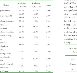
factors of heart diseases, prevalence of diabetes was 23.3% and 9.9% in the groups with resolutions less than 50% and more than 50%, respectively that was significantly higher in the first group (P: 0.052). The prevalence of hypertension in the groups with the resolutions less and more than 50% were 44.2% and 18.3% respectively and it was significantly higher in the first group (P" 0.003). In terms of the smoking history, the prevalence of smoking in the groups with resolutions less and more than 50% were 37.2 and 35.2%, respectively that no difference was seen in those groups (P: 0.829). In terms of using opium in the groups with resolutions less and more than 50%, it was 11.6% and 8.5% respectively and without a difference between those groups, as well (P: 0.745). In terms of hyperlipidemia, the prevalence of this complication in the groups with resolutions less and more than 50% was 11.6% and 7%, respectively and without a difference between those groups, as well (P: 0.402). Prevalence of MI in the groups with resolutions less and more than 50% was 14% and 4.2%, respectively and without a difference between those groups, as well (P: 0.07). Prevalence of pulmonary diseases in the groups with resolutions less and more than 50% was 4.7% and 5.6.2%, respectively and without a difference between those groups, as well (P: 0.999). In terms of familial cardiovascular diseases, this history in the groups with resolutions less and more than 50% was 4.7% and 12.7%, respectively and without a difference between those groups, as well (P: 0.209). Also, in terms of prevalence of peripheral vascular disease, this prevalence in the groups with resolutions less and more than 50% was 4.7% and 0%, respectively and without a difference between those groups, as well (P: 0.14).
Table 1: Basic details of patients with ST resolution of less and more than 50%
Details Resolution
less than 50%
Resolution
more than 50% p-value
Frequency of men 69.8% 90.1% 0.006
Mean age (years old) 62.07 ±12.96
56.32
±9.29 0.007
Prevalence of diabetes 23.3% 9.9% 0.052
Prevalence of
hypertension 44.2% 18.3% 0.003
Prevalence of smoking 37.2% 35.2% 0.829
Prevalence of using
opium 11.6% 8.5% 0.745
Prevalence of
hyperlipidemia 11.6% 7.0% 0.402
Prevalence of
pulmonary diseases 4.7% 5.6% 0.999
History of cardiac
diseases 4.7% 12.7% 0.209
History of peripheral
vascular diseases 4.7% 0% 0.140
Using of statins 16.3% 2.8% 0.026
Using β blockers 0 4.2% 0.289
Using ACE inhibitors 18.6% 9.9% 0.181
Using Ca blockers 0 8.5% 0.082
Using anti-diabetic
drugs 14.0% 5.6% 0.174
Frequency of using
diuretic drugs 0 1.4% 0.999
Mean of MPV 9.42
±0.77
9.59
±1.00 0.377
Mean of WBC count 10027.91 ±2433.04
10520.00
±2800.03 0.343
In terms of using the cardiovascular drugs, the prevalence of using statins in the groups of resolution less and more than 50% was 16.3% and 2.8%, respectively that in the first group was significantly higher than the second group (P:0.026). Frequency of beta blockers in the groups of resolution less and more than 50% was 0% and 4.2%, respectively that there was no difference between both groups (P: 0.289). Frequency of using ACE inhibitors in the groups of resolution less and more than 50% was 18.6% and 9.9%, respectively and without any difference between two groups (P: 0.181). Similarly, using the Ca blockers in the groups of resolution less and more than 50% was evaluated as 0% and 8.5%, respectively and with no difference between two groups, as well (P: 0.082). Frequency of using anti-diabetic drugs in the groups of resolution less and more than 50% was 14% and 5.6%, respectively that the difference between the groups was not significant (P: 0.174). Also, frequency of using diuretic drugs in the groups of resolution less and more than 50% was 0% and 1.4%, respectively and without any difference between two groups, again (P: 0.999) (Table 1).
Mean of MPV in patients with resolution less than 50% was 9.42±0.77 as well as 9.59±1.00 in the patients with resolution more than 50% that the difference between two groups was not significant (P: 0.377). Means of WBC count in patients with resolution less and more than 50% were 1027.91±2433.04 and 10520.00±2800.03, respectively and the difference between two groups was not significant (P: 0.343) (Table 1).
In the evaluation with multiple logistic regression, the only predictor of ST resolution less than 50% was using statins so that the history of using statin increased the probability of ST resolution less than 50% for about 7 times (P: 0.028, OR: 7.306).
Table 2: Logistic regression model in determining the factors related to resolution less than 50%
Index Beta coefficient SE p-value OR
Gender -0.584 0.630 0.354 0.558
Journal of Advanced Pharmacy Education & Research | Oct-Dec 2017 | Vol 7 | Issue4 495
diabetes 0.299 0.651 0.646 1.349
Hypertension 0.837 0.517 0.105 2.310
Using statin 1.989 0.902 0.028 7.306
In the evaluation by ROC curve analysis, it was found that determination of MPV is not considered as an accurate and efficient indicator in predicting ST resolution after thrombolysis (area under curve: 0.574). Determining WBC count was not an applicable indicator for predicting ST resolution after thrombolysis, as well (area under curve: 0.660) (charts 1, 2).
Chart 1:AUC-ROC for MPV to differentiate the resolutions
less and more than 50%
Chart 2: AUC-ROC for MPV to differentiate the resolutions
less and more than 50%
Discussion
and conclusion
First of all, the purpose of this study was to evaluate the difference between MPV and WBC indices in two groups of patients with ST resolution less and more than 50% after thrombolysis. In fact, the objective was to find if these two measures have an acceptable value to differentiate ST resolutions less and more than 50% after thrombolysis or not or on the other hand, whether a cut-off point is obtained that considering that cut-off point for two MPV and WBC indicators, ST resolution less and more than 50% can be differentiated with acceptable sensitivity and specificity or not. Despite the observed numerical difference in two MPV and WBC indicators between two groups of ST resolution less and more than 50%, this difference was not statistically significant according to this study. Also, in the evaluation of area under ROC curve (AUC-ROC), it was shown that none of these indicators are valuable enough to differentiate ST resolutions less and more than 50% and therefore a cut-off point with acceptable sensitivity and specificity for such this differentiation ability cannot be determined. Unlike some conducted studies, our study was not able to determine the relationship between MPV as well as WBC with ST resolution. In Ghaffari et al. study, patients with MPV higher than 8.2 fl had lower ST resolution and fewer acute heart failures. In the evaluation with regression model, MPV was the predictor for ST resolution as well as MACE occurrence [35] that was completely contrary to
our study. In the study of Kirbas et al., MPV value in patients with ST resolution less than 50% was lower than it in patients with ST resolution more than 50% and based on ROC curve analysis, the cut-off point of 9.3 fl for MPV was the strong predictor for predicting ST resolution with the sensitivity of 66.7% and 77.9% [41] that was not contrary to our study, as
well. In Varasteh et al. study, patients with ST resolution less than 70% had higher values of MPV, PDW and WBC count. The best cut-off point of MPV for predicting ST resolution less
than 70% was 10.05 fl with the sensitivity and specificity of 71.8% and 80.9%, respectively. Also, the best cut-off point of WBC count for predicting ST resolution less than 70% was 12.65 per thousand with the sensitivity and specificity of 42.9% and 82.7%, respectively [42]. Of course, the studies
conducted about the relationship between ST resolution and values of the laboratory indicators are very few. In general, it seems that the disapproval reason of the relationship between ST resolution and values of MPV and WBC indicators can be the following items. First, determination of cut-off point for ST resolution has been different in various studies so that in some studies the cross section of 50% was considered and in the others, 70% was considered that can be very effective in the significance of differences between the groups. Second, the time range was mainly 1 to 4 hours in order to determine resolution after thrombolysis which affects the laboratory indices and occurrence of a complete resolution (which was less than 6 hours in our study). Third, type of thrombolysis therapy as well as its associated treatments may be effective on the occurrence of complete resolution after thrombolysis. It is important to mention that in the above studies, the history of using aspirin and clopidogrel was not included in the exclusion criteria while in our study, patients with history of using these drugs excluded from the study and this point can be effective in the results of the study. Also, type of using technique and its accuracy estimating the laboratory indices will be an important and effective factor in determining the relationship among the indices and occurrence of a complete resolution. So, with the presence of possible contingency indicators mentioned above, it is not possible to find a significant relationship between two MPV and WBC laboratory indices and the occurrence of a complete resolution.
In this regard, history of using statin was only related to resolution occurrence among all basic indices of patients and multiple logistic regression. The obtained result was the reverse finding of Varasteh et al. in which among all patients' indices, only the history of using statins was different between two groups of with and without resolution was different, but the group with the use of statin ST resolution more than 50%
[42]. Of course, the cause of this relationship was not evaluated
in their study, as well. In some studies, using statins was associated with the improvement of ST resolution [42].
Although, in some other studies, there was not a significant relationship between the history of using statins and occurrence of improving complete ST resolution [44,43]. Maybe, the
496 Journal of Advanced Pharmacy Education & Research | Oct-Dec 2017 | Vol 7 | Issue4
Therefore, there are not enough evidences that show the assessment of these indicators have the enough value for predicting ST resolution after thrombolysis based on this study. According to our study, patients with history of using statins have lower prevalence of complete ST resolution (more than 50%).
References
1. Schröder R, Dissmann R, Bruggemann T; Extent of early ST segment elevation resolution. a simple but strong predictor of outcome in patients with acute myocardial infarction. J Am Coll Cardiol. 24 1994:384-391.
2. Schröder R, Wegscheider K, Schroder K, Dissmann R, Meyer-Sabellek W, for the INJECT Trial Group. Extent of early ST segment elevation resolution: a strong predictor of outcome in patients with acute myocardial infarction and a sensitive measure to compare thrombolytic regimens. A substudy of the International Joint Efficacy Comparison of Thrombolytics (INJECT) trial. J Am Coll Cardiol 1995; 26:1657–64.
3. Ito H, Tomooka T, Sakai N; Lack of myocardial perfusion immediately after successful thrombolysis. a predictor of poor recovery of left ventricular function in anterior myocardial infarction. Circulation. 85 1992:1699-1705. 4. Ito H, Maruyama A, Iwakura K; Clinical implications of
the “no reflow” phenomenon. a predictor of complications
and left ventricular remodeling in reperfused anterior wall myocardial infarction. Circulation. 93 1996:223-228. 5. Ohman E.M, Kleiman N.S, Gacioch G; Combined
accelerated tissue-plasminogen activator and platelet glycoprotein IIb/IIIa integrin receptor blockade with integrilin in acute myocardial infarction. Circulation. 95 1997:846-854.
6. Antman E.M, Giugliano R.P, Gibson C.M; Abciximab facilitates the rate and extent of thrombolysis. results of TIMI 14 trial. Circulation. 99 1999:2720-2732.
7. Strategies for Patency Enhancement in the Emergency Department (SPEED) Group. Trial of abciximab with and without low-dose reteplase for acute myocardial infarction. Circulation. 101 2000:2788-2794.
8. Neumann F.J, Blasini R, Schmitt C; Effect of glycoprotein IIb/IIIa receptor blockade on recovery of coronary flow and left ventricular function after the placement of coronary-artery stents in acute myocardial infarction. Circulation. 98 1998:2695-2701.
9. De Lemos J.A, Antman E.M, Gibson C.M; Abciximab improves both epicardial flow and myocardial reperfusion in ST elevation myocardial infarction. observations from the TIMI 14 trial. Circulation. 101 2000:239-243. 10. Schröder R, Zeymer U, Wegscheider K, Neuhaus K.L;
Comparison of the predictive value of ST segment
elevation resolution at 90 and 180 min after start of streptokinase in acute myocardial infarction. a substudy of the Hirudin for Improvement of Thrombolysis (HIT)-4 study. Eur Heart J. 20 1999:1563-1571.
11. Purcell I.F, Newall N, Farrer M; Change in ST segment elevation 60 minutes after thrombolytic initiation predicts clinical outcome as accurately as later electrocardiographic changes. Heart. 78 1997:465-471.
12. Carlsson J, Kamp U, Hartel D; Resolution of ST-segment elevation in acute myocardial infarction—early prognostic significance after thrombolytic therapy. results from the COBALT trial. Herz. 241999:440-447.
13. De Lemos J.A, Antman E.M, Giugliano R.P; Very early risk stratification after thrombolytic therapy with a
bedside myoglobin assay and the 12-lead
electrocardiogram. Am Heart J. 140 2000:373-378. 14. De Lemos J.A, Antman E.M, Giugliano R.P; Comparison
of a 60- versus 90-minute determination of ST-segment resolution after thrombolytic therapy for acute myocardial infarction. Am J Cardiol. 86 2000:1235-1237.
15. Matetzky S, Freimark D, Chouraqui P; The distinction between coronary and myocardial reperfusion after thrombolytic therapy by clinical markers of reperfusion. J Am Coll Cardiol. 32 1998:1326-1330.
16. Saran R, Been M, Furniss S, Hawkins T, Reid D; Reduction in ST segment elevation after thrombolysis predicts either coronary reperfusion or preservation of left ventricular function. Br Heart J. 64 1990:113-117. 17. Matetzky S, Novikov M, Gruberg L; The significance of
persistent ST elevation versus early resolution of ST segment elevation after primary PTCA. J Am Coll Cardiol. 34 1999:1932-1938.
18. Shah A, Wagner G.S, Granger C.B; Prognostic implications of TIMI flow grade in the infarct related artery compared with continuous 12-lead ST-segment
resolution analysis. reexamining the “gold standard” for
myocardial reperfusion assessment. J Am Coll Cardiol. 35 2000:666-672.
19. Andrews J, Straznicky I.T, French J.K; ST-segment recovery adds to the assessment of TIMI 2 and 3 flow in predicting infarct wall motion after thrombolytic therapy. Circulation. 101 2000:2138-2143.
20. Van’t Hof A, Liem A, de Boer M, Zijlstra F; Clinical value
of 12-lead electrocardiogram after successful reperfusion therapy for acute myocardial infarction. Lancet. 350 1997:615-619.
Journal of Advanced Pharmacy Education & Research | Oct-Dec 2017 | Vol 7 | Issue4 497
22. Claeys M.J, Bosmans J, Veenstra L, Jorens P, De Raedt H, Vrints C.J; Determinants and prognostic implications of persistent ST-segment elevation after primary angioplasty for acute myocardial infarction. importance of microvascular reperfusion injury on clinical outcome. Circulation. 99 1999:1972-1977.
23. Dissmann R, Linderer T, Goerke M, von Ameln H, Rennhak U, Schroder R; Sudden increase of the ST segment elevation at time of reperfusion predicts extensive infarcts in patients with intravenous thrombolysis. Am Heart J. 126 1993:832-839.
24. Kondo M, Tamura K, Tanio H, Shimono Y; Is ST segment re-elevation associated with reperfusion an indicator of marked myocardial damage after thrombolysis?. J Am Coll Cardiol. 21 1993:62-67. 25. Miida T, Oda H, Toeda T, Higuma N; Additional
ST-segment elevation immediately after reperfusion and its effect on myocardial salvage in anterior wall acute myocardial infarction. Am J Cardiol. 73 1994:851-855. 26. Morrow D, Antman A, Charlesworth A; The TIMI risk
score for ST elevation myocardial infarction. a convenient, bedside, clinical score for risk assessment at presentation: An InTIME II substudy. Circulation. 102 2000:2031-2037.
27. Ohman E.M, Armstrong P, Christenson R.H; Cardiac troponin T levels for risk stratification in acute myocardial ischemia. N Engl J Med. 335 1996:1333-1341.
28. Stubbs P, Collinson P, Moseley D, Greenwood T, Noble M; Prognostic significance of admission troponin T concentrations in patients with myocardial infarction. Circulation. 94 1996:1291-1297.
29. Ohman E.M, Armstrong P.W, White H.D; Risk stratification with a point-of-care cardiac troponin T test in acute myocardial infarction. Am J Cardiol. 84 1999:1281-1286.
30. Van der Loo B, Martin JF. A role for changes in platelet production in the cause of acute coronary. Arterioscler Thromb Vasc Biol. 1999; 19:672–9.
31. Huczek Z, Kochman J, Filipiak KJ, Horszczaruk GJ, Grabowski M, Piatkowski R, et al. Mean platelet volume on admission predicts impaired reperfusion and long-term mortality in acute myocardial infarction treated with primary percutaneous coronary intervention. J Am Coll Cardiol. 2005; 46:284–90.
32. Massberg S, Schulz C, Gawaz M. Role of platelets in the pathophysiology of acute coronary syndrome.Semin Vasc Med. 2003;3:147–62.
33. Sabatine MS, Morrow DA, Cannon CP, Murphy SA, Demopoulos LA, DiBattiste PM, et al. Relationship between baseline white blood cell count and degree of coronary artery disease and mortality in patients with acute coronary syndromes: A TACTICS-TIMI 18 (Treat
angina with aggrastat and determine cost of therapy with an invasive or conservative strategy- thrombolysis in myocardial infarction 18 trial) substudy. J Am Coll Cardiol. 2002; 40:1761–8.
34. Vagdatli E, Gounari E, Lazaridou E, Katsibourlia E, Tsikopoulou F, Labrianou I. Platelet distribution width: A simple, practical and specific marker of activation of coagulation. Hippokratia. 2010; 14:28–32.
35. Khademvatani K, Basiri M, Alinejad V, Seyed Mohammad Zad MH, Prevalence and correlates of aortic root dilatation in patients with essential hypertension admitted to Seyedoshohada Hospital, Urmia 2012-2013. Journal of Global Pharma Technology. 2016; 02(8):01-06.
36. Seyed Mohammad Zad MH, khalili N, Alinejad V, Khadem Vatani K, Is There a Correlation Between Coronary Artery Ectasia and Neutrophil-Lymphocyte Ratio? Journal of Global Pharma Technology. 2016; 02(8):01-06.
37. Khademvatan K, Alinejad V, Eghtedar S, Rahbar N, Agakhani N. Survey of the relationship between metabolic syndrome and myocardial infarction in hospitals of Urmia University of medical sciences. Glob J Health Sci. 2014 Sep 18;6(7 Spec No):58-65. doi: 10.5539/gjhs. v6n7p58. 38. Heris SO, Rahimi B, Faridaalaee G, Hajahmadi M, Sayyadi H, Naghipour B, QT dispersion after thrombolytic therapy. International Cardiovascular Research Journal, Volume 8, Issue 4, 1 December 2014, Pages 161-165 39. Rahimi Darabad B, Vatandust J, Pourmousavi Khoshknab
MM,Hajahmadi Poorrafsanjani M. Survey of the effect of opioid abuse on the extent of coronary artery diseases. Global journal of health science, Volume 6, Issue 7, 2014, Pages 83-91.
40. Hajahmadi Poorrafsanjani M, Rahimi Darabad B, Evaluate the sensitivity and specificity echocardiography in trans-Doppler and tissue trans-Doppler method in the estimation of left ventricular end-diastolic pressure. Global journal of health science, Volume 6, Issue 7, 2014, Pages 92-97.
41. Kırbaş Ö1, Kurmuş Ö, Köseoğlu C, Duran Karaduman
B, Saatçi Yaşar A, Alemdar R,et al. Association between admission mean platelet volume and ST segment resolution after thrombolytic therapy for acute myocardial infarction. Anadolu Kardiyol Derg. 2014 Dec;14(8):728-32.
498 Journal of Advanced Pharmacy Education & Research | Oct-Dec 2017 | Vol 7 | Issue4
43. Kim JS, Kim J, Choi D, Lee CJ, Lee SH, Ko YG, et al. Efficacy of high-dose atorvastatin loading before primary percutaneous coronary intervention in ST-segment elevation myocardial infarction: the STATIN STEMI trial. JACC Cardiovasc Interv. 2010;3:332–339
44. Woo JS1, Cho JM, Kim SJ, Kim MK, Kim CJ Combined Assessments of Biochemical Markers and ST-.
Segment Resolution Provide Additional