Lidia Usnarska-Zubkiewicz
1, a, c, d, Jadwiga Hołojda
2, b, c, Michał Jeleń
3, b,
anna Zubkiewicz-Zarębska
4, b, g, Jakub dębski
1, b,
Kazimierz Kuliczkowski
1, a, fThe Occurrence of AL Amyloidosis
(Light-Chain Amyloidosis) in Patients
with Multiple Myeloma in Lower Silesia Region, Poland
1 department of Hematology, blood Neoplasms and bone Marrow Transplantation, Wroclaw Medical University,Poland
2 Hematology department of district Specialist Hospital, Legnica, Poland
3 division of Pathomorphology and Oncological cytology, Wroclaw Medical University, Poland 4 department of Infectious diseases, Hepatology and acquired Immune deficiencies, Wroclaw Medical
University, Poland
A – research concept and design; B – collection and/or assembly of data; C – data analysis and interpretation;
D – writing the article; E – critical revision of the article; F – final approval of article; G – other
Abstract
Background. The incidence of amyloidosis is difficult to determine because the disease is often undiagnosed or diagnosed incorrectly. In Polish studies, there are no statistics and analyses of the factors that may influence the development of amyloidosis in patients with multiple myeloma
Objectives. The goal of this study was to estimate the incidence of aL amyloidosis in MM patients in Lower Silesia region.
Material and Methods. 70 patients treated at the department of Hematology, Provincial Hospital in Legnica and the department of Hematology, blood Neoplasm and bone Marrow Transplantation, Medical University in Wroclaw were enrolled in the survey. 37 patients were newly diagnosed, 33 had been treated for 2–34 months. The basis for the diagnosis of amyloidosis was the presence of green colored amyloid deposits in the polarized light microscope in the adipose tissue (received from abdominal fold and stained with congo red).
Results. amyloidosis was diagnosed in 18 (25.7%) patients with MM, 9/9 f/M, aged 47–83 years. 6 (33%) pts with amyloidosis had newly diagnosed MM, in 12 (67%) progression of the disease was diagnosed. amyloidosis occurred significantly more often (p = 0.048) in already treated patients. The odds ratio (OR) was 2.95. amyloidosis occurred most frequently in patients with Igg myeloma (67%), (OR = 1.98), was more often found in patients with kappa light chain versus lambda, respectively 67% and 33%. The probability of amyloidosis in patients with clini-cal stage III was 1.5 times higher (p = 0.05) than in other stages (OR = 1.5), in persons with renal dysfunction was twice as high (OR = 2.4) compared to the renal competence group (p = 0.05).
Conclusions. aL amyloidosis in the course of MM occurs in Lower Silesia region with a comparable rate to other regions of the world. It is significantly more often diagnosed in patients with relapsed or refractory disease, in per-sons with clinical stage III and with renal failure (Adv Clin Exp Med 2014, 23, 2, 235–244).
Key words: amyloidosis, multiple myeloma, epidemiology, Lower Silesia, Poland.
adv clin Exp Med 2014, 23, 2, 235–244 ISSN 1899–5276
ORIgINaL PaPERS
© copyright by Wroclaw Medical University
amyloidosis is an inherited or acquired sys-temic storage disease in which pathologic, amor-phous substance produced as a result of abnormal protein metabolism and resistant to proteolysis is deposited in the extracellular space of various tissues (intracellular deposits occur rarely). This leads to the destruction of normal tissue structure
and disturbs its function. The disease may affect one or several organs simultaneously and is always fatal [1-3].
in the course of myeloma (and other monoclo-nal gammapathies). Kyle demonstrated that 85% of patients diagnosed with aL amyloidosis meet the criteria of various forms of plasma cell dyscra-sias, while the remaining 15% lack clinical symp-toms of plasma cells clonal growth. [2] according to abraham, all patients with aL amyloidosis re-veal monoclonal gammapathy on the basis of se-rum free light chains evaluation. [4] aL amyloido-sis affects organs originating from the mesoderm tissue (the heart, digestive tract, peripheral ner-vous system, skin, tongue).
The incidence of amyloidosis is difficult to de-termine because the disease is often undiagnosed or diagnosed incorrectly. In order to determine aL amyloidosis prevalence, Kele et al. analyzed the oc-currence of the disease in one district in Minne-sota in the period from 1952 to 1992. They found the annual incidence of amyloidosis in 8.9 per mil-lion in the region. [5] This was the basis for the estimates, according to which, every year, in the United States, 1275–3200 new cases of aL amyloi-dosis are recognized, which is 5.1–12.8 cases per million people. Studies have shown that in the U.S. and Western Europe, systemic aL amyloidosis is more common. [6] The incidence of this disease is estimated at 0.8–1/100 000 persons / year, patients aged 50–70 years. [7]
There is no center in Europe that would re-cord cases of amyloidosis. The data published in each country determines the incidence of amyloi-dosis in the particular region. [8] The incidence of aL amyloidosis in the UK is 600 per year. Occa-sionally, there is a concomitant amyloidosis with IgM paraproteinemia. according to the report of the UK National amyloidosis centre, in the peri-od from 1988 to 2006, this kind of incidence was observed in 103 patients.
Studies have shown that in 10–20% of patients with multiple myeloma contributes to the develop-ment of aL amyloidosis. [9,10] Primary aL amy-loidosis also occurs in 7% of patients with nonhe-matologic cancers. [11]
amyloidosis is a rare disease which affects pa-tients before the age of 40 and is more common in men (50–65%). Those aged 50–70 years accounted for 60% of patients. [7] The primary amyloidosis confirmatory diagnostic test is a biopsy of adipose tissue from the abdominal fold and a demonstra-tion in the downloaded material (after staining with congo red light) a characteristic birefractive polarized light, or unbranched fibrous structures with a diameter of 10 nm by an electron micros-copy. If the test result is negative, the biopsies of salivary gland, rectal mucosa, gums or bone are suggested. Percentage identification of amyloid in other locations is varied: biopsies of kidney, spleen
or liver are positive in more than 90%, of the as-pirations of abdominal fat in 60–80%, rectal bi-opsy in 50–70%, bone marrow bibi-opsy in 50–55%, whereas skin biopsy is positive in 50–80%. [12] To visualize the amyloid deposits, radioisotope meth-ods are used e.g. technetium Tc 99m which proves particularly useful in cardiac amyloidosis identifi-cation. It is applied in a very advanced stage of car-diomyopathy and, along with echocardiography, provides a diagnosis basis. Scintigraphy is anoth-er diagnostic method of amyloid deposits. It takes advantage of the co-existence of amyloid deposits with normal serum protein SaP, which can be la-beled with technetium or iodine (I123).
The most common clinical manifestations of amyloidosis include loss of body weight, ankle swelling, hoarseness, paresthesia, fatigue, orthos-tatic blood pressure drops, heart failure, enlarged liver or tongue and carpal tunnel syndrome [13]. In the course of aL amyloidosis, kidneys and the heart are mostly affected, and the development of peripheral neuropathy as well as damage of the central nervous system are observed. [6] according to Meller, kidney damage in the form of nephrotic syndrome occurs in 28% of patients, heart failure and peripheral neuropathy in 17%, carpal tunnel syndrome in 21%, orthostatic hypotension in 11%, and orbital purpura in 15% of patients [14].
The goal of this study was to estimate the inci-dence of aL amyloidosis in patients with multiple myeloma in the province of Lower Silesia, Poland.
Material and Methods
The study was approved by the Ethics com-mittee of the Medical University of Wroclaw. Each patient received detailed information about the purpose of the tests and signed a consent form. 70 patients with multiple myeloma treated at the department of Hematology, Provincial Hospital in Legnica and the department of Hematology, blood Neoplasms and bone Marrow Transplanta-tion, Medical University in Wroclaw were enrolled in the survey. among them, 37 pts were newly di-agnosed and had not been treated and 33 per-sons had been treated for 2–34 months with Vad, cVMbP, cTd or MP.
tissue (received from abdominal fold and stained with congo red).
basic descriptive statistics were used to de-scribe all quantitative variables. The arithmetic mean, standard deviation, minimum and max-imum values were used. a comparison of data among the groups was performed using the exact fischer test, the chi-square test, as well as the t-stu-dent test. The Kaplan-Meyer curve was used for timating the overall survival (OS). In order to es-timate the thresholds of quantitative variables for patient differentiation, Receiver Operating char-acteristic (ROc) analysis was performed. The odds ratio (OR) was calculated for each threshold. In all analyses carried out, P value 0.05 was considered statistically significant. Statistical evaluation was performed using Microsoft Office EXcEL and Sta-tistica software.
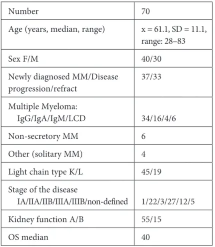
The characteristics of the study group are pres-ent in Table 1.
Test of Amyloid
in the Adipose Tissue
aspirate material was mottled on a slide and after 1 h of drying, it was congo red stained and then evaluated in a polarized light microscope.
Results
The Occurrence of Amyloidosis
in the Study Group
amyloidosis was diagnosed in 18 (25.7%) pa-tients with multiple myeloma, nine women and nine men, aged 47–83 years, Me = 66, x = 63.5 years, Sd = 9.4.
Example of green glowing amyloid plaque in polarizing microscope light is presented in fig. 1.
Table 1. clinical and laboratory data of the patients
Number 70
age (years, median, range) x = 61.1, Sd = 11.1, range: 28–83
Sex f/M 40/30
Newly diagnosed MM/disease
progression/refract 37/33 Multiple Myeloma:
Igg/Iga/IgM/Lcd 34/16/4/6 Non-secretory MM 6 Other (solitary MM) 4 Light chain type K/L 45/19 Stage of the disease
Ia/IIa/IIb/IIIa/IIIb/non-defined 1/22/3/27/12/5 Kidney function a/b 55/15
OS median 40
* Lcd – light chain disease.
Material
The adipose tissue samples were harvested by needle aspiration of the abdominal area, and 10 mL of blood were collected to clot to obtain serum.
The analysis used the results of routine testing in all patients to determine the disease stage and was performed according to the standards of the hospital laboratory. Historical data of co-existent diseases were used and, in the case of refractory or relapse, multiple myeloma patients, information about prior causative treatment was exploited.
Fig. 1. amyloid deposits stained with congo red in polarizing microscope light in a multiple myeloma patient
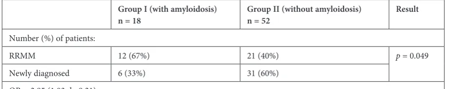
Six (33%) patients with amyloidosis had new-ly diagnosed MM, in the remaining 12 (67%) pro-gression or relapse of the disease was diagnosed. In the study group, amyloidosis occurred signif-icantly more often (p = 0.048) in already treated patients with the disease refract or progression. In the patients with refractory MM the odds ratio (OR) was 2.95 (95% confidence interval: from 1.03 to 9.21.) This means that the probability of amyloi-dosis in patients with relapsed and refractory dis-ease was 3 times greater than in the case of newly diagnosed ones (Table 2).
In 52 (74.3%) patients with myeloma, includ-ing 31 women aged 28–81 years, Me – 62 years,
the group of multiple myeloma without amyloido-sis. among MM patients without amyloidosis, there were 31 (60%) persons with newly diagnosed and 21 (40%) with refractory/relapsed MM (RRMM).
Comparison of Selected
Clinical Parameters in Patients
with Multiple Myeloma
with Amyloidosis
and Without this Complication
In the study group, amyloidosis occurred most frequently in Igg MM pts (67%). The probabili-ty of amyloidosis in patients with this probabili-type of my-eloma was two times higher than in other patients (OR = 1.98) (Table 3). Quite similarly, amyloi-dosis was more often found in patients with kap-pa light chain versus lambda, respectively 67% and 33%. The probability of amyloidosis in patients with clinical stage III was one and a half times higher (p = 0.05) than in other stages (OR = 1.5). The per-centage of patients with renal failure in amyloid and no amyloidosis groups was respectively 28% and 19% The probability of amyloidosis presence in patients with renal dysfunction was twice as high (OR = 2.4) compared to the renal competence group (p = 0.05). In patients with multiple myeloma com-plicated by renal failure and amyloidosis, renal amy-loidosis should be considered as a cause of renal fail-ure. during the study, however, no renal biopsy was performed revealing glomerular lesions.
There was no statistically significant correla-tion between amyloidosis prevalence and patients’ age and sex.
Analysis of Amyloidosis
Symptoms in the Study Group
abnormalities characteristic for amyloidosis were considered. The incidence of symptoms in patients with MM complicated by amyloidosis and without this complication was compared. No sig-nificant differences were analyzed in the manifes-tation of symptoms. Some of the symptoms, such
as periorbital ecchymosis or large tongue specta-cles, were present only in the group with amyloi-dosis (Table 4).
Occurrence of Amyloidosis in
Patients with Refractory/
/Relapsed Myeloma Depending
on the Duration and the Type
of Cytostatic Therapy
a separate analysis was made for a group of 33 patients with refractory and/or relapsed multi-ple myeloma, including 12 patients with amyloi-dosis and 21 without this complication. The dura-tion of cytostatic treatment, the number of lines as well as the type of treatment carried out was taken into account. It revealed the tendency (p = 0.06) of a higher incidence of amyloidosis (OR 2.81, 3.03) in patients who received more than two lines of Vad treatment. Treatment with cVMbP, cTd or MP was not associated with a higher risk of amyloi-dosis (OR respectively 1.57, 0.37 and 1.04.) Thor-ough analysis of amyloidosis prevalence in respect to the cytostatic therapy is presented in Table 5.
Occurrence of Amyloidosis
in Patients with Multiple
Myeloma Depending
on Comorbidity and Treatment
In the whole group of patients with multiple myeloma, comorbidities were found including heart disease and cardiovascular disease, diabetes or chronic inflammatory conditions. an analysis was carried out to determine these diseases and their treatment impact on the formation of aL amyloidosis. There was no significantly higher in-cidence of amyloidosis in myeloma patients with ischaemic heart disease with or without myocardi-al infarction, hypertension, diabetes or chronic in-flammatory. adjusted odds ratio ranged from 0.45 to 1.84 for the selected comorbidities.
Quite similarly, analysis of the effect of selected
Table 2. Relationship between duration of the disease and risk of amyloidosis
Group I (with amyloidosis)
n = 18 Group II (without amyloidosis)n = 52 Result
Number (%) of patients:
RRMM 12 (67%) 21 (40%) p = 0.049
Table 3. Relationship between type of myeloma, type of light chain as well as renal function and risk of amyloidosis
Group I
(with amyloidosis) Group II(without amyloidosis) Result
Number n: 18 (100%) 52 (100%)
MM type:
Igg 12 (66.6%) 22 (42.3%) p = 0.2278
OR = 1.98 (0.66 do 5.92)
other 6 (33.4%) 30 (57.7%)
kappa (κ) 12 (67%) 33 (63%) χ2
ν=2 = 2.44
p = 0.295 lambda (λ) 6 (33%) 13 (25%)
Non secretory 0 (0%) 6 (12%)
disease stage:
stage Ia 0 (0%) 1 (2%) χ2
5=ν = 16.2
p = 0.761 stage IIa 8 (44%) 14 (26%)
stage IIb 0 (0%) 3 (6%)
stage IIIa 5 (28%) 22 (42%) stage IIIb 5 (28%) 7 (14%)
undefined 0 (0%) 5 (10%)
comparison of stages
in stage III 10 (56%) 29 (56%) p = 0.5928
OR = 1.51 (0.53 do 4.46) in stage I or II 8 (44%) 23 (44%)
with renal failure 5 (28%) 10 (19%) p = 0.05
OR = 2.39 (0.71 do 8.05) without renal failure 13 (72%) 42 (81%)
Table 4. The incidence of some symptoms in MM patients with diagnosed amyloidosis (group I) and without this complica-tion (group II)
Number of patients (%) Test result
Symptoms Group I
n = 18 Group IIn = 52 Total n = 70 P
Oedema
Orthostatic hypotension Ecg findings
Enlargement of the heart (Ecg, ultrasound) Hepatomegaly
Spleen enlargement diarrhea
Skin changes
chronic inflammation Tongue enlargement Polyneuropathy Hemorrhages Proteinuria Paresthesia cachexia
Periorbital ecchymoses Impotence
4 (22%) 4 (8%) 5 (28%) 2 (11%) 1 (6%) 0 (0%) 0 (0%) 1 (6%) 2 (11%) 1 (6%) 4 (22%) 1 (6%) 4 (22%) 0 (0%) 5 (28%) 1 (6%) 2 (4%)
6 (12%) 1 (6%) 24 (46%) 5 (10%) 3 (6%) 2 (4%) 5 (10%) 2 (4%) 6 (12%) 0 (0%) 4 (8%) 3 (6%) 7 (13%) 5 (10%) 8 (15%) 0 (0%) 0 (0%)
10 (14%) 5 (7%) 29 (41%) 7 (10%) 4 (6%) 2 (3%) 5 (7%) 3 (4%) 8 (11%) 1 (1%) 8 (11%) 4 (6%) 11 (16%) 5 (7%) 13 (19%) 1 (1%) 2 (3%)
patients using such treatment as insulin, oral hy-poglycemic agents, acE inhibitors, diuretics and inotropic agents – odds ratios ranged from 0.38 to 3.0 for the different classes of drugs. detailed data is presented in Table 6 and 7.
Comparison of BMI in Patients
with Multiple Myeloma
Based on the Occurrence
of Amyloidosis
In the group of amyloidosis myelomas, bMI ranged from 20.8 to 27.4 (x = 23.6, Sd = 2.3) and was not significantly different from the values re-ported in patients without amyloidosis: from 17.2 to 29.6, (x = 23.7, Sd = 2.6).
Amyloidosis Influence
on Survival Time in Patients
with Multiple Myeloma
There was no statistically significant differ-ence in overall survival time between multiple my-eloma patients with amyloidosis and without one (p = 0.49) (fig. 4).
Discussion
congo red staining of adipose tissue taken from the abdominal fold and the characteristic
bi-Fig. 2. Periorbital ecchymoses in a patient with Igg MM and aL amyloidosis
Fig. 3. Ecchymosis in a patient with Igg myeloma complicated by aL amyloidosis
Table 5. Relationship between cytostatic therapy and the risk of mayloidosis
Therapy line MM Patients with
amyloidosis n = 12
MM Patients without amyloidosis n = 21
OR (95% confidence
interval) Chi-square test or Fisher’s exact test
Treatment > 12 months 14 (77.8%) 37 (71.2%) 1.42 (0.40 ÷ 5.02) fisher’s exact test:
p = 0.4154 Treatment ≤ 12 months 4 (22.2%) 15 (28.8%) 1 (ref.)
Number of lines > 2 13 (72.2%) 25 (48.1%) 2.81 (0.87 ÷ 9.01) fisher’s exact test:
p = 0.0658 Number of lines ≤ 2 5 (27.8%) 27 (51.9%) 1 (ref.)
Treated with Vad 13 (72.2%) 24 (46.2%) 3.03 (0.94 ÷ 9.74) fisher’s exact test:
p = 0.0658 Not treated with Vad 5 (27.8%) 28 (53.8%) 1 (ref.)
Treated with cVMbP 7 (38.9%) 15 (28.8%) 1.57 (0.51 ÷ 4.82) χ2 = 0.247
Not treated with cVMbP 11 (61.1%) 37 (71.2%) 1 (ref.) p = 0.619 Treated with cTd 9 (50.0%) 38 (73.1%) 0.37 (0.12 ÷ 1.12) χ2 = 2.266
Not treated with cTd 9 (50.0%) 14 (26.9%) 1 (ref.) p = 0.132
Treated with MP 5 (27.8%) 14 (26.9%) 1.04 (0.31 ÷ 3.47) fisher’s exact test:
p = 1.000 Not treated with MP 13 (72.2%) 38 (73.1%) 1 (ref.)
Table 6. The occurrence of amyloidosis in patients with multiple myeloma based on comorbidities
Number (%) of patients: Group I (with amyloidosis)
n = 18 Group II (without amyloidosis)n = 52 Test result
With coronary artery disease 5 (28%) 10 (19%) χ2
ν=1 = 0.18
p = 0.668 Without coronary artery disease 13 (72%) 42 (81%)
OR = 0.09 (0.03 do 0.32)
With myocardial infarction 3 (17%) 4 (8%) χ2
ν=1 = 0.41
p = 0.523 Without myocardial infarction 15 (83%) 48 (92%)
OR = 2.40 (0.48 do 11.95)
With heart failure 2 (11%) 4 (8%) χ2
ν=1 = 0.002
p = 0.967 Without heart failure 16 (89%) 48 (92%)
OR = 1.50 (0.25 do 8.98)
With artery hypertension 9 (50%) 23 (44%) χ2
ν=1 = 0.02
p = 0.882 Without artery hypertension 9 (50%) 29 (56%)
OR = 0.79 (0.27 do 2.32)
With Ecg changes 5 (28%) 24 (46%) χ2
ν=1 = 1.18
p = 0.277 Without Ecg changes 13 (72%) 28 (54%)
OR = 0.45 (0.14 do 1.44)
With diabetes 5 (28%) 9 (17%) χ2
ν=1 = 0.38
p = 0.538 Without diabetes 13 (72%) 43 (83%)
OR = 1.84 (0.52 do 6.46)
With chronic inflammation 2 (11%) 6 (12%) χ2
ν=1 = 0.15
p = 0.704 Without chronic inflammation 16 (89%) 46 (88%)
OR = 0.96 (0.18 do 5.24)
Table 7. Occurrence of amyloidosis in myeloma patients depending on the treatment of concomitant diseases
Number (%) of patients: Group I
n = 18 Group IIn = 52 Test result
With acE inhibitors 3 (22%) 13 (12%) χ2
ν=1 = 0.16
p = 0.689 Without acE inhibitors 15 (78%) 39 (88%)
OR = 0.60 (0.15 do 2.41)
With diuretics 5 (28%) 25 (48%) χ2
ν=1 = 1.50
p = 0.221 Without diuretics 13 (72%) 27 (52%)
OR = 0.42 (0.13 do 1.33)
With inotropes 3 (17%) 18 (35%) χ2
ν=1 = 1.29
p = 0.257 Without inotropes 15 (83%) 34 (65%)
OR = 0.38 (0.10 do 1.48)
With insulin 1 (17%) 1 (35%) χ2
ν=1 = 0.001
p = 0.981 Without insulin 17 (83%) 51 (65%)
OR = 3.00 (0.18 do 50.6)
With diaprel 2 (22%) 7 (12%) χ2
ν=1 = 0.02
p = 0.879 Without diaprel 16 (78%) 45 (88%)
refractive polarized light shining is, for more than 40 years, the gold standard in the diagnosis of am-yloidosis and is the most preferred method to show the presence of amyloid deposits. [15]
despite the simple and good diagnostic tool, amyloidosis is not recognized in all patients, and its occurrence is considered to be undervalued. There are no statistics and analyses of the factors that may influence the development of amyloidosis in patients with multiple myeloma in Polish stud-ies. In the mid-nineties, in research carried out on the U.S. population, Rajkumar estimated the oc-currence of aL amyloidosis in 5–10% of cases with newly diagnosed plasma cell dyscrasias. [16] Ve-la – Ojeda in 2009 in the Mexican material found amyloidosis in 68/201 (34%) of newly diagnosed myelomas. [17] On the other hand, a retrospective analysis conducted at the Mayo clinic found that 40% of patients with aL amyloidosis had more than 10% plasma cells in bone marrow. [18]
Studies carried out on patients with plasma cells dyscrasias in Lower Silesia region revealed the presence of amyloidosis in 18/70 (25.7%) pa-tients and similarly to the studies of the amer-ican group, in 8.6% of people with newly diag-nosed disease. amyloidosis was found significantly more often in patients with relapsed or refracto-ry disease treated, especially with Vad, for more than 12 months. In our material, amyloidosis oc-curred most frequently in patients with Igg kap-pa myeloma, but kap-patients with this type of myelo-ma were most strongly represented group. In the study conducted by desikan, including 81 patients with multiple myeloma, amyloidosis was found in 32 (38%) patients, and just like in our group, there was no dominance of lambda chain. [19] The study made by Madan involving 47 patients with myelo-ma complicated by amyloidosis, ratio of kappa to
lambda was 1:2 and was closer to that of aL am-yloidosis running without symptomatic myelo-ma [20]. In amyloidosis without evidence of pro-liferation of plasma cells, lambda chain is more common (1:3), which according to some authors, suggests the existence of embryonic Vλ gene as-sociated with amyloid. In this study, amyloido-sis was found in 9/40 women and 9/30 men and, as in other studies, a slight superiority in num-ber of men with this complication was found. [17] In the study by Madan, aL amyloidosis was diag-nosed in 47/4318 patients with multiple myelo-ma, including 29 (62%) of men. [20] amyloidosis, such as multiple myeloma, is rare in patients under 40 years of age. among 800 patients with amyloi-dosis, both women and men, seen in the National Health Service and the National amyloidosis cen-tre in the UK, 66% were at the age of 50–70, 17% were younger than 50 years and only 3% were un-der the age of 30. Our study also showed no differ-ence in age between the group with amyloidosis and without this complication, patients with amyloido-sis were at the average age of 63.2. clinical mani-festations of amyloidosis occurred in 12 patients, including seven with acute illness and five patients with relapsed one. In 7 patients, an enlarged heart and/or changes in the Ecg were found, and these were the most common complications. Proteinu-ria, oedema, and peripheral neuropathy occurred in 4 patients. Thus, the heart, kidneys and ner-vous systems were, like in aL amyloidosis without myeloma, most commonly affected organs in the course of the disease.
In patients with enlarged heart, or changes in Ecg, other reasons for the deviations were exclud-ed and it was assumexclud-ed that they could be second-ary to amyloidosis, although not confirmed by bi-optic examination and troponin level control. The
occurrence of cardiac amyloidosis is one of the most unfavorable prognostic factors, with an estimated overall survival time of 1.1 years after the diagno-sis of amyloidodiagno-sis and 0.75 years after the onset of heart failure [21]. In one patient, a large tongue, hematomas and periorbital ecchymoses character-istic for amyloidosis were observed. Noteworthy is the fact that 6/18 (33%) patients showed no clinical signs of amyloidosis, while a body fat test proved positive, and the presence of amyloidosis was con-firmed by the study of free light chains in serum.
In our study, amyloidosis was significant-ly more common in patients with refractory/re-lapsed multiple myeloma. The treatment course was analyzed in 12 patients with amyloidosis and compared to 21 patients free of this complication. There were no significant differences in the treat-ment in both compared groups. There was on-ly a trend (p = 0.06), which showed that patients treated with more than two lines, and treated with Vad, presented amyloidosis more frequently. In the available literature, there are no similar studies. These results deserve further attention especially due to the fact that the study group is small.
chronic inflammation, chronic infection and diseases associated with activation of b lympho-cytes, with polyclonal hypergammaglobulinaemia and increased concentration of polyclonal fLcs may contribute to the development of amyloido-sis, especially aa one.
The distinction between aL amyloidosis aa in certain clinical conditions may be difficult. Some examples of diagnosis of aL amyloidosis in patients with suspected aa amyloidosis were presented [22]. additionally, certain chronic inflammatory diseas-es proceed with the prdiseas-esence a monoclonal protein and are complicated by aa or aL amyloidosis [23]. This led some investigators to recommend testing both in the light chain and aa amyloidosis, espe-cially in renal amyloidosis [24]. Our tested materi-al was anmateri-alyzed in terms of the coexistence of other chronic diseases that could contribute to the devel-opment of aL amyloidosis in patients with multi-ple myeloma. None of the patients had autoimmune disease. Patients with amyloidosis were burdened with comorbidities no more than those with myelo-mas without amyloidosis.
also, medications, most often acE inhibitors, diuretics and inotropic drugs were inconsequential
in the development of amyloidosis. Moreover, they were recommended in the treatment of cardiovas-cular disease in the course of cardiac amyloidosis.
demonstration of amyloidosis in patients with multiple myeloma is an unfavorable prog-nostic factor, regardless of the presence or absence of symptoms at the time of amyloidosis diagno-sis [17]. abraham showed that the median surviv-al time of patients with multiple myeloma compli-cated by amyloidosis was 1.1 years compared to 2.9 years without amyloidosis. [25]
In our study, however, in a 39 month follow-up, the survival time of patients with multiple my-eloma both complicated by amyloidosis and with-out amyloidosis did not differ significantly. also in the analysis presented by Madan, in 2010, sur-vival times in patients with myeloma complicat-ed with amyloidosis were not different from those in patients without one [20]. also, the studies by desikan, Vela Ojeda and bahlis are noteworthy. The authors demonstrated that the prevalence of amyloidosis does not affect the results of assisted megachemotherapy followed by single or tandem autologous transplantation in myeloma patients [17, 19, 26]. The authors emphasize, however, that cardiac amyloidosis and multi-organ changes may be associated with high peritransplantation mor-tality. among our patients, none of them under-went megachemotherapy and autologous stem cell transplant.
In conclusion, it should be noted that aL amy-loidosis in the course of multiple myeloma occurs in Lower Silesia with a comparable rate as in other regions of the world, where similar analyses were performed. cytostatic therapy conducted over two lines, progression of the disease, can promote the formation of amyloid. diabetes melitus, chronic heart disease and drugs used in the treatment of these conditions do not affect the formation of am-yloid fibres in patients with plasma cell dyscrasias. However, these observations require confirmation in a larger group of patients.
It is noteworthy that in some patients with plasma cell dyscrasias, despite the development of amyloidosis, no clinical signs of amyloidosis were demonstrated, that is why testing for the presence of fat amyloid in all patients with newly diagnosed multiple myeloma and in relapsed disease should be considered.
References
Merlini G, Belloti V:
[1] Molecular mechanismus of amyloidosis N Engl J Med 2003, 349, 583–596.
Kyle RA, Gertz MA:
[2] Primary systemic amyloidosis: clinical and laboratory features in 474 cases. Semin Hematol 1995, 32, 45–59.
Pepys, MB:
Abraham RS, Katzmann JA, Clark RJ, Bradwell AR, Kyle RA, Gertz MA:
[4] Quantitative analysis of serum free
light chains. a new marker for the diagnostic evaluation of primary systemic amyloidosis. am J clin Pathol 2003, 119, 274–278.
Kyle RA, Linos A, Beard CM:
[5] Incidence and natural history of primary systemic amyloidosis in Olmstead county, Minnesota, 1950 through 1989. blood 1992, 79, 1817–1822.
Gertz MA, Lacy MQ, Dispenzieri A:
[6] amyloidosis: diagnosis and management. clin Lymphoma Myeloma 2005,
6, 208–219.
Mayo MM, Johns GS:
[7] Serum free light chains in the diagnosis and monitoring of patients with plasma cell dyscra-sias. contrib Nephrol. basel, Karger 2007, 153, 44–65.
Gertz MA, Merlini G, Treon SP:
[8] amyloidosis and Waldenström’s macroglobulinemia. Hematology. am Soc Hematol Educ Program 2004, 257–282.
Kurusu A, Hamada T, Yamaji K:
[9] a case of primary immunoglobulin light chain amyloidosis with a delayed appearance of bence Jones protein in urine. Nephrology 2004, 9, 122–125.
Barosi G, Boccarodo M, Cavo M:
[10] Management of multiple myeloma and related-disorders: guidelines from the Italian Society of Hematology (SIE), Italian Society of experimental Hematology (SIES) and Italian group for bone Marrow Transplantation (gITMO). Haematologica 2004, 89, 717–741.
Iwahashi N, Tome E, Nagasaka T:
[11] Massive hemorrhagie and pseudo-obstruction of the small intestine caused by primary aL amyloidosis associated with gastric cancer. Surg Todayic report and cause 2004, 34, 871–874.
Duston MA, Skinner M, Shraham T, Cohen AS:
[12] diagnosis of amyloidosis by abdominal fat aspiration: analysis
of 4 years experience. am J Med 1987, 82, 412–441.
Merlini G, Stone MJ:
[13] dangerous small-b cell clones. blood 2006, 349, 583–596.
Müller AMS, Geibel A, Neumann HPH:
[14] Primary (aL) amyloidosis in Plasma cell disorders. The Oncologist
2006, 11, 824–830.
Puchtler H, Sweat F:
[15] congo red as a stain for fluorescence microscopy of amyloid. cytochem 1965, 13, 693–684.
Rajkumar SV, Gertz MA, Kyle RA:
[16] Primary systemic amyloidosis with delayed progression to multiple myeloma. cancer 1998, 82, 1501–1505.
Vela-Ojeda J, Garcia-Ruiz MA, Padilla-Gonzales V:
[17] Multiple myeloma associated amyloidosis is an independent
high-risk prognostic factor. ann Hematol 2009, 88, 59–66.
Gertz MA, Greipp GR:
[18] Hematological malignancies: multiple myeloma and related plasma cell disorders. Springer Verlag 2004.
Desikan KR, Dhodapkar MV, Hough A:
[19] Incidence and impact of light chain associated (aL) amyloidosis on
the prognosis of patients with multiple myeloma treated with autologous transplantation. Leuk Lymp 1997, 27, 315–319.
Madan S, Dispenzieri A, Lacy MQ:
[20] clinical features and treatment response of light chain (aL) amyloidosis diag-nosed in patients with previous diagnosis of multiple myeloma. Mayo clin Proc 2010, 85, 232–328.
Dubrey SW, Cha K, Anderson J:
[21] The clinical features of immunoglobulin light chain (aL) amyloidosis with heart involvement. QJM 1998, 91, 141–157.
Kracker D, Litbarg N, Picken MM:
[22] amyloidosis in ankylosing spondylitis: unexpected findings underscoring the importance of typing amyloid deposits. amyloid 2006, 13 Suppl 1, 38a.
Quinton R, Siersema PD, Michiels JJ, Ten Kate FJWW:
[23] Renal aa amyloidosis in a patient with bence Jones
proteinuria and ankylosing spondylitis. J clin Pathol 1992, 45, 934–946.
Satoskar AA, Burdge K, Cowden DJ, Nadasdy GM, Hebert LA, Nadasdy T:
[24] Typing of amyloidosis in renal
biop-sies: diagnostic pitfalls. arch Pathol Lab Med 2007, 1319, 17–22.
Abraham RS, Geyer SM, Price-Troska TL, Allmer C, Kyle R, Gertz MA:
[25] Immunoglobulin light chain variable(V)
region genes influence clinical presentation and outcome in light chain – associated amyloidosis (aL). blood 2003, 101, 3801–3808.
Bahlis NJ, Lazarus HM:
[26] Multiple myeloma associated aL amyloidosis: is distinctive therapeutic approach war-ranted?bone Marrow Transpl 2006, 38, 7–15.
Address for correspondence:
Lidia Usnarska-Zubkiewicz department of Haematology
blood Neoplasms and bone Marrow Transplantation Wroclaw Medical University
Pasteura 4 51-137 Wrocław Poland
E-mail: [email protected]
conflict of interest: None declared