Michał Szpinda, Marcin Wiśniewski, Łukasz Rolka
The Biceps Femoris Muscle in Human Fetuses
– A Morphometric, Digital and Statistical Study
Mięsień dwugłowy uda u płodów człowieka – badania morfometryczne,
cyfrowe i statystyczne
Department of Normal Anatomy, Ludwik Rydygier Collegium Medicum in Bydgoszcz, Nicolaus Copernicus University in Torun, Poland
Abstract
Background. The biceps femoris muscle, the posterolateral hamstring of the thigh is relevant in both knee stabili-zation and reconstructive surgery.
Objectives. The present study aimed to compile the normative data for dimensions of the biceps femoris muscle at varying fetal age.
Material and Methods. Using anatomical dissection, digital image analysis (Multiscan v. 14.02) and statistical analysis (ANOVA and regression analysis) a range of measurements (length and width) for either head of the biceps femoris muscle and its common tendon length in 30 spontaneously aborted fetuses aged 17–30 weeks was examined.
Results. No significant sex differences were found (P > 0.05). All the parameters were found to increase in a linear fashion during gestation and significant positive correlations were found. There were significant laterality differ-ences in relation only to either parameter of the short head of the biceps femoris. The following linear models were generated: y = –25.27+3.61x (r = 0.90) for the length of the long head, y = –2.75 + 0.35x (r = 0.77) for the width of the long head, y = –10.09 + 1.86x (r = 0.79) and y = –4.45 + 1.58x (r = 0.77) for the length of the short head on the right and left respectively, y = –0.80 + 0.12x (r = 0.54) and y = 0.73 + 0.04x (r = 0.25) for the width of the short head on the right and left respectively, and y = –9.85 + 1.41x (r = 0.90) for the common tendon length.
Conclusions. The study has found that the developmental dynamics of the biceps femoris muscle follows a linear regression (Adv Clin Exp Med 2011, 20, 5, 575–582).
Key words: biceps femoris muscle, measurements, length, width, regression analysis.
Streszczenie
Wprowadzenie. Mięsień dwugłowy uda jest tylno-bocznym zginaczem uda, bardzo ważnym zarówno w stabilizacji kolana, jak i chirurgii rekonstrukcyjnej.
Cel pracy. Opracowanie danych normatywnych dotyczących rozmiarów mięśnia dwugłowego uda w różnym wieku płodowym.
Materiał i metody. U 30 płodów człowieka w wieku 17–30 tygodni, pochodzących z poronień samoistnych, zba-dano zakres długości i szerokości dla obu głów mięśnia dwugłowego uda i długość jego ścięgna końcowego za pomocą dysekcji anatomicznej, cyfrowej analizy obrazu (Multiscan v. 14.02) i analizy statystycznej (ANOVA, ana-liza regresji).
Wyniki. Nie stwierdzono różnic płciowych (P > 0.05). Podczas ciąży wszystkie badane parametry wzrastały zgod-nie z funkcją liniową i wykazywały istotne dodatzgod-nie wskaźniki korelacji. Stwierdzono istotne różnice lateralne jedynie w odniesieniu do głowy krótkiej mięśnia dwugłowego uda. Opracowano następujące modele liniowe: y = –25.27+3.61x (r = 0.90) dla długości głowy długiej, y = –2.75 + 0.35x (r = 0.77) dla szerokości głowy długiej, y = –10.09 + 1.86x (r = 0.79) i y = –4.45 + 1.58x (r = 0.77) dla długości głowy krótkiej, odpowiednio po stronie prawej i lewej, y = – 0.80 + 0.12x (r = 0.54) i y = 0.73 + 0.04x (r = 0.25) dla szerokości głowy krótkiej odpowiednio po stronie prawej i lewej, i y = –9.85 + 1.41x (r = 0.90) dla długości jego ścięgna.
Wnioski. Badania wykazały, że dynamika rozwojowa mięśnia dwugłowego uda następuje zgodnie z funkcją linio-wą (Adv Clin Exp Med 2011, 20, 5, 575–582).
Słowa kluczowe: mięsień dwugłowy uda, pomiary, długość, szerokość, analiza regresji.
Adv Clin Exp Med 2011, 20, 5, 575–582 ISSN 1230-025X
ORIgINAL PAPERS
al hamstring in the posterior compartment of the thigh and has two heads of origin [1]. This muscle is a strong flexor of the knee joint, an extensor of the hip joint and a lateral rotator of the flexed knee, which can additionally increase activity in anterior cruciate ligament-deficient knees [2, 3]. The long head starts in common with the semitendinosus muscle from the inferomedial surface of the ischial tuberosity, whereas the short head originates from the lateral lip of the linea aspera [4]. The muscle bel-ly of the long head passes obliquebel-ly from medial to lateral, and fuses distally with the short head. These combined heads then form their common tendon, which inserts into the lateral aspect of the head of the fibula, crural fascia and proximal tibia [5, 6].
The biceps femoris muscle is relevant in knee stabilization, so injuries to the biceps femoris ten-don are associated with anterolateral-anteromedial rotatory instability of the knee [5, 14]. Due to the vascular anatomy of the hamstrings, the long head of the biceps femoris can be transposed to form the anal neosphincter in fecal incontinence [7, 8], whereas the short head of the biceps femoris may be used as the neurovascularized musculoseptal flap to repair defects around the knee [9, 10].
To date, little is known about the detailed mor-phometric parameters of the biceps femoris mus-cle. Therefore, the following objectives were set to examine: the normal values for the length and width of the two heads of the biceps femoris, and its common tendon length as well at varying gesta-tional ages, the growth curves for normal develop-ment of the parameters examined, the influence of sex and laterality on the value of the parameters studied.
Material and Methods
The examinations were performed on 30 hu-man fetuses of both sexes (14 males, 16 females) from spontaneous abortions or stillbirths. The present study was approved by the University Re-search Ethics Committee (KB/402/2009). All spec-imens included were diagnosed as normal, with-out any anatomical malformations. gestational ages of the fetuses were determined by the crown-rump length (CRL) [11] and ranged from 17 to 30 weeks of gestation. For fixation, the specimens were immersed in a 10% neutral formalin solution for 12–24 months. Using standard autopsy tech-niques, dissection of the posterior compartment of the thigh was performed, thereby the biceps femo-ris muscle was exposed. In each fetus, the biceps femoris muscle in situ with the millimeter scale was placed vertically to the optical lens axis, then
digitalized in 1600 × 1200 pixel resolution to IPEg images. After that, digital pictures of the biceps femoris muscle underwent quantitative analysis using the Multiscan v. 14.02 digital image analysis system, which semi-automatically estimated the parameters studied.
For each fetus the five following measurements (in mm) on both sides were performed: the length of the long head, measured from the ischial tuberos-ity to the origin of the common tendon; the length of the short head, measured from the top of its proximal attachment to the origin of the common tendon; the width of the long head, measured at its widest level; the width of the short head, measured at its widest level; the common tendon length, mea-sured from its origin to the head of the fibula.
The digital method allowed authors to estimate precisely all the measurements with an accuracy of 0.1 mm. The parameters obtained were correlated to fetal age so as to establish their growth. The re-sults were evaluated by a one-way ANOVA test for unpaired data and post hoc RIR Tukey’s test. Regression analysis was used to derive the line of best fit for the plot for each morphometric feature against gestational age.
Results
All the biceps femoris muscles (Figs. 1, 2) ex-tended typically: from the ischial tuberosity (long head) and the lateral lip of the linea aspera (short head) to the head of the fibula (common tendon). The individual results obtained have been present-ed in Table 1. The statistical analysis of the param-eters obtained did not reveal any sex differences (P > 0.05). Therefore, the results were presented collectively in Fig. 3–9, without regard to sex. The findings showed a statistically significant correla-tion (P < 0.05) between the parameters examined and fetal age. The values of all the parameters ap-peared to be linearly related to advanced fetal age.
Long Head
Table 1. Individual parameters of the biceps femoris muscle in human fetuses under examination Tabela 1. Szczegółowe parametry mięśnia dwugłowego u badanych płodów człowieka Foetal age –
weeks (Wiek
płodu
–
tygodnie)
Sex ♂♀ płeć
Right biceps femoris muscle (Mięsień dwugłowy uda – strona prawa) Left biceps femoris muscle (Mięsień dwugłowy uda – strona lewa) long head (głowa długa) short head (głowa krótka) tendon (ścięgno) long head (głowa długa) short head (głowa krótka) tendon (ścięgno)
length (długość) mm
width
(szerokość)
mm
length (długość) mm
width
(szerokość)
mm
length (długość) mm length (długość) mm
width
(szerokość)
mm
length (długość) mm
width
(szerokość)
mm
length (długość) mm
Table 1. Individual parameters of the biceps femoris muscle in human fetuses under examination – cont. Tabela 1. Szczegółowe parametry mięśnia dwugłowego u badanych płodów człowieka – cd. Foetal age –
weeks (Wiek
płodu
–
tygodnie)
Sex ♂♀ płeć
Right biceps femoris muscle (Mięsień dwugłowy uda – strona prawa) Left biceps femoris muscle (Mięsień dwugłowy uda – strona lewa) long head (głowa długa) short head (głowa krótka) tendon (ścięgno) long head (głowa długa) short head (głowa krótka) tendon (ścięgno)
length (długość) mm
width
(szerokość)
mm
length (długość) mm
width
(szerokość)
mm
length (długość) mm length (długość) mm
width
(szerokość)
mm
length (długość) mm
width
(szerokość)
mm
length (długość) mm
Short Head
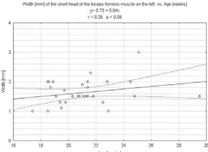
There were significant laterality differences in either parameter of the short head of the biceps femoris muscle (P < 0.05). The values of the length of the short head on the right and left sides ranged from 20.0 to 39.5 mm and from 18.0 to 39.0 mm, respectively. Plotted against the fetal age these val-ues generated the linear functions: y = –10.09 +
1.86x (r = 0.79) on the right (Fig. 5) and y = –4.45
+ 1.58x (r = 0.77) on the left side (Fig. 6). The width
of the short head increased from 1.0 to 2.8 mm for
Fig. 1. The biceps femoris muscle (in situ) in a female fetus aged 29.5 weeks: A – ischial tuberosity, B – head of the fibula, 1 – long head, 2 – common tendon Ryc. 1. Mięsień dwugłowy uda (in situ) u płodu żeń-skiego w wieku 29,5 tygodni: A – guz kulszowy, B – głowa kości strzałkowej, 1 – głowa długa, 2 – wspólne ścięgno
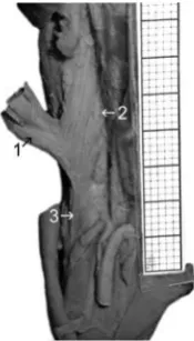
Fig. 2. The biceps femoris muscle (in situ) in a male fetus aged 24.8 weeks: 1 – long head, 2 – short head, 3 – common tendon
Ryc. 2. Mięsień dwugłowy uda (in situ) u płodu płci męskiej w wieku 24,8 tygodni: 1 – głowa długa, 2 – głowa krótka, 3 – wspólne ścięgno
Fig. 3. Regression line for the length (y) of the long head (right and left) versus fetal age (x)
Ryc. 3. Wykres regresji liniowej dla długości (y) głowy długiej (prawej i lewej) względem wieku
Fig. 4. Regression line for the width (y) of the long heads (right and left) versus fetal age (x)
Ryc. 4. Wykres regresji liniowej dla szerokości (y) głowy długiej (prawej i lewej) w zależności od wieku (x)
Fig. 5. Regression line for the length (y) of the short head on the right side versus fetal age (x)
the right biceps femoris and from 1.0 to 2.3 mm for the left one. Their growth was dependent on fetal age in accordance with the linear functions:
y = –0.80 + 0.12x (r = 0.54) on the right (Fig. 7)
and y = 0.73 + 0.04x (r = 0.25) on the left (Fig. 8).
Common Tendon
There were no laterality differences in relation to the common tendon length of the biceps femoris muscle (P > 0.05). During the study period, the val-ues of the common tendon length of the biceps fem-oris ranged from 13.5 to 28.3 mm on the right, and from 13.8 to 28.7 mm on the left. With regard to fe-tal age, the common tendon length (Fig. 9) showed a proportional increase with fetal age, according to the function: y = –9.85 + 1.41x (r = 0.90).
Discussion
From a developmental point of view, the biceps femoris muscle consists of the two different parts: the short head is derived from the gluteal muscula-ture, whereas the long head comes from flexors of the leg [12, 13]. In the opinion of some authors the biceps femoris varies little [6]. The short head of the biceps femoris may be absent. Instead, additional slips may originate from the ischial tuberosity, linea aspera or medial supracondylar ridge to form accessory heads of the biceps femoris. Sinav et al. [14] described two accessory muscle slips, deriving from the long head of the biceps femoris. The one is inserted into the crural fascia, whereas the other inserts into the semitendinosus muscle. Stelmasiak [12, 13] demonstrated that all of the 95 lower limbs of fetuses, neonates and adults were characterized
by the two typical heads of the biceps femoris. These results correspond to present observations, because the two heads of the biceps femoris were found to occur typically. A lack of statistically significant sex differences for the dimensions of the biceps femo-ris muscle was emphasized in this study. In this respect, the present results are in close accordance with Stelmasiak’s statement concerning the extent of the proximal attachment of the short head of the biceps femoris.
Having reviewed the professional literature on the biceps femoris muscle, the authors failed to find reference data for dimensions of the biceps femoris muscle. Therefore, in this study a digital-image analysis system was used to gather detailed normative data on the developing biceps femo-ris muscle at varying gestational ages from 17 to
Fig. 6. Regression line for the length (y) of the short head on the left side versus fetal age (x)
Ryc. 6. Wykres regresji liniowej dla długości (y) głowy krótkiej po stronie lewej w zależności od wieku (x)
Fig. 7. Regression line for the width (y) of the short head on the right side versus fetal age (x)
Ryc. 7. Wykres regresji liniowej dla szerokości (y) głowy krótkiej po stronie prawej w zależności od wieku (x)
Fig. 8. Regression line for the width (y) of the short head on the left side versus fetal age (x)
30 weeks. In the present study, it was found that during the study period both the lengths of the two heads and the common tendon length in-creased approximately 2-fold, whereas the widths of the long head – as many as 3-fold. The width of the short head was found to increase more on the right side (a 3-fold increase) than on the left side (a 2-fold increase).
The findings present the linear growth of the parameters studied of the biceps femoris plotted against the gestational age together with the ap-propriate correlation coefficients, the lines of best fit, and the 3rd and 97th percentile lines. Plots
show-ing the parameters examined were modeled on the right and left sides, respectively by the following linear functions: y = –25.27 + 3.61x (r = 0.90) for the length of the long head, y = –2.75 + 0.35x (r = = 0.77) for the width of the long head, y = –10.09 +
+ 1.86x and y = –4.45 + 1.58x for the length of the
short head, y = – 0.80 + 0.12x and y = 0.73 + 0.04x
for the width of the short head, and y = –9.85 +
+ 1.41x for the common tendon length.
The laterality differences were found to be significant only in relation to either parameter of the short head of the biceps femoris muscle. The strongest correlations were related to the length of the head of the biceps femoris and the common tendon length (r = 0.90), the intermediate – to the length of the short head (r = 0.77, 0.79) and the width of the long head (r = 0.77), and the weak-est – to the width of the short head on the left (r = 0.25) and right (r = 0.54) sides.
The lack of information concerning the pa-rameters studied limits discussion on this subject. The normative data of the biceps femoris muscle obtained in this study provide the background for future autopsy studies.
Fig. 9. Regression line for the length (y) of the tendon (right and left) of the biceps femoris muscle versus fetal age (x)
Ryc. 9. Wykres regresji liniowej dla długości (y) ścięgna dwugłowego (po stronie prawej i lewej) w zależności od wieku (x)
References
[1] Covey DC: Injuries of the posterolateral corner of the knee. J Bone Joint Surg Am 2001, 83, 106–118.
[2] Marshall JL, Girgis FG, Zelko RR: The biceps femoris tendon and its functional significance. J Bone Joint Surg Am 1972, 54, 1444–1450.
[3] Sebastianelli WJ, Hanks GA, Kalenak A: Isolated avulsion of the biceps femoris insertion. A case report. Clin Ortho Rel Res 1990, 259, 200–203.
[4] Terry GC, LaPrade RF: The biceps femoris muscle complex at the knee. Its anatomy and injury patterns associ-ated with acute anterolateral-anteromedial rotatory instability. Am J Sports Med 1996, 24, 2–8.
[5] Sneath RS: The insertion of the biceps femoris. J Anat 1955, 89, 550–553.
[6] Tubbs RS, Caycedo FJ, Oakes WJ, Salter EG: Descriptive anatomy of the insertion of the biceps femoris muscle. Clin Anat 2006, 19, 517–521.
[7] Rab M, Mader N, Kamolz LP, Hausner T, Gruber H, Girsch W: Basic anatomical investigation of semitendi-nosus and the long head of biceps femoris muscle for their possible use in electrically stimulated neosphincter formation. Surg Radiol Anat 1997, 19, 287–291.
[8] Shanahan D, Jordan RK, Coulthard A, Cooper PN, Varma J: The intramuscular arterial anatomy of the long head of biceps femoris muscle. J Anat 1997, 190, 467–472.
[9] Hayashi A, Maruyama Y: Lateral intermuscular septum of the thigh and short head of the biceps femoris muscle: an anatomic investigation with new clinical applications. Plast Reconstr Surg 2001, 108, 1646–1654.
[10] Hayashi A, Maruyama Y: Neurovascularized free short head of the biceps femoris muscle transfer for one-stage reanimation of facial paralysis. Plast Reconstr Surg 2005, 115, 394–405.
[11] Iffy L, Jakobovits A, Westlake W, Wingate MB, Caterini H, Kanofsky P, Menduke H: Early intrauterine devel-opment: I. The rate of growth of caucasian embryos and fetuses between the 6th and 20th weeks of gestation. Pediatrics 1975, 56, 173–186.
[12] Stelmasiak M: The biceps femoris muscle in fetuses, neonates and adult humans. Ann Univ Mariae Curie Skłodowska, Sectio D: Med 1946, 1, 63–76.
[13] Stelmasiak M: The short head of the biceps femoris muscle, Petit’s and grynfeldt’s triangles in adult humans from an constitutional point of view. Ann Univ Mariae Curie Skłodowska, Sectio D: Med 1946, 1, 231–281.
Michał Szpinda
Department of Normal Anatomy
Ludwik Rydygier Collegium Medicum in Bydgoszcz Karłowicza 24
85-092 Bydgoszcz Poland
Tel.: +48 52 585 37 05 E-mail: [email protected]
Conflict of interest: None declared