R E S E A R C H A R T I C L E
Open Access
Molecular pathology of vertebral deformities in
hyperthermic Atlantic salmon (Salmo salar)
Elisabeth Ytteborg
1,2, Grete Baeverfjord
1, Jacob Torgersen
1, Kirsti Hjelde
1, Harald Takle
1,3*Abstract
Background:Hyperthermia has been shown in a number of organisms to induce developmental defects as a result of changes in cell proliferation, differentiation and gene expression. In spite of this, salmon aquaculture commonly uses high water temperature to speed up developmental rate in intensive production systems, resulting in an increased frequency of skeletal deformities. In order to study the molecular pathology of vertebral
deformities, Atlantic salmon was subjected to hyperthermic conditions from fertilization until after the juvenile stage.
Results:Fish exposed to the high temperature regime showed a markedly higher growth rate and a significant higher percentage of deformities in the spinal column than fish reared at low temperatures. By analyzing phenotypically normal spinal columns from the two temperature regimes, we found that the increased risk of developing vertebral deformities was linked to an altered gene transcription. In particular, down-regulation of extracellular matrix (ECM) genes such ascol1a1, osteocalcin,osteonectinanddecorin, indicated that maturation and mineralization of osteoblasts were restrained. Moreover, histological staining andin situhybridization visualized areas with distorted chondrocytes and an increased population of hypertrophic cells. These findings were further confirmed by an up-regulation ofmef2candcol10a, genes involved in chondrocyte hypertrophy.
Conclusion:The presented data strongly indicates that temperature induced fast growth is severely affecting gene transcription in osteoblasts and chondrocytes; hence change in the vertebral tissue structure and composition. A disrupted bone and cartilage production was detected, which most likely is involved in the higher rate of deformities developed in the high intensive group. Our results are of basic interest for bone metabolism and contribute to the understanding of the mechanisms involved in development of temperature induced vertebral pathology. The findings may further conduce to future molecular tools for assessing fish welfare in practical farming.
Background
Industrial fish farming makes use of intensive produc-tion regimes in an effort to decrease producproduc-tion time and costs. Elevated water temperatures are commonly applied, often without explicit control of factors like nutrition, water quality, densities and vaccination. The intensive rearing systems are unfortunately correlated with deformities affecting both skeletal and soft tissues [1,2]. In teleosts, hyperthermia can induce vertebral deformities both during the embryonic development and after the vertebral column has been established [3-5]
The teleost vertebral body is built using a minimal bone mass to reduce negative buoyancy [6]. In salmon, the vertebral body comprises four mineralized or ossi-fied layers. Formation of the different layers involves the balanced and highly regulated formation of bone and cartilaginous structures through patterns of mineraliza-tion and matrix deposimineraliza-tion [7]. The specialized architec-ture makes it vulnerable to alterations in its tissue composition. Intramembranous ossification occurs by coordinated processes of production, maturation and mineralization of osteoid matrix [8]. Initially osteoblasts produce a thickening osteoid seam by collagen deposi-tion without mineralizadeposi-tion. This is followed by an increase in the mineralization rate and the final stage where collagen synthesis decreases and mineralization * Correspondence: harald.takle@nofima.no
1Nofima Marin, Norwegian Institute of Food, Fisheries and Aquaculture
Research, P.O. Box 5010, NO-1432 Ås, Norway
continues until the osteoid seam is fully mineralized. As part of the process, mineralization time lag appears to be required for allowing modifications of the osteoid so that it is able to support mineralization [9]. Indeed, fast growing Atlantic salmon has been shown to exhibit low vertebral mineral content and mechanical strength, together with an increased risk of developing vertebral deformities [10,11].
Skeletal growth depends upon the dynamic equili-brium between cartilage production and bone apposition rate [12]. Ontogeny and growth of the vertebral column is under control of regulatory mechanisms involving transcription factors, signaling molecules and extracellu-lar matrix proteins. The pathways of chondrocyte and osteoblast differentiation are interconnected during ver-tebral formation and must be coordinated. In particular, regulatory proteins, like the transcription factors Sox9, Runx2, Osterix, Twist and Mef2c have distinct functions both in the establishment of the vertebral bodies and later in the differentiation and maturation of specific skeletal cell types (review [13]). Similarly, signaling molecules like bone morphogenetic proteins (Bmp2 and Bmp4), and hedgehog proteins (Ihh and Shh) plays dif-ferent roles both during cell difdif-ferentiation and skeletal tissue ontogeny [14-16]. Osteoblasts and chondrocytes secrete the collagen fibers and ground substances of bone and cartilage. These cells are also responsible for the mineralization of the matrix through secretion of specialized molecules, such as Alkaline phosphatase (ALP), Osteocalcin and Osteonectin that binds inorganic minerals [17,18]. A widely accepted view is that the spa-tial restriction of ECM mineralization to bone is explained by osteoblast-specific gene products that initi-ate the formation of hydroxyapatite crystals (Ca10[PO4]6
[OH]2) [19]. The requirement for specifically expressed genes in osteoblasts (e.g.col1, osteocalcinand osteonec-tin) and chondrocytes (e.g.col2andcol10) to initiate the formation of matrix or control the growth of hydroxy-apatite crystals is supported by numerous studies [18,20,21]. Furthermore, Matrix metalloproteinases (MMPs) and Tartrate-resistant acid phosphatase (TRAP) are involved in degradation of ECM and in the bone remodeling process performed by the osteoclasts [22].
In this work, 20 skeletal genes were used to study the effect of long term hyperthermic exposure on vertebral development and growth in Atlantic salmon. Fish exposed to high temperature (high intensive regime) had a significant higher incidence of deformities than fish from the same origin reared under a conservative temperature regime (low intensive regime). The study was aimed at exposing differences in risk level between the groups, rather than elaborating the pathologies of deformed vertebrae, hence, the study concentrated on phenotypically normal fish from both temperatures.
Significant changes in gene transcription were found between phenotypically normal vertebrae of both groups, including down-regulation of genes encoding proteins important for mineralization. Further, in situ
hybridization (ISH) and histological staining revealed phenotypical and functional changes in the arch centra. Our results are of basic interest for understanding bone metabolism and deformities, as well as a tool for asses-sing fish welfare in practical farming.
Results
In the present study we analyzed and compared Atlantic salmon vertebrae from high and low temperature inten-sity regimes. Rate of development and growth was influ-enced by temperature regime as observed through SGR and time of sampling. The development from fertiliza-tion to first feeding lasted 5 months in the low intensive regime at 6°C, compared to 3 months in the high inten-sive regime at 10°C. Juveniles of the high inteninten-sive group also grew more rapidly after start-feeding than the low intensive group, where the former reached 2 g in 6 weeks after first feeding, 15 g in 3 months and 60 g in 7 months after first feeding, at a rearing temperature of 16°C. In comparison, the low intensive group at rear-ing temperature of 10°C reached similar sizes in 11 weeks, 5 months and 10 months, respectively. Accord-ingly, after start-feeding fish from the high intensive temperature regime displayed a higher SGR than the low temperature fish, 2.82 and 1.96 respectively.
Radiography, morphology and mineral analyses
On radiography analysis, the incidence of fish with ske-letal abnormalities at 2 g size was 4.0 ± 2.8% and 10.0 ± 1.7% in the low and high intensive groups, respectively (n.s.; not significant). At 15 g size, the difference was more pronounced, 3.4 ± 2.0% and 17.9 ± 1.3% (p < 0.001). At the final sampling at 60 g size, 8 ± 1.4% of the fish in the low intensive group displayed some degree of skeletal pathology compared to 28.1 ± 2.3% in the high intensive group (p < 0.0001), results are shown in figure 1.
with normal phenotype from the high and low intensive group at 15 g are shown in figure 2.
Due to the built-in image contrast enhancement pro-cedures of the semi-digital X-ray system, evaluation of skeletal mineralization as judged by radio density in images was impaired. Nevertheless, a lower contrast in skeletal structures was observed in the high intensity fish, in particular at the 15 g sampling, indicative of a lower mineralization rate at this stage.
Whole body mineral content at the end of the experi-ment (60 g size) showed low values for Ca, P and Zn content for both temperature regimes (Table 1), with no significant differences between treatments. There was a small, but significant lower level of whole body Fe and Na in the high intensive group. All Fe and Na values were lower than reference values [23], but in correspon-dence with Ca, P and Zn values, they were within a range which is commonly seen in commercially reared salmon.
Quantitative vertebral mRNA expression
The skeletal genes were divided into three groups according to function; ECM constituents, transcription factors, and signaling molecules (Figure 3A-C).
ECM constituents included genes involved in bone matrix production and mineralization and 7 out of 9 of these genes were found to be down-regulated in high intensive group at 2 and 15 g (Figure 3A). Tran-scription of col1a1, osteocalcin, decorin, osteonectin, mmp9and mmp13 were reduced in the high intensive group compared to the low intensive group. Col2a1
transcription was also down-regulated at both develop-mental stages, however the values were insignificant.
Osteocalcin was severely down-regulated in 2 g high intensive group. Converse transcription profiles could be observed for col10a1and alpbetween 2 g and 15 g fish; col10a1 was down-regulated at 2 g and up-regu-lated at 15 g whereas alpwas up-regulated at 2 g and down-regulated at 15 g.
0 5 10 15 20 25 30 35
2g 15g 60g
Sampling size
Pe
rc
e
n
ta
g
e
d
e
fo
rm
e
d
(%
)
Low intensive High intensive a
b
a b
a
a
Figure 1Frequency (%) of deformities in the vertebral column based on radiographic examination of Atlantic salmon sampled from the low temperature intensive group (white bars) and the high temperature intensive group (black bars) from fertilization until 60 g. Each bar represents the total number of deformities, scored as present or absent. Data are given in percentage ± st.dev, different letters indicate significant differences (P≤0.01) within the same size group, n = 4 tanks per treatment.
Temporal changes in transcription factor mRNA expression were found between high and low tempera-ture group, and all genes exceptsox9showed opposite expression at 2 and 15 g (Figure 3B). In the high intensive group,sox9was down-regulated at 2 g (n.s.) and 15 g, but more pronounced in the latter. Investigation of the two osteoblast markersrunx2andosterix, revealed opposite mRNA expression levels at 2 and 15 g.Runx2was up-regulated at 2 g (n.s), but down-up-regulated at 15 g. On the contrary,osterixwas down-regulated (n.s.) at 2 g, but up-regulated at 15 g.Mef2candtwistwas also down-regu-lated at 2 g, while up-regudown-regu-lated at 15 g (twistn.s.).
Signaling molecules included bmp2, bmp4, shh and
ihh. Expression analysis of mRNA for signaling
mole-A. ECM components
0 1 2 3 Co l1 a1 Co l2 a1 Co l1 0a 1 O ste oc al cin AL
P Deco
rin Os te on ec tin Mm p9 Mm p1 3 Vi m en tin R el at iv e m R N A ex pr es si on * * * * * * * * * * * * * * * * *
B. Signaling molecules
0 1 2 3 4 5 6 BM P4 BM P2 Ihh Sh h PD G F rb Re la tiv e m R NA e xp re ss io n * * * * * * * * * *
C. Transcription factors
0 1 2 So x9 Os te rix Runx Me f2 c Tw is t Re la tiv e m R NA e xp re ss io n * * * * * *
Low temperature 2g high temperature 15g high temperature
Figure 3Relative gene transcription of A. ECM components, B. Signaling molecules and C. Transcription factors in phenotypical normal spinal columns from 2 g (grey bars) and 15 g (black bars) high temperature intensive group compared to the low temperature intensive group (white bars). Data are given as mean values + SE, n = 15 and significant differences (P = 0.05) are indicated by *. Expression ratios are shown in relative mRNA expression along the y-axis, genes along the x-axis.
Table 1 Mineral analysis
Low intensive group High intensive group
P 3828 ± 162 3886 ± 285
Ca 3655 ± 341 3700 ± 677
Mg 302.3 ± 8 299 ± 7
Na 1303 ± 70 1084 ± 87*
Fe 9.7 ± 1.2 8.1 ± 0.2*
Mn 0.86 ± 0.05 0.8 ± 0.09
Zn 31.8 ± 2.6 33.5 ± 1.7
Cu 0.8 ± 0.12 0.7 ± 0.09
cules showed statistically significant differences in expression levels between the temperature regimes and all transcripts were found more abundant in the 15 g group when compared to 2 g vertebrae. Bmp2was the only up-regulated signaling molecule at 2 g, while all signaling genes were up-regulated at 15 g (Figure 3C).
To further examine changes in chondrocyte recruit-ment and structure between the temperature regimes, we included platelet derived growth factor receptor b
(pdgfrb) andvimentin, because of their importance in proliferation and the cytoskeleton, respectively [24,25]. Both transcripts were significantly down-regulated in 2 g, while significantly up-regulated at 15 g (Figure 3A, B).
In summary, we found that out of the 20 genes we analyzed, 8 were down-regulated in both temperature groups (col1a1, col2a1, osteocalcin, sox9, decorin, osteo-nectin, mmp9and mmp13), 9 genes were up-regulated in the 15 g high intensive group, but down-regulated at 2 g (bmp4, col10a1, osterix, ihh, shh, mef2c, twist, vimen-tin andpdgfrb). And finally, alp andrunx2 were up-regulated at 2 g but down-up-regulated at 15 g.
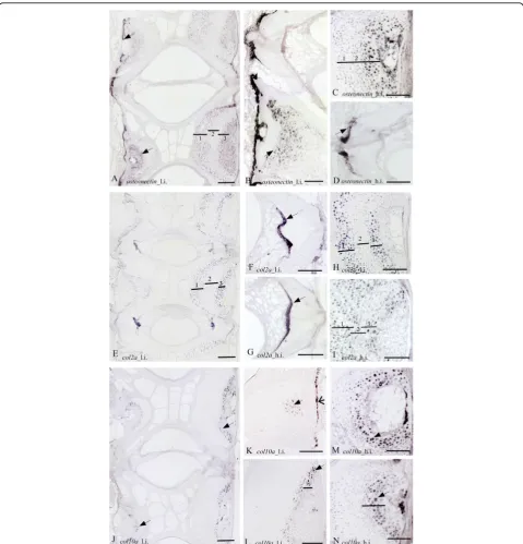
Vertebral tissue morphology and spatial mRNA expression
In areas where osteoblasts secrete the osteoid matrix, a generally strongerISH signals was apparent in the low intensive group for all probes. The osteogenic marker genecol1a showed distinct staining to osteoblasts at the growth zone of the endbones of the vertebral bodies
from fish of both temperature regimes (Figure 4A-C). Moreover,col1a signal was identified in the bone lining osteoblast cells situated at the lateral surfaces of the tra-beculae and along the rims of the vertebral bodies. Investigation of osteocalcinmRNA revealed an expres-sion pattern similar tocol1a, with staining of cells in the osteogenous areas and in bone lining osteoblasts and apical surfaces of the trabeculae (Figure 4D-G). Specifi-cally high osteocalcinsignal was detected in the prolif-erative osteoblast growth zones on the endbones of the vertebral bodies. OsteonectinmRNA was detected in the osteogenic growth zone of the endbones and lining the exterior part of the vertebral body (Figure 5A-D). The chondrocytic marker col2a, hybridized heavily to chordoblasts in the notochord (Figure 5E-G), whereas
col10awas detected in a continuous layer of cells along the rims of the vertebral body (Figure 5J-L).
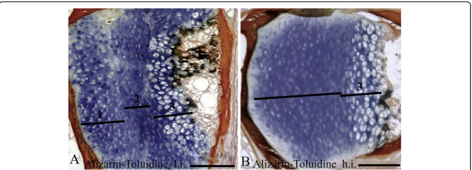
Alizarin red S and toluidine blue stained chondrocytes in the arch centra and revealed distinct morphological differences between vertebrae from the two temperature groups. The low intensive group was defined by distinct sub-groups of chondrocytes in the different maturational stages i.e. resting, proliferating and hypertrophic. In con-trast, the equivalent chondrocytes were more distorted in the high intensive group (Figure 6A, B). ISHanalysis ofcol2a,col10aandosteonectin enabled classification of the different chondrocytes into distinct sub-populations of maturational development. Col2ahybridized to rest-ing and pre-hypertrophic chondrocytes in two distinct
bands of both low and high intensive group, but the mRNA expression was more evenly distributed in all cells of the latter group (Figure 5H, I). There were also generally less proliferating chondrocytes that tended to be less compact in this group. In proliferating chondro-cytes we detected strong col2amRNA expression in the high intensive group, but no expression in the low intensive group. Analysis ofcol10ashowed restriction to the pre-hypertrophic and hypertrophic chondrocytes located in the deep cartilage zone (Figure 5M, N). Osteo-nectinwas also expressed in chondrocytes and the signal increased towards the hypertrophic chondrocytes (Figure 5A-C). The pre-hypertrophic chondrocyte zone was found to be expanded in the high intensive fish and both col10a1 and osteonectin showed an expanded expression domain corresponding to an increased hyper-trophic zone. No signal was detected in any of the sam-ples hybridized with sense probes (data not shown).
In normal spinal columns from the low intensive group, positive TRAP staining was detected at the ossi-fying boarders of the hypertrophic chondrocytes in the arch centra. No positive staining was detected in sam-ples from the high intensive group (Figure 7A, B).
Discussion
The presented study aims at describing the molecular pathology underlying the development of vertebral deformities in Atlantic salmon reared at a high tempera-ture regime that promotes fast growth during the early life stages. Within the period investigated, vertebral bodies form and develop and the skeletal tissue minera-lizes. Rearing at high temperatures resulted in higher frequencies of vertebral deformities, as expected. The vertebral pathology observed in this study was most
likely induced both during the embryonic development and after start-feeding, since the incidence of deformi-ties continued to increase throughout the experiment after the first radiographic examination at 2 g. Similar temperature regimes before and after start-feeding have independently been shown to induce vertebral defects in juvenile salmon [1,26]. However, whereas high tempera-tures during embryonic development is commonly related to somitic segmentation failure, deformities later in development may possibly be linked to fast growth induced by elevated temperatures and the impact this might have on the natural maturation and ontogeny of the vertebral bodies [3,10,11]. This causative relation has been shown for fast growing underyearling smolt that has a higher incidence of vertebral deformities than slower growing yearling smolt [27]. Further, morpho-metric analyses showed that elevated water temperature and faster growth is manifested by a difference in length-height proportion of vertebrae between fish from the two temperature regimes. Similar decrease in length-height proportion was described for the rapid-growing underyearling smolt [27]. Radiographic observa-tions indicated a lower level of mineralization of osteoid tissues in the high temperature fish. However, we could not find any pronounced altered mineral content between the two temperature regimes. The observed values were low compared to reference values [23], but in a range commonly observed in commercially reared salmon. Apparently, whole body mineral analysis seems insufficient to assess problems related to the develop-ment of spinal deformities.
we examined the expression of selected skeletal mRNAs in phenotypical normal salmon fry at 2 and 15 g. Histo-logical examination of 15 g fish was included to improve interpretation of the transcriptional data. The selected genes showed conservation and similar spatial expres-sion with those examined in other vertebrates, support-ing that most of the factors and pathways that control skeletal formation are highly conserved in vertebrates.
The lower transcription of ECM genes such ascol1a1,
osteocalcin, osteonectinanddecorin suggests a defect in the late maturation of osteoblasts [17,21,28]. The corre-lation to impaired mineralization is supported by the shorter vertebral bodies in the high intensive groups throughout the study, as well as the impaired minerali-zation indicated by low contrast observed on X-ray.
Col1a1 is the primary ECM component secreted by osteoblasts in the trabecular bone and growth plate and defects in the synthesis of col1or type 1 procollagen have been found in several heritable disorders of con-nective tissue. Likewise, defects in the assembly of Col1 fibrils have been reported to cause abnormally thin and branched structures [29]. Decreased diameter and cross-link density of the collagen fibers have been suggested to reduce thermal stability of collagen and thereby the tissues ability to support load during elevated tempera-tures [30]. In chum salmon, Oncorhynchus keta, the denaturation temperature of collagen type 1 from skin has been reported to be about 19°C [31]. The collagen fibres are further organized and stabilized by a range of non-collagenous proteins, which functions by linking other proteins and minerals to the ECM scaffold.
Decorin, which belongs to the small leucine rich repeat proteoglycan (PG) group (SLRPs) is involved in deter-mining the mature collagen fibril structural phenotype and tissue function by facilitating protein-protein inter-action with a range of other matrix components (mainly collagen fibres) and with the mineral phase during the formation of calcified tissues [32]. As a result, decorin has been shown to increase tensile strength of the col-lagen-decorin fiber [33]. Further, osteonectin is a phos-phorylated glycoprotein that binds to collagen fibrils, calcium, and hydroxyapatite, linking the bone mineral and collagen phases and perhaps initiating active miner-alization in normal skeletal tissue [17,34].Osteonectin -null mice display decreased trabecular bone volume and have bone of lesser stiffness than control mice [35].
part in the bone matrix and mineralization strongly sup-ports an assumption that disturbances of these processes constitute an important part of the mechanisms of development of vertebral deformities.
As for the ECM genes involved in osteoblast develop-ment and mineralization, high intensive temperature treatment had a significant effect on the transcription of transcription factors and signaling molecules involved in these processes. Intriguingly, Runx2 and Osterix, known as master regulators of osteoblast dif-ferentiation [39,40], exhibited opposite mRNA expres-sion levels at 2 and 15 g. Runx2-null mice have osteoblast differentiation arrested [41], while osterix -null mice embryos have a significant reduction of col1
expression and do not express the late osteoblast speci-fic marker osteocalcin [39]. In addition, we analyzed the bHLH transcription factor twist. This gene works as a negative regulator of osteoblastogenesis by inhibit-ing expression of genes downstream ofrunx2 [42]. At 2 g when osterix and twist was down-regulated while
runx2was up-regulated, osteocalcin was heavily down-regulated as wascol1a1. The mRNA expression pattern was inverted at 15 g. Then osterix andtwist was up-regulated and runx2down-regulated, whileosteocalcin
andcol1a1were weakly down-regulated. Linking these results to the pathways involved in osteoblast develop-ment, the required simultaneous activation ofosterix
and runx2did not appear at 2 g or at 15 g. However, Osterix function downstream of Runx2 during osteo-blast differentiation, but may be regulated by Bmp2 in a Runx2-independent pathway [43]. Bmp2 can induce ectopic bone and cartilage formation in adult verte-brates [44]. Spinella-Jaegle et al [16] found that coop-eration between Bmp2 and Shh was necessary to promote a strong induction of the osteoblast marker
alpin human mesenchymal cell lines. At both 2 and 15 g,bmp2 was highly up-regulated in the high inten-sive group, possibly as a response to the low ECM mRNA expression and under-mineralized tissue. In addition, osterixand shh was up-regulated at 15 g, as was bmp4. Bmp4 treatment has been shown to stimu-late new bone formation and is also expressed in osteo-blasts prior to formation of mineralized bone nodules [45,46]. However, in comparison to Spinella-Jaegles
in vitro findings, we did not detect an increase inalp
mRNA expression. Further, we detected a weaker sig-nal of osteocalcin and osteonectin in osteoblasts from the ISH of the high intensive group at 15 g. Hence, despite the possible attempt of bmp2to restore bone formation and mineralization, there was still lower transcription of ECM components in the high intensive group at 15 g. Summarized, our results may indicate that osteoblast proliferation and mineralization were restrained in the fast growing group.
The percentage of deformities significantly increased in the high intensive group from 2 g till 15 g, while the percentage was stable in the low intensive group. Hence, this period seems to involve important steps for the developmental fate of deformities. Between these two size stages we observed a change in expression pattern, from a downregulated to an upregulated transcription, of 9 genes, where 8 of them are involved in chondrogen-esis. This suggested that chondrocytes go through changes in this period that could be important for the development of the observed pathologies.
In vertebrates as mouse and human [47,48], the growth zones of long bones consists of well defined layers of progenitor, proliferative and hypertrophic chondrocytes [49]. These chondrocytes differ in their morphology, proliferation abilities and secretion of ECM components. For example, transcription of col2a1 is characteristic for the proliferative state whereas col10a1
is restricted to the hypertrophic state [47,50]. ISHof these genes revealed that 15 g Atlantic salmon raised at the low intensive regime also had distinct sub-popula-tions of progenitor, proliferative and hypertrophic chon-drocytes at the growth zone of the neural and haemal arches. On the contrary, more distorted layers were found in Atlantic salmon raised at the high intensive regime. Moreover, an increased zone of hypertrophic chondrocytes was found in the proximity of the minera-lized bone matrix in the high intensive group. Once these hypertrophic chondrocytes are fully differentiated, matrix calcification would normally be initiated [12]. However, we could not identify any variance in minera-lization at the ossifying borders of the hypertrophic chondrocytes when examined by histological Alizarin red S staining.
The increased zone of hypertrophic chondrocytes in the high intensive group and the up-regulated transcrip-tion of hypertrophic marker genes suggest an arrest prior to the final maturation of chondrocytes. Thus, these chondrocytes seems unable to initiate mineraliza-tion. The chondrocyte hypertrophy marker col10a1and its activatormef2c [51] were both up-regulated at 15 g in the high intensive group. Moreover, ihh, a repressor of terminal hypertrophic differentiation [52], was found to be highly up-regulated, whereas sox9, which is involved in early chondrocyte differentiation, and its downstream structural proteincol2a [53], were down-regulated. The severely down-regulation of runx2 at 15 g is of interest, sincerunx2-null mice embryos have a narrow zone of proliferating chondrocytes and a wide zone of hypertrophic chondrocytes [54]. In addition,
[25] have reported that chondrocytes respond to PDGF by enhancing proliferation and cartilage matrix produc-tion while maintaining the cells in a less mature pheno-type; corroborating our findings that the chondrocytes are some how arrested in the late hypertrophic stage at 15 g with a reduced possibility of completing the endo-chondral ossification process with calcified bone as end product. Similar findings have also been shown in rat ulnae, where loading was associated with an increased hypertrophic zone in the growth plate [56], but minera-lization rate was suppressed [57]. Another interesting comparative pathological condition to our findings in salmon is tibial dyschondroplasia (TD), a metabolic dis-ease of young poultry that affects the growth of bone and cartilage. The lesion is morphologically character-ized by an accumulation of chondrocytes that appear to be unable to differentiate past a pre-hypertrophic stage [58]. TD often occurs in broilers and other poultry that have been bred for fast growth rates. The tibial cartilage does not mature enough to ossify, which leaves the growth plate prone to fracture, infection, and deformed bone development.
The observed shorter phenotype of vertebral bodies from the high intensive group might have been a conse-quence of higher mechanical load in fast growing fish coincidental with a lower transcription of supportive ECM components. Together with the up-regulation of hypertrophic genes in high intensive fish at 15 g, we also found increased transcription ofvimentin. Vimentin filaments have been shown to regulate the swelling pres-sure of chondrocytes [59] and strengthen resistance to mechanical stress [60]. Hence, the increased activation of vimentin and the increased proportion of hyper-trophic chondrocytes in the high intensive temperature group at 15 g may reflect an adaptation to the fast growth by prioritizing maturation of chondrocytes that are more resistant to mechanical stress. At 2 g, however, the reduced level of vimentinmRNAs might possibly be linked to the mal-adaptive down-regulation of chondro-cytic genes in high intensive group. Indeed, disruption of vimentin filaments has been shown to result in loss of cell contact with the surrounding matrix which may alter the signaling dynamics of the cell and in effect shut down transcriptional events [24].
Mineralizing hypertrophic chondrocytes acquire and express most of the phenotypic characteristics of osteo-blasts, including high Alp activity and expression of
osteonectinandosteocalcin [61]. These phenotypic traits shared with osteoblasts may be needed to bring about the final phase of endochondral ossification and replace mineralized cartilage with bone [62]. They may also per-mit mineralized cartilage to act as bone-like structural tissue and allow for a transition from cartilage to bone. In contrast to the down-regulated transcription of
osteonectinand osteocalcin, as determined by real time qPCR, we observed an increased transcription pattern of these genes in the arch centra in the high intensive group by ISH. We also observed a tendency of lower transcription of the same genes in osteoblasts of the high intensive group. However, establishment of a calci-fiable matrix requires degradation of some matrix mole-cules. Endochondral bone formation includes the participation of MMPs, which degrade cartilage matrix and allow vascular invasion [63]. At least two proteases are involved in this process; MMP13 which regulates remodeling of the hypertrophic cartilage matrix and MMP9 which has a role in vascularisation of the growth plate [64,65]. When analyzing these MMPs in salmon vertebral columns, a significant down-regulation of both
mmp9 and mmp13 in the high intensive group at 2 g were observed. At 15 g, mmp13 mRNA expression decreased even more, whilemmp9was significantly up-regulated. Indeed, MMP13 is known as the dominant collagenase in cartilage and its absence cause delay in endochondral ossification [65]. Further supporting the hypothesis that endochondral ossification was in some way delayed in the spinal columns from the high inten-sive group, runx2deficiency has been shown to inhibit
mmp expression [66] and lead to mild disturbances of chondrocyte differentiation, as discussed above. In addi-tion, TRAP activity, essential for completing endochon-dral ossification [22], was absent in the erosive front of cartilage in neural and heamal arches of spinal columns from the high temperature group.
Conclusion
transcriptional changes in several genes that correlated with the higher risk of developing deformities later in ontogeny. Hence, this article reveals the potential use of gene transcription profiling as a prognostic approach in aquaculture.
Methods
Experimental design
The fish experiment was done at Nofima Marine at Sunndalsøra, Norway, in 2007 with Atlantic salmon from the Salmobreed strain. Two experimental tempera-ture regimes were set up; a high intensive temperatempera-ture group and a low intensive temperature group (Figure 8). Pooled batches of unfertilized eggs and milt were trans-ported on ice to the hatchery and were fertilized, rinsed and disinfected according to standard procedures. The eggs were incubated in a hatchery designed for incuba-tion of small egg volumes, with approximately 0.2 liters of eggs per unit in six units per temperature regime. During egg rearing water supply was continuous from two temperature controlled tanks stabilized at 10 ± 0.3° C and 6 ± 0.3°C, respectively, monitored twice daily. At 850 d° (day degrees = sum of daily temperature), a selec-tion of fry were mixed and transferred to 150 liter tanks for start-feeding, four tanks per temperature regime. The number of fry per tank was 400. Water flow in the tanks was adjusted throughout the experimental period to secure oxygen supply in excess. The fish were fed commercial diets and the light was continuous. The temperature for the high intensive tanks was gradually increased at first feeding to 16 ± 0.3°C and the tempera-ture for the low intensive tanks was gradually increased to 10 ± 0.3°C (1°C per day). These temperatures were
kept stable until the average size in each group reached 20 g. At this size, the differentiated temperature treat-ment was ended. 100 fish per tank were selected ran-domly, and were tagged individually with pit-tags in the abdominal cavity. Fish from the four tanks on same temperature regime were mixed in a larger tank, and reared at ambient temperature until termination at 60 g. Specific growth rates (SGR) in the period between start-feeding and 60 g were measured according to equation SGR = ((endweight/startweight)^(1/days)-1) × 100).
Tissue sampling, radiography, morphology and mineral analyses
Vertebral columns of phenotypically normal specimens from both temperature groups were sampled for gene expression analysis at 2 and 15 g size and histological analysis at 15 g size. The term phenotypically normal was defined as vertebral columns without any obvious aberrations or deformities when imaged by radiography at sampling. For this purpose, fish were heavily sedated in MS 222 (Tricaine methane sulphate, Pharmaq, Over-halla, Norway) (150 mg/litre) and imaged with an IMS Giotto mammography system (model number 6020/3, IMS Giotto, Bologna, Italy) equipped with a FCR Profect phosphorus film plate (Fuji Medical Inc., Japan). The resulting 20 pixels/mm images were enhanced with digi-tal software (Fuji Computed Radiography Console) and evaluated manually concurrent with sampling. Fish with-out any specific pathology of the vertebral column were identified for sampling, and killed by an anesthetic over-dose. Approximately ~5 vertebral bodies (~1 cm) were carefully dissected from the area under the dorsal fin. For gene expression analyses, samples were flash-frozen
in liquid nitrogen and transported on dry-ice to a -80°C freezer for storage. For histological analysis, vertebrae were fixated in 4% PFA for 24 h at 4°C, dehydrated in ethanol (25, 50 and 70%) and stored at 70% ethanol at -20°C. At 2 g size, 350 fish were screened and a total of 40 were sampled for this study. At 15 g size, 900 fish were screened, and 70 were sampled. Fish that were not selected for sampling following radiography were trans-ferred to clean water and returned to the rearing tank. At 60 g size, following an on-growing period on ambient temperatures, 800 fish were radiographed, 100 per origi-nal first feeding tank.
Incidence of skeletal deformities was recorded on radiographs from all samplings, and the presence or absence of vertebral pathology was recorded. It should be noted that fish with deviant vertebral morphology, mainly those with fusion type changes, were heavily sampled on basis of live X-ray at 2 g and 15 g (Ytteborg, manuscript in progress). This gives an underestimation of the differences between the two groups. In order to quantify differences observed in proportions of vertebral bodies, length and height of vertebral bodies were mea-sured on X-rays (ImageJ 1.39, NIH, USA), The length (craniocaudal) and height (dorsoventral) of 5 vertebral bodies under the dorsal fin was measured in 12 indivi-duals from each group at 2, 15 g and 60 g, and the length: height ratio was calculated.
At termination of the experiment, fish were sampled for analysis of whole body mineral content. Four sam-ples per treatment were taken, one per each of the origi-nal first feeding tanks. Each sample consisted of 10 fish, which were pooled before analysis. The samples were stored frozen at -20°C, and were homogenized prior to analysis. The dry matter of samples was determined after drying at 104°C for 16 h. For mineral analysis, samples were prepared as described [67,68] before analyzed by inductive coupled plasma (ICP) mass-spectroscopy.
Statistical analyses
A one-way analysis of variance model on incidence of deformities were carried out by SAS 9.1 software (SAS Institute Inc., USA), including the fixed effect of tem-perature regime. Statistics for gene transcription analysis are described in the real time qPCR section.
RNA isolation and cDNA synthesis
Tissue homogenization from 15 replicates from each treatment and developmental stage was achieved in a mortar with liquid nitrogen. Total RNA from the pow-dered vertebrae was isolated by using TRIzol™ and Micro to Midi Kit® (Invitrogen, MD, USA). Samples were treated with DNase1 (Invitrogen) before cDNA synthesis using oligo(dT) and Taqman Gold RT-PCR kit
(Applied Biosystems, CA, USA). The cDNA synthesis was performed with 10 min primer incubation at 25°C, 60 min RT step at 48°C and 5 min RT inactivation at 95°C in accordance to the manufacturer’s protocol. All reactions were performed in accordance to the manufac-turer’s protocol.
Sequence information and primer design
Primers for expression analysis were based on known Atlantic salmon sequences or on conserved regions of known teleost sequences paralogues. Primers were designed using the Vector NTI Advance 10 (Life tech-nologies, MD, USA), and NetPrimer (PREMIER Biosoft, CA, USA) software. All PCR products were cloned using pGEM T-easy (Promega, WI, USA) and sequenced with Big Dye Terminator chemistry and the ABI 3730 auto-mated sequencer, both delivered by Applied Biosystems. The obtained Atlantic salmon sequences were analyzed by BLAST and deposited in the Genbank database (Table 2).
Real time PCR
Triplicate real-time qPCR reactions were performed using the Light cycler 480 and SYBR Green chemistry (Roche, Switzerland) at the following thermal cycling conditions: 95°C for 10 min, followed by 45 cycles at 95°C for 15 s, 60 ± 1°C for 15 s and 72°C for 15 s. Further, specificity was assessed by the melting curves, determined post PCR (95°C for 15 s, 60°C for 1 min and 97°C continuous). PCR efficiencies for each target and the three housekeeping genes; elongation factor 1a
(el1a), heat shock protein 90b(hsp90b) and glyceralde-hyde 3-phosphate dehydrogenase(gapdh) were tested as endogenous controls. Relative target gene mRNA was normalized to relativeel1amRNA levels for all sample, as recommended by Olsvik et al. [69]. The transcription ratios of the 20 genes in all individual vertebrae from the two developmental stages were tested by using the Relative Expression Software Tool, REST, according to Pfaffl et al. [70]. Differences between the transcription ratios were tested for significance by the Pair Wise Fixed Reallocation Randomization Test© [70].
In situhybridization and histology
Table 2 Primers used for Real time qPCR (RT) and probes for in situ hybridization (ISH)
Gene Orientation Genbank Tm Use Sequence (5’-3’)
Extracellular Matrix constituents
Col1a1 Forward FJ195608 68.6 RT AGAGAGGAGTCATGGGACCCGT
Reverse 68.8 RT GGGTCCTGGAAGTCCCTGGAAT
Forward 69.8 ISH TAGCCGTGGTTTCCCTGGTT
Reverse 68.7 ISH CCGGGAGGTCCAAATCTACC
Col2a1 Forward FJ195613 62.6 RT TGGTCGTTCTGGAGAGACT
Reverse 62.7 RT CCTCATGTACCTCAAGGGAT
Forward 64.4 ISH GCTGGCGAGACAGGAGAGA
Reverse 63.4 ISH GCCTCATCAGCCCTCATGTA
Col10a1 Forward EG837148 66.4 RT TGGTGCTCTTTGACTGCCTGTAA
Reverse 65.1 RT CATCCTGTGTGTTGCAATATCACA
Forward 61.9 ISH AACAAGGGCTTCTTGGATCA
Reverse 60.9 ISH CATAATGCATCCTCAGGCAT
ALP Forward FJ195609 62.6 RT CTAGTTTGGGTCGTGGTATGT
Reverse 62.2 RT TGAGGGCATTCTTCAAAGTA
Osteocalcin Forward FJ195611 62.9 RT GTGAACCAACAGCAAAGAGA
Reverse 62.8 RT CCAGGTCCTTCTTAACAAACA
Forward 59.6 ISH CTCATACTTGTTGATCGTCCAG
Reverse 60.2 ISH TCTTTCTCTCTCGCTCTCCC
Osteonectin Forward FJ195614 65.3 RT ATTACTGAGGAGGAGCCCATCATT
Reverse 65.9 RT CCTCATCCACCTCACACACCTT
Forward 64.6 ISH CTGAACGATGAGGGTGTGGA
Reverse 67.2 ISH CGAGTGGTGCAGTGCTCCAT
Decorin Forward DQ452069 65.0 RT GAACCTGGCTAAGCTGGGTCTAA
Reverse 66.0 RT GAACAGGCTGATGCCAGAGTACAT
MMP9 Forward CA342769 65.9 RT AGTCTACGGTAGCAGCAATGAAGGC
Reverse 65.7 RT CGTCAAAGGTCTGGTAGGAGCGTAT
MMP13 Forward DW539943 66.9 RT TGATGTCCAAGTCAGCCGCTTC
Reverse 63.6 RT AAGGAGGCAGGAGGAAGAGG
Vimentin Forward GE618371 62.5 RT CAAGATCCTGTTAGCAGAGCT
Reverse 67.8 RT TGGTCTGCCACTTGCGATTGTC
Transcription factors
Sox9 Forward EU344852 62.8 RT CCTGCAAACAAGACAAGGT
Reverse 62.8 RT GGGTCGAGTAGATTCATACGA
Runx2 Forward FJ195615 64.8 RT CCACCAGGGACAGACACAGAT
Reverse 64.0 RT GAACGGACTGAGATCTGACGAA
Osterix Forward FJ195612 63.0 RT TCCCATAGACTTTCCCACA
Reverse 62.9 RT TGCCTCAGGACATGTACAA
Mef2c Forward GU252207 62.5 RT CACCGTAACTCGCCTGGTCT
Reverse 62.3 RT GCTTGCGGTTGCTGTTCATA
Twist* Forward AY546100 61.8 RT GCTTCAAAAGTGGAGACCGTTT
Reverse 61.6 RT GGGAGAACTTGAGCCCTCTTC
Signalling molecules:
BMP4 Forward FJ195610 62.2 RT TCAAGTTGCCCATAGTCAGT
Reverse 62.6 RT CACCTGAACTCTACCAACCA
BMP2 Forward BT059611 62.5 RT ATGTGGTATTGCACCCATT
Reverse 62.9 RT ATGGACAGTTTCCCAATGA
Shh* Forward AY370830 61.7 RT CCGGCTCATGACTCAGAGATG
Reverse 62.2 RT TATCCCTGGCCACTGGTTCA
Ihh Forward FJ195617 63.2 RT GGCTCAATCTCCTCTCTCCAAT
Inc., PA, USA) and mounted on pre-coated slides (0.01% Poly-L-lysine (Sigma) and 2% polyvinyl acetate glue (Casco, Arnheim, Germany)). ISHwas carried out with digoxigenine labeled (DIG RNA Labeling Kit, Roche) probes as described [71]. A total of five ECM producing genes were analyzed, including col1a, col2a, col10a,
osteocalcinandosteonectin.
Histological examination of vertebrae with toluidine blue and alizarin red S double staining was carried out on deplastified and rehydrated sections. Briefly, the sec-tions were stained for 2-3 min at RT in 0.1% toluidine blue (Sigma-Aldrich) (pH 2.3) and rinsed in distilled H2O followed by alizarin red (Sigma-Aldrich) (pH 4.2)
staining for 5 min. Prior to microscopy, the stained sec-tions were dehydrated in ethanol and mounted with Cytoseal 60 (Electron Microscopy Science, Fort Washington, PA, USA). Bright field microscopic ana-lyses were performed on a Zeiss Axio Observer equipped with an AxioCam MRc5 camera and AxioVi-sion software (Carl Zeiss Microimaging GmbH, Göttin-gen, Germany).
Specimens for paraffin embedding were stepwise rehy-drated in ethanol (50 and 25%) and decalcified for 7 days in 10% EDTA solution buffered with 0.1 M Tris base (Merck) at pH 7.0. The decalcified specimens were rinsed in PBS and stepwise dehydrated in ethanol (50, 70 and 100%), before being embedded in paraffin. We used 3 paraffin infiltration steps carried out at 60°C for 2 × 2 h and 1 × 3 h. The specimens were embedded in paraffin, stiffened at room temperature and hardened over night at 4°C. 5 μm serial sections were prepared using a Microm HM 355S. Paraffin sections were floated on demineralised water (25°C), mounted on uncoated slides and dried ON at 37°C. Prior to staining the sec-tions were de-waxed with Clear Rite (Richard-Allan, MI, USA), followed by 2× washes in xylene (Merck) for 5 min each. Sections were then rehydrated before rinsed in dH2O. To demonstrate TRAP activity, the Acid
phos-phatase leukocyte kit No. 387 (Sigma-Aldrich) was used and followed according to the manufacturer’s protocol; except that incubation lasted for 2 h at 37°C.
Subsequently, slides were rinsed in dH2O. Specimens
were counterstained with Mayers hematoxylin (Sigma-Aldrich) for 30 s and rinsed in running tap water before dehydrated, cleared and mounted with Cytoseal 60 (Electron Microscopy Science). Controls were incubated without substrate.
Abbreviations
ALP: alkaline phosphatase; BCIP/NBT: 5-bromo-4-chloro-3 indolyl phosphate p-toluidine salt/nitro blue tetrazolium chloride; bHLH: basic helix loop helix; BMP: bone morphogenetic proteins; dl: deciliter, Gla: y-carboxyglutamic acid; Ihh: indian hedge hog; MMP: matrix metalloproteinase; PBS: phosphate-buffered saline; PDGFrb: platelet derived growth factor receptor b; PFA: paraformaldehyde; Runx2: runt related transcription factor 2; shh: Sonic hedge hog; Sox9: sex determining region Y-box 9; TRAP: Tartrate-resistant acid phosphatase.
Acknowledgements
The authors are grateful to Asbjørn Valset, Britt Seljebø and Kjellrun Gannestad for technical assistance during the project. The study was supported by The Norwegian Research Council (project no 172483) and EU (COLL-CT-2005-012451, FINE FISH).
Author details 1
Nofima Marin, Norwegian Institute of Food, Fisheries and Aquaculture Research, P.O. Box 5010, NO-1432 Ås, Norway.2Norwegian University of Life Sciences, NO-1432 Ås, Norway.3AVS Chile SA, Imperial 0655, Of. 3A, Puerto Varas, Chile.
Authors’contributions
EY carried out the molecular studies, participated in sampling and drafted the manuscript. GB carried out the radiological diagnostics, accompanied in statistical analysis and participated in the experimental design. KH radiographed the fish and participated in the diagnostics and statistical analysis. JT participated in the design of theISHexperiments, sequence alignment and probe design, microscopic analyzes and acquisition and interpretation of data. HT conceived the study and its experimental and molecular design, coordinated the sampling, and participated in acquisition and interpretation of data and in the drafting of the manuscript. All authors read and approved the final manuscript.
Received: 29 June 2009 Accepted: 6 July 2010 Published: 6 July 2010
References
1. Ornsrud R, Gil L, Waagbo R:Teratogenicity of elevated egg incubation temperature and egg vitamin A status in Atlantic salmon, Salmo salar L.
Journal of Fish Diseases2004,27:213-223.
2. Takle H, Baeverfjord G, Lunde M, Kolstad K, Andersen O:The effect of heat and cold exposure on HSP70 expression and development of deformities during embryogenesis of Atlantic salmon (Salmo salar).
Aquaculture2005,249:515-524.
Table 2: Primers used for Real time qPCR (RT) and probes for in situ hybridization (ISH)(Continued)
PDGFrb Forward BT071824 62.2 RT TAGGACCAGCCGATGTTACTGC
Reverse 62.9 RT TCTCTGAGCCTCTCGATGT
Housekeeping genes:
EF1a** Forward DQ834870 71.3 RT CACCACCGGCCATCTGATCTACAA
Reverse 69.4 RT TCAGCAGCCTCCTTCTCGAACTTC
GAPDH*** Forward BT043825.1 72.4 RT TGGTGCAGAACCTCATGGTCCTCA
Reverse 70.2 RT ATCCCGGATGATTCCAAAGTCGTC
Hsp90b*** Forward BX074486 59.7 RT AATGGGTAACCTGGTCAGTG
Reverse 60.6 RT GACTCAGGCAAAGGAACCTT
3. Wargelius A, Fjelldal PG, Hansen T:Heat shock during early somitogenesis induces caudal vertebral column defects in Atlantic salmon (Salmo salar).Dev Genes Evol2005,215:350-357.
4. Ornsrud R, Gil L, Waagbo R:Teratogenicity of elevated egg incubation temperature and egg vitamin A status in Atlantic salmon, Salmo salar L.
Journal of Fish Diseases2004,27:213-223.
5. Georgakopoulou E, Angelopoulou A, Kaspiris P, Divanach P, Koumoundouros G:Temperature effects on cranial deformities in European sea bass, Dicentrarchus labrax (L.).Journal of Applied Ichthyology2007,23:99-103.
6. Casadevall M, Casinos A, Viladiu C, Ontanon M:Scaling of Skeletal Mass and Mineral-Content in Teleosts.Zoologischer Anzeiger1990,225:144-150. 7. Nordvik K, Kryvi H, Totland GK, Grotmol S:The salmon vertebral body
develops through mineralization of two preformed tissues that are encompassed by two layers of bone.J Anat2005,206:103-114. 8. Boivin G, Meunier PJ:Changes in bone remodeling rate influence the
degree of mineralization of bone.Connective Tissue Research2002, 43:535-537.
9. Hernandez CJ, Beaupre GS, Carter DR:A model of mechanobiologic and metabolic influences on bone adaptation.Journal of Rehabilitation Research and Development2000,37:235-244.
10. Fjelldal G, Hansen TJ, Berg AE:A radiological study on the development of vertebral deformities in cultured Atlantic salmon (Salmo salar L.).
Aquaculture2007,273:721-728.
11. Fjelldal PG, Nordgarden U, Berg A, Grotmol S, Totland GK, Wargelius A, Hansen T:Vertebrae of the trunk and tail display different growth rates in response to photoperiod in Atlantic salmon, Salmo salar L., post-smolts.Aquaculture2005,250:516-524.
12. Breur GJ, Vanenkevort BA, Farnum CE, Wilsman NJ:Linear Relationship Between the Volume of Hypertrophic Chondrocytes and the Rate of Longitudinal Bone-Growth in Growth Plates.Journal of Orthopaedic Research1991,9:348-359.
13. Karsenty G:Transcriptional control of skeletogenesis.Annual Review of Genomics and Human Genetics2008,9:183-196.
14. Karp SJ, Schipani E, St-Jacques B, Hunzelman J, Kronenberg H,
McMahon AP:Indian hedgehog coordinates endochondral bone growth and morphogenesis via Parathyroid Hormone related-Protein-dependent and -independent pathways.Development2000,127:543-548.
15. Hogan BLM:Bone morphogenetic proteins in development.Current Opinion in Genetics & Development1996,6:432-438.
16. Spinella-Jaegle S, Rawadi G, Kawai S, Gallea S, Faucheu C, Mollat P, Courtois B, Bergaud B, Ramez V, Blanchet A, Baron R, Roman-Roman S: Sonic hedgehog increases the commitment of pluripotent mesenchymal cells into the osteoblastic lineage and abolishes adipocytic
differentiation.Journal of Cell Science2001,114:2085-2094.
17. Termine JD, Kleinman HK, Whitson SW, Conn KM, Mcgarvey ML, Martin GR: Osteonectin, A Bone-Specific Protein Linking Mineral to Collagen.Cell 1981,26:99-105.
18. Cowles EA, Derome ME, Pastizzo G, Brailey LL, Gronowicz GA: Mineralization and the expression of matrix proteins during in vivo bone development.Calcified Tissue International1998,62:74-82. 19. Glimcher MJ:Recent Studies of the Mineral Phase in Bone and Its
Possible Linkage to the Organic Matrix by Protein-Bound Phosphate Bonds.Philosophical Transactions of the Royal Society of London Series B-Biological Sciences1984,304:479.
20. Glimcher MJ:Mechanism of Calcification - Role of Collagen Fibrils and Collagen Phosphoprotein Complexes Invitro and Invivo.Anatomical Record1989,224:139-153.
21. Boskey AL:Mineral-Matrix Interactions in Bone and Cartilage.Clinical Orthopaedics and Related Research1992, 244-274.
22. Witten PE, Huysseune A:A comparative view on mechanisms and functions of skeletal remodelling in teleost fish, with special emphasis on osteoclasts and their function.Biological Reviews2009,84:315-346. 23. Shearer KD, Asgard T, Andorsdottir G, Aas GH:Whole-Body Elemental and
Proximate Composition of Atlantic Salmon (Salmo-Salar) During the Life-Cycle.Journal of Fish Biology1994,44:785-797.
24. Blain EJ, Gilbert SJ, Hayes AJ, Duance VC:Disassembly of the vimentin cytoskeleton disrupts articular cartilage chondrocyte homeostasis.Matrix Biology2006,25:398-408.
25. Kieswetter K, Schwartz Z, Alderete M, Dean DD, Boyan BD:Platelet derived growth factor stimulates chondrocyte proliferation but prevents endochondral maturation.Endocrine1997,6:257-264.
26. Siemien MJ, Carline RF:Effect of Temperature on the Growth of 1St-Feeding Atlantic Salmon Fry.Progressive Fish-Culturist1991,53:11-14. 27. Fjelldal PG, Lock EJ, Grotmol S, Totland GK, Nordgarden U, Flik G, Hansen T:
Impact of smolt production strategy on vertebral growth and mineralisation during smoltification and the early seawater phase in Atlantic salmon (Salmo salar, L.).Aquaculture2006,261:715-728. 28. Mochida Y, Duarte WR, Tanzawa H, Paschalis EP, Yamauchi M:Decorin
modulates matrix mineralization in vitro.Biochemical and Biophysical Research Communications2003,305:6-9.
29. Ishida Y, Kubota H, Yamamoto A, Kitamura A, Bachinger HP, Nagata K:Type I collagen in Hsp47-null cells is aggregated in endoplasmic reticulum and deficient in N-propeptide processing and fibrillogenesis.Molecular Biology of the Cell2006,17:2346-2355.
30. Naimark WA, Waldman SD, Anderson RJ, Suzuki B, Pereira CA, Lee JM: Thermomechanical analysis of collagen crosslinking in the developing lamb pericardium.Biorheology1998,35:1-16.
31. Kimura S, Zhu XP, Matsui R, Shijoh M, Takamizawa S:Characterization of Fish Muscle Type-I Collagen.Journal of Food Science1988,53:1315-1318. 32. Weber IT, Harrison RW, Iozzo RV:Model structure of decorin and
implications for collagen fibrillogenesis.Journal of Biological Chemistry 1996,271:31767-31770.
33. Pins GD, Christiansen DL, Patel R, Silver FH:Self-assembly of collagen fibers. Influence of fibrillar alignment and decorin on mechanical properties.Biophysical Journal1997,73:2164-2172.
34. Bolander ME, Young MF, Fisher LW, Yamada Y, Termine JD:Osteonectin Cdna Sequence Reveals Potential Binding Regions for Calcium and Hydroxyapatite and Shows Homologies with Both A Basement-Membrane Protein (Sparc) and A Serine Proteinase-Inhibitor
(Ovomucoid).Proceedings of the National Academy of Sciences of the United States of America1988,85:2919-2923.
35. Delany AM, Amling M, Priemel M, Howe C, Baron R, Canalis E:Osteopenia and decreased bone formation in osteonectin-deficient mice.Journal of Clinical Investigation2000,105:915-923.
36. Furie B, Furie BC:Molecular-Basis of Gamma-Carboxylation - Role of the Propeptide in the Vitamin K-Dependent Proteins.Annals of the New York Academy of Sciences1991,614:1-10.
37. Klein BY, Gal I, Segal D:Studies of the Levamisole Inhibitory Effect on Rat Stromal-Cell Commitment to Mineralization.Journal of Cellular
Biochemistry1993,53:114-121.
38. Wennberg C, Hessle L, Lundberg P, Mauro S, Narisawa S, Lerner UH, Millan JL:Functional characterization of osteoblasts and osteoclasts from alkaline phosphatase knockout mice.Journal of Bone and Mineral Research 2000,15:1879-1888.
39. Nakashima K, Zhou X, Kunkel G, Zhang ZP, Deng JM, Behringer RR, de Crombrugghe B:The novel zinc finger-containing transcription factor Osterix is required for osteoblast differentiation and bone formation.Cell 2002,108:17-29.
40. Ducy P, Zhang R, Geoffroy V, Ridall AL, Karsenty G:Osf2/Cbfa1: A transcriptional activator of osteoblast differentiation.Cell1997, 89:747-754.
41. Komori T, Yagi H, Nomura S, Yamaguchi A, Sasaki K, Deguchi K, Shimizu Y, Bronson RT, Gao YH, Inada M, Sato M, Okamoto R, Kitamura Y, Yoshiki S, Kishimoto T:Targeted disruption of Cbfa1 results in a complete lack of bone formation owing to maturational arrest of osteoblasts.Cell1997, 89:755-764.
42. Bialek P, Kern B, Yang XL, Schrock M, Sosic D, Hong N, Wu H, Yu K, Ornitz DM, Olson EN, Justice MJ, Karsenty G:A twist code determines the onset of osteoblast differentiation.Developmental Cell2004,6:423-435. 43. Liu TJ, Gao YH, Sakamoto K, Minamizato T, Furukawa K, Tsukazaki T,
Shibata Y, Bessho K, Komori T, Yamaguchi A:BMP-2 promotes differentiation of osteoblasts and chondroblasts in Runx2-deficient cell lines.Journal of Cellular Physiology2007,211:728-735.
45. Chen D, Harris MA, Rossini G, Dunstan CR, Dallas SL, Feng JQ, Mundy GR, Harris SE:Bone morphogenetic protein 2 (2) enhances 3, BMP-4, and bone cell differentiation marker gene expression during the induction of mineralized bone matrix formation in cultures of fetal rat calvarial osteoblasts.Calcified Tissue International1997,60:283-290. 46. Harris SE, Sabatini M, Harris MA, Feng JQ, Wozney J, Mundy GR:Expression
of Bone Morphogenetic Protein Messenger-Rna in Prolonged Cultures of Fetal-Rat Calvarial Cells.Journal of Bone and Mineral Research1994, 9:389-394.
47. Castagnola P, Dozin B, Moro G, Cancedda R:Changes in the Expression of Collagen Genes Show 2 Stages in Chondrocyte Differentiation Invitro.
Journal of Cell Biology1988,106:461-467.
48. Long FX, Zhang XYM, Karp S, Yang YZ, McMahon AP:Genetic manipulation of hedgehog signaling in the endochondral skeleton reveals a direct role in the regulation of chondrocyte proliferation.
Development2001,128:5099-5108.
49. Hunziker EB, Schenk RK:Physiological-Mechanisms Adopted by Chondrocytes in Regulating Longitudinal Bone-Growth in Rats.Journal of Physiology-London1989,414:55-71.
50. Kielty CM, Kwan APL, Holmes DF, Schor SL, Grant ME:Type-X Collagen, A Product of Hypertrophic Chondrocytes.Biochemical Journal1985, 227:545-554.
51. Arnold MA, Kim Y, Czubryt MP, Phan D, McAnally J, Qi X, Shelton JM, Richardson JA, Bassel-Duby R, Olson EN:MEF2C transcription factor controls chondrocyte hypertrophy and bone development.
Developmental Cell2007,12:377-389.
52. Colnot C, de la Fuente L, Huang S, Hu D, Lu CY, St-Jacques B, Helms JA: Indian hedgehog synchronizes skeletal angiogenesis and perichondrial maturation with cartilage development.Development2005,
132:1057-1067.
53. Bell DM, Leung KKH, Wheatley SC, Ng LJ, Zhou S, Ling KW, Sham MH, Koopman P, Tam PPL, Cheah KSE:SOX9 directly regulates the type-II collagen gene.Nature Genetics1997,16:174-178.
54. Inada M, Yasui T, Nomura S, Miyake S, Deguchi K, Himeno M, Sato M, Yamagiwa H, Kimura T, Yasui N, Ochi T, Endo N, Kitamura Y, Kishimoto T, Komori T:Maturational disturbance of chondrocytes in Cbfa1-deficient mice.Developmental Dynamics1999,214:279-290.
55. Shum L, Wang XB, Kane AA, Nuckolls GH:BMP4 promotes chondrocyte proliferation and hypertrophy in the endochondral cranial base.
International Journal of Developmental Biology2003,47:423-431.
56. Robling AG, Duijvelaar KM, Geevers JV, Ohashi N, Turner CH:Modulation of appositional and longitudinal bone growth in the rat ulna by applied static and dynamic force.Bone2001,29:105-113.
57. Ohashi N, Robling AG, Burr DB, Turner CH:The effects of dynamic axial loading on the rat growth plate.Journal of Bone and Mineral Research 2002,17:284-292.
58. Farquharson C, Jefferies D:Chondrocytes and longitudinal bone growth: The development of tibial dyschondroplasia.Poultry Science2000, 79:994-1004.
59. Durrant LA, Archer CW, Benjamin M, Ralphs JR:Organisation of the chondrocyte cytoskeleton and its response to changing mechanical conditions in organ culture.Journal of Anatomy1999,194:343-353. 60. Trickey WR, Vail TP, Guilak F:The role of the cytoskeleton in the
viscoelastic properties of human articular chondrocytes.Journal of Orthopaedic Research2004,22:131-139.
61. Lian JB, Mckee MD, Todd AM, Gerstenfeld LC:Induction of Bone-Related Proteins, Osteocalcin and Osteopontin, and Their Matrix Ultrastructural-Localization with Development of Chondrocyte Hypertrophy Invitro.
Journal of Cellular Biochemistry1993,52:206-219.
62. Kirsch T, Nah HD, Shapiro IM, Pacifici M:Regulated production of mineralization-competent matrix vesicles in hypertrophic chondrocytes.
Journal of Cell Biology1997,137:1149-1160.
63. Vu TH, Shipley JM, Bergers G, Berger JE, Helms JA, Hanahan D, Shapiro SD, Senior RM, Werb Z:MMP-9/gelatinase B is a key regulator of growth plate angiogenesis and apoptosis of hypertrophic chondrocytes.Cell 1998,93:411-422.
64. Engsig MT, Chen QJ, Vu TH, Pedersen AC, Therkidsen B, Lund LR, Henriksen K, Lenhard T, Foged NT, Werb Z, Delaisse JM:Matrix
metalloproteinase 9 and vascular endothelial growth factor are essential for osteoclast recruitment into developing long bones.Journal of Cell Biology2000,151:879-889.
65. Stickens D, Behonick DJ, Ortega N, Heyer B, Hartenstein B, Yu Y, Fosang AJ, Schorpp-Kistner M, Angel P, Werb Z:Altered endochondral bone development in matrix metalloproteinase 13-deficient mice.Development 2004,131:5883-5895.
66. Pratap J, Javed A, Languino LR, van Wijnen AJ, Stein JL, Stein GS, Lian J.B: The Runx2 osteogenic transcription factor regulates matrix
metalloproteinase 9 in bone metastatic cancer cells and controls cell invasion.Mol Cell Biol2005,25:8581-8591.
67. Denstadli V, Skrede A, Krogdahl A, Sahstrom S, Storebakken T:Feed intake, growth, feed conversion, digestibility, enzyme activities and intestinal structure in Atlantic salmon (Salmo salar L.) fed graded levels of phytic acid.Aquaculture2006,256:365-376.
68. Helland S, Denstadli V, Witten PE, Hjelde K, Storebakken T, Skrede A, Asgard T, Baeverfjord G:Hyper dense vertebrae and mineral content in Atlantic salmon (Salmo salar L.) fed diets with graded levels of phytic acid.Aquaculture2006,261:603-614.
69. Olsvik PA, Lie KK, Jordal AEO, Nilsen TO, Hordvik I:Evaluation of potential reference genes in real-time RT-PCR studies of Atlantic salmon.Bmc Molecular Biology2005,6.
70. Pfaffl MW, Horgan GW, Dempfle L:Relative expression software tool (REST (c)) for group-wise comparison and statistical analysis of relative expression results in real-time PCR.Nucleic Acids Research2002,30. 71. Torgersen JS, Takle H, Andersen O:Localization of mRNAs and Proteins in
Methyl Methacrylate (MMA) Embedded Tissues.Journal of Histochem Cytochem2009, 825-830.
doi:10.1186/1472-6793-10-12
Cite this article as:Ytteborget al.:Molecular pathology of vertebral
deformities in hyperthermic Atlantic salmon (Salmo salar).BMC
Physiology201010:12.
Submit your next manuscript to BioMed Central and take full advantage of:
• Convenient online submission
• Thorough peer review
• No space constraints or color figure charges
• Immediate publication on acceptance
• Inclusion in PubMed, CAS, Scopus and Google Scholar • Research which is freely available for redistribution