Serum/Plasma Proteome
Selected Expression Profiling of Full-Length Proteins
and Their Variants in Human Plasma
Urban A. Kiernan, Dobrin Nedelkov, Kemmons A.Tubbs,
Eric E. Niederkofler and Randall W. Nelson*
*Intrinsic Bioprobes, Inc, 625 S. Smith Road Suite 22, Tempe, AZ 85281
Copyright ©Humana Press Inc.
All rights of any nature whatsoever are reserved. ISSN 1542-6416/04/01:7–16/$25.00
*Author to whom all correspondence and reprint requests should be addressed: E-mail: [email protected]
Abstract
With increased interest in clinical pro-teomics—the comparative investigation of dif-ferential protein expression patterns for use in the diagnostic and prognostic assessment of disease states—the demand for techniques that can readily identify changes in select proteome components is greater than ever before. This article describes a targeted proteomics approach to recover and quantify C-reactive protein (CRP) directly from human plasma. CRP, a putative biomarker for cardiac health, was isolated from microliter volumes of human plasma by using novel proteomics tools that combine micro-scale affinity capture with matrix assisted laser desorption/ionization time-of-flight mass spectrometry (MALDI-TOF MS) detection. Native CRP was analyzed along with serum amyloid P component (SAP) and retinol
Key Words:Relative quantification; MALDI-TOF MS; C-reactive protein; human plasma; protein expression profiles; immunoassay.
Introduction
Clinical proteomics focuses on the use of rapid fractionation approaches in combination with mass spectrometry (MS) to directly profile human biofluids, plasma in particular. By design, these studies are meant to establish protein signatures, in the form of mass spec-tra, that are able to distinguish between healthy and diseased states in individuals. Indeed, numerous pioneering investigations have shown clinical proteomics to be one of the most promising new approaches for defining diagnostic signatures related to ovarian, breast, prostate and lung cancers (1–3). However, a particular concern of these studies is that many of the profiling approaches distinguish healthy from diseased states based on computer learn-ing algorithms that may not identify a land-mark mass-to-charge (m/z) value as actually being a protein species. Such an approach may be misleading if the m/z values used have no corresponding peptide signals. Moreover, to date no clinical proteomics study has indepen-dently “rediscovered” known biomarkers of various cancers (e.g., prostate specific antigen [PSA] is not detected in individuals suffering from prostate cancer). In missing, or not specif-ically targeting these proteins as part of the pro-filing, these studies do not take full advantage of past clinical/diagnostic studies, the results of which are the basis of numerous current diagnostic assays (4). Accordingly, there exists a growing demand to discover and identify pro-teins for use as putative biomarkers, and at the same time detect proteins that are presently used (and validated) as diagnostic species (5).
Given that the identity of the biomarker is known (either old or newly found), it is
clearly more efficient to construct assays that target them directly, rather than to continue to assay samples using the global discovery methodologies. A foremost advantage to using a targeted approach in combination with mass spectrometry is the ability to significantly improve the limit of detection of the assay. Specifically, affinity approaches are able to selectively concentrate target proteins prior to mass spectrometry, the result of which is a highly purified form of the target protein entering the mass spectrometer without any accompanying interferences stemming from the presence of other proteins (e.g., ion suppression or overlapping signals). A second advantage is the use of targeted high-performance mass spectrometric assays to discern and character-ize genomic and epigenomic differences that are found in proteins. Because of genetic and posttranslational variations, any protein under investigation has the potential of existing within or between individuals as multiple species. Mass spectrometry of highly purified pro-teins has been used for many years to both detect and characterize and/or identify these variations (6). This long-standing ability is ide-ally complimented by a simple fractionation approach that can be used to selectively con-centrate chosen proteins directly from biologi-cal milieu.
Overview of the Mass Spectrometric
Immunoassay Process
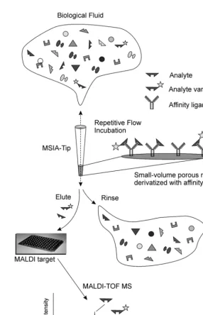
Previous works have described a hybrid protein analysis technology—mass spectromet-ric immunoassay (MSIA)—that is used to selec-tively retrieve target proteins from complex biological milieu for mass spectrometric characterization and quantification (7–10). Figure 1 gives a brief overview of the process. As applied here, a human plasma sample is repeatedly drawn and expelled through a pipettor tip containing a stationary phase to which an affinity ligand is covalently bound (termed MSIA-Tip). After rinsing, MALDI matrix is drawn into the MSIA-Tip, breaking the affinity interaction. The resulting matrix/ analyte mixture is deposited directly onto a mass spectrometer target for subsequent matrix-assisted laser desorption/ionization time-of-flight mass spectrometry (MALDI-TOF MS). Analyte detection/characterization is essen-tially a combination of affinity isolation via immobilized antibodies and characterization of retained species by MALDI-TOF MS. In this sense, target proteins and their variants are selectively concentrated from plasma, and MALDI-TOF MS is used to (1) unambiguously identify the wild-type target protein by detec-tion at a known molecular weight and (2) rec-ognize the presence of variants retained by the antibody at masses shifted away from the wild-type protein. This ability forms the basis of an assay capable of identifying protein variants resulting from posttranslational modifications
(9), pointmutations (10), or truncations (11–13). The simultaneous analysis of mass-resolved species lays the foundation for the rigorous quantification of a target protein (14) or the development of multiple-analyte assays (7).
Presented here is the development of a multi-analyte MSIA for use in the
expres-sion profiling of a select group of proteins— C-reactive proteins (CRP), retinol binding pro-tein (RBP), and serum amyloid P component (SAP) (and their variants)—present in human plasma. The central target in these analyses is CRP, a known clinical protein biomarker of inflammation that has been determined to have short- and long-term predictive value of patient outcomes with cardiovascular disease and the development of coronary events that can be used to identify the onset and recur-rence of myocardial infarction (MI) (15–18), stroke (19–20), and angina (21–22). The two other protein species, RBP and SAP were chosen as complement proteins (for use in normalizing the MSIA profiles between indi-viduals) based on a few simple criteria. First, for optimization of MALDI-TOF MS condi-tions (e.g., matrix, delayed-extraction), the proteins must have similar, but fully resolved molecular weights. Second, the complement proteins are found in healthy populations at roughly equivalent and reasonably stable con-centrations. Lastly, MSIA conditions for the complement proteins (e.g., extraction and rinses) are the same as for the target protein.
Materials and Methods
Samples
Plasma samples from eight individuals (five males and three females betweent the ages of 26 and 48) were acquired following a procedure approved by the IBI’s Institutional Review Board (IRB), and only after each subject had signed an Informed Consent form. Sam-ples were acquired, under sterile conditions, through a lancet-punctured finger using two nonheparinized (75 µL volume) microcolumns (Drummond Scientific Co., Broomall, PA). Each
150 µL whole blood sample collected was
buffered saline (10 mMHEPES, 150 mMNaCl,
pH 7.4 [HBS]) containing 50 mM
ethylenedi-amine tetraacetic acid (EDTA) and 2 µL of a protease inhibitor cocktail set II (Calbiochem, San Diego, CA), and centrifuged for 2 min (at 7000 rpm/2500g) to pellet red blood cells. The
supernatant (diluted plasma, 250 µL) was
decanted from each sample and stored at –70°C until ready for use.
For the standard addition quantification experiments, a single plasma sample was dis-persed into eight 25 µL aliquots. Increasing
amounts (0 through 7 aliquots of 14 µL) of 0.0183 mg/mL standard solution containing highly purified human C-reactive protein (Cal-biochem, San Diego, CA) were added to the plasma aliquots. Each aliquot was then brought to 300 µL with HBS-EDTA buffer.
MSIA Analysis
The plasma samples were addressed in par-allel using a multi channel pipettor, using CDI (1,1’-carbonyldiimidazole)-activated MSIA-Tips prepared and derivatized with polyclonal anti-bodies specific towards retinol binding protein, C-reactive protein and serum amyloid P com-ponent (DakoCytomation, Carpinteria, CA), as previously described (8). Sample incubation consisted of 50 cycles (aspiration and
dispens-ing) of 150 µL of the sample through each
MSIA-Tip. After incubation, the MSIA-Tips were thoroughly rinsed using HBS, 10 cycles of 150 µL; double distilled water, 5 cycles of 150 µL; 20% acetoniltrile/2Mammonium acetate, 10 cycles of 150 µL; and finally with double distilled water, 15 cycles of 150 µL. Retained proteins were eluted by drawing 4 µL of MALDI matrix solution (saturated aqueous solution of sinapic acid (SA), in 33% (v/v) acetonitrile, 0.4% (v/v) trifluoroacetic acid) into each tip and depositing the eluates directly onto a 96-well formatted hydrophobic/hydrophilic contrasting MALDI-TOF target (14). Samples were allowed to air dry prior to insertion of the MALDI target into the mass spectrometer. The total time required for preparation of the samples was less than 10 min.
MALDI-TOF MS
Intact protein expression profiling was performed on a Bruker Biflex III MALDI-TOF mass spectrometer operating in linear delayed-extraction mode with 25.50 kV full accelerating potential. Draw-out pulses of 2.175 kV (900 ns delay) were used for parent protein analysis. Mass spectra were manually acquired. Five spectra from each spot were
acquired and averaged for relative quantita-tive measurements.
Results
Individual and Multi-Protein MSIA Assays
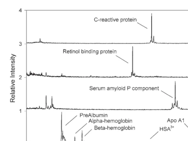
Assays for the individual proteins were developed and investigated to gauge compati-bility and to check for any unforeseen interfer-ences. Figure 2 shows a comparison between the MSIA analyses of human plasma for CRP, RBP, SAP, and a MALDI-TOF MS analysis of the stock plasma (diluted 100-fold with water, taken prior to any MSIA extraction). In each case, the target species was selectively isolated to the exclusion of higher concentration plasma proteins that are generally observed in the anal-ysis of human plasma with no prior fractiona-tion (i.e., albumin, apolipoproteins) (Fig. 2D). Additionally, there is no signal overlap between the chosen proteins (i.e., the three proteins reg-ister at distinct m/zregions in the mass spectra), nor are any interferences observed in the spec-tra, owing to, for example, nonspecific binding.
Based on the compatibility of the individ-ual analyses, a multi-analyte MSIA was con-structed by linking antibodies targeting the three species to the same MSIA-Tip. Figure 3 shows the results of using the assay on a plasma sample taken from a healthy 29-yr-old male (Individual No. 5). Observed in the profile are, from low-to-high mass: RBP-L (C-terminally truncated RBP missing one leucine residue, m/zobs= 20,951; m/zcalc= 20,953); RBP (m/zobs = 21,065; m/zcalc = 21,066); CRP-P (CRP missing its C-terminal proline residue,
m/zobs = 22,930; m/zcalc = 22,932); CRP (m/zobs
=23,028; m/zcalc = 23,029); SAP-V&sialic acid (SAP missing one sialic acid and its C-terminal valine, m/zobs = 25,068; m/zcalc = 25,073);
SAP-sialic acid (m/zobs = 25,168; m/zcalc = 25,172);
Individ-ual No. 5 plasma, as well as in the subsequent application of the assay to the all eight plasma samples screened throughout the remainder of the study.
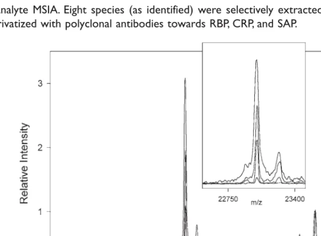
Validation of CRP Response
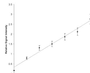
As the foremost purpose of the assay was that of viewing modulation in CRP, a standard addition approach was used to confirm a direct relationship between CRP signal inten-sity and concentration. Eight samples (using the Individual No. 5 sample) were prepared to contain increasing amounts of CRP standard. Figure 4 shows four (of the eight) CRP-MSIA profiles performed on the standard addition samples. The spectra are normalized to the SAP signal height. When the ratio of the CRP/SAP signal intensities for each aliquot were plotted against the CRP concentration, linear relationship (correlation r2 = 0.984) is
observed in the concentration range of 0.0 to 6.0 mg/L (Fig. 5), thus verifying that an
ele-vated CRP response denotes an increase in concentration. The endogenous concentration of CRP in the individual was determined to be 0.069 mg/L from the line equation derived from the data:
ICRP/ISAP= 0.463|[CRP]| + 0.319
This value is consistent with literature values of basal CRP levels in healthy individ-uals at < 1 mg/L(23).
Profiling Relative to Medical History
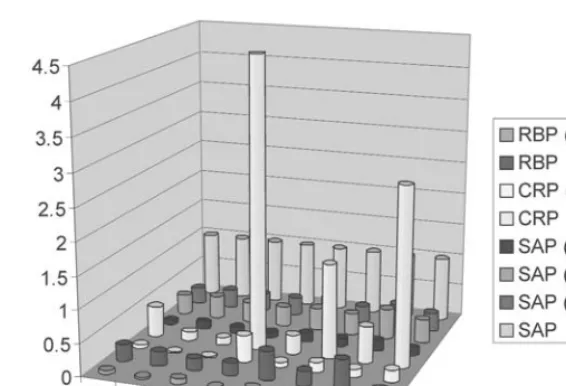
Plasma samples from eight individuals— with knowledge of their medical disposition— were screened using the RBP–CRP–SAP assay. Profiles comparable in detail to that shown in Figure 3 were obtained for each individual. Figure 6 shows a simple comparison of the profiles where ion signals for the eight species were normalized to the wt-SAP signal, seven data points for each individual with the eighth serving to normalize between individuals.
Signal for each species can be evaluated on its own, or as part of the collective profile, and put into perspective with regard to the origin of the protein species and the medical history
of the individual. For instance, three individu-als, Nos. 4, 6, and 8, exhibited exceedingly high levels of CRP. The histories of these individu-als revealed that subject No. 4 was 48 h
post-Fig. 3. Multi-analyte MSIA. Eight species (as identified) were selectively extracted from plasma using a single MSIA-Tip derivatized with polyclonal antibodies towards RBP, CRP, and SAP.
surgery when the plasma sample was col-lected, whereas subject No. 6 suffered from chronic rheumatoid arthritis and No. 8 had recently suffered an acute myocardial infarc-tion. The elevated levels of CRP in these indi-viduals is in agreement with the fact that CRP is an established acute-phase protein known to respond to both immediate and chronic inflammation (24), as well as a marker of car-diovascular health (25). Although not previ-ously cataloged as an endogenous species, the truncated CRP variant mimics the CRP response in these individuals, which may be expected considering that the two species are linked biologically. Individual No. 7 was noted to have elevated levels of all the signals in the profile, with the exception of the wt-SAP (normalization) signal. This observation was consistent with the individual having a
below average level of the fully glycosylated SAP, the cause of which remains unclear.
Conclusion
The need for targeted mass spectrometric analysis of human proteins increases dramati-cally as the field of clinical proteomics evolves from the discovery phase to validation and application. Given here has been the develop-ment and use of a proteomics approach for the expression profiling a select group of pro-teins from human plasma. Importantly, the approach is able to yield information towards a specific question. The RBP–CRP–SAP MSIA was designed to recognize, and if chosen rigorously quantify, changes in the plasma concentration of CRP. Notwithstanding this ability, the profiling approach also contains an element of discovery. As a result of genetic
Fig. 5. Response curve generated from the CRP standard addition series. A linear response (r2 = 0.984) is
polymorphism, splice variants, and posttrans-lational modifications, any particular protein has the potential of being produced as one of numerous different variants. Moreover, additional variants can be created by the natural metabolism/catabolism of the protein after expression. As a case in-point, the three proteins under investigation, RBP–CRP–SAP, actually register as eight identifiable and con-sistently observed species in the expression profiling. The ability to readily monitor such variants clearly adds an additional dimension of performance to an otherwise routine anal-ysis, which in turn allows subtle changes in protein structure to be discovered and investi-gated. Given these abilities and their general application in number, MSIA is viewed as an exceptional approach for the expression profil-ing of select protein targets in human plasma to determine qualitative and quantitative changes between individuals. Such selected expression profiling stands to find use in population
screening and validation efforts subsequently derived from discoveries made through ongo-ing comparative proteomics studies.
Acknowledgments
We would like to thank Dr. Allan Bieber for the critical reading of this manuscript. This publication was supported in part by grant numbers 4 R44 CA099117-02 and 4 R44HL072671–02, from the National Insti-tutes of Health. Its contents are solely the responsibility of the authors and do not nec-essarily represent the official views of the National Institutes of Health.
References
1. Yanagisawa K, Shyr Y, Xu BJ, et al. Proteomic patterns of tumour subsets in non-small-cell lung cancer. Lancet 2003;362(9382):433–439. 2. Petricoin EF, Ardekani AM, Hitt BA, et al. Use
of proteomic patterns in serum to identify ovarian cancer. Lancet 2002;359(9306):572–577.
Fig. 6. Expression profiling of eight individuals. Given are relative intensity values of the eight protein species identified in Fig. 3. All values are normalized to the wt-SAP signal. Relatively high levels of CRP (and truncated variant) are observed for individuals Nos. 4, 6, and 8, who are noted to be ailing from post-surgical inflam-mation, chronic rheumatoid arthritis, and acute MI, respectively. Color image available for viewing at www.humanapress.com
1
3. Conrads TP, Zhou M, Petricoin EF, 3rd, Liotta L, Veenstra TD. Cancer diagnosis using pro-teomic patterns. Expert Rev Mol Diagn 2003; 3(4):411–420.
4. Anderson NL, Anderson NG. The human plasma proteome: history, character, and diag-nostic prospects. Mol Cell Proteomics 2002; 1(11):845–867.
5. Diamandis EP. Point: Proteomic patterns in biological fluids: do they represent the future of cancer diagnostics? Clin Chem 2003;49(8): 1272–1275.
6. Mann M, Hendrickson RC, Pandey A. Analy-sis of Proteins and Proteomes By Mass Spec-trometry. Annu Rev Biochem 2001;70:437–473. 7. Nelson RW, Krone JR, Bieber AL, Williams P. Mass-Spectrometric Immunoassay. Anal Chem 1995;67(7):1153–1158.
8. Niederkofler EE, Tubbs KA, Kiernan UA, Nedelkov D, Nelson RW. Novel mass spectro-metric immunoassays for the rapid structural characterization of plasma apolipoproteins. J Lipid Res 2003;44(3):630–639.
9. Tubbs KA, Nedelkov D, Nelson RW. Detection and quantification of beta-2-microglobulin using mass spectrometric immunoassay. Anal Biochem 2001;289(1):26–35.
10. Kiernan UA, Tubbs KA, Gruber K, et al. High-Throughput Protein Characterization Using Mass Spectrometric Immunoassay. Anal Bio-chem 2002;301(1):49–56.
11. Kiernan UA, Tubbs KA, Nedelkov D, Nieder-kofler EE, Nelson RW. Comparative pheno-typic analyses of human plasma and urinary retinol binding protein using mass spectro-metric immunoassay. Biochem Biophys Res Commun 2002;297(2):401–405.
12. Kiernan UA, Tubbs KA, Nedelkov D, Nieder-kofler EE, Nelson RW. Detection of novel trun-cated forms of human serum amyloid A protein in human plasma. FEBS Lett 2003; 537(1–3):166–170.
13. Kiernan UA, Tubbs KA, Nedelkov D, Nieder-kofler EE, McConnell E, Nelson RW. Compar-ative urine protein phenotyping using mass spectrometric immunoassay. J Proteome Res 2003;2(2):191–197.
14. Niederkofler EE, Tubbs KA, Gruber K, et al. Determination of beta-2 microglobulin levels in plasma using a high-throughput mass
spec-trometric immunoassay system. Anal Chem 2001;73(14):3294–3299.
15. Haverkate F, Thompson SG, Pyke SD, Gal-limore JR, Pepys MB. Production of C-reactive protein and risk of coronary events in stable and unstable angina. European Concerted Action on Thrombosis and Disabilities Angina Pectoris Study Group. Lancet 1997;349(9050): 462–466.
16. Liuzzo G, Baisucci LM, Gallimore JR, et al. Enhanced inflammatory response in patients with preinfarction unstable angina. J Am Coll Cardiol 1999;34(6):1696–1703.
17. Ridker PM, Buring JE, Shih J, Matias M, Hen-nekens CH. Prospective study of C-reactive protein and the risk of future cardiovascular events among apparently healthy women. Cir-culation 1998;98(8):731–733.
18. Tommasi S, Carluccio E, Bentivoglio M, et al. C-reactive protein as a marker for cardiac ischemic events in the year after a first, uncom-plicated myocardial infarction. Am J Cardiol 1999;83(12):1595–1599.
19. Rost NS, Wolf PA, Kase CS, et al. Plasma con-centration of C-reactive protein and risk of ischemic stroke and transient ischemic attack: the Framingham study. Stroke 2001;32(11): 2575–2579.
20. Curb JD, Abbott RD, Rodriguez BL, et al. C-reactive protein and the future risk of thromboembolic stroke in healthy men. Circu-lation 2003;107(15):2016–2020.
21. Kuller LH, Tracy RP, Shaten J, Meilahn EN. Relation of C-reactive protein and coronary heart disease in the MRFIT nested case-control study. Multiple Risk Factor Intervention Trial. Am J Epidemiol 1996;144(6):537–547.
22. Ferreiros ER, Boissonnet CP, Pizarro R, et al. Independent prognostic value of elevated C-reactive protein in unstable angina. Circu-lation 1999;100(19):1958–1963.
23. Kushner I. The phenomenon of the acute phase response. Ann N Y Acad Sci 1982;389: 39–48.
24. Zimmerman MA, Selzman CH, Cothren C, Sorensen AC, Raeburn CD, Harken AH. Diag-nostic implications of C-reactive protein. Arch Surg 2003;138(2):220–224.
25. Kushner I. C-reactive protein and atheroscle-rosis. Science 2002;297(5581):520–521.