Phd Dissertation
Novel data-driven analysis methods for
real-time fMRI and simultaneous
EEG-fMRI neuroimaging
Author:
Nicola Soldati
Supervisors:
Dr. Jorge Jovicich Prof. Lorenzo Bruzzone
A thesis submitted for the degree of
PhilosophiæDoctor (PhD)
Doctoral School in Cognitive and Brain Sciences
XXIV cycle
Acknowledgements
A journey coming to an end always leaves behind itself a long path of moments and people filling them. This PhD path permitted to me to live plenty of good (and less good) moments, in which I encountered several people who have been very important to me. Starting from the beginning I need to be very grateful to my PhD advisors Jorge Jovicich and Lorenzo Bruzzone, whose mentoring always followed me in all the steps toward this goal. I also need to thank Christina Tryantafillou and Oliver Hinds, for having introduced me to the practice of real-time fMRI in my firsts uncertain steps. Great efforts into improving my expertises on ICA knowledge have been sustained by Vince Calhoun, whose help has been a key feature of this work. Finally a special mention goes to Andrzej Cichocki for the warm hospitality that he made me feel, the precious help and good time in a far away country and the high level profile of the research done at his laboratory. A special thank is for Sara Assecondi, who always generously offered her expertise and data to support my research.
In all the above mentioned laboratories I also found always very good friends, who made the journey less difficult and helped me a lot with practi-cal issues and theoretipracti-cal discussions. Although mentioning everyone would be impossible I would like to particularly thanks Elena Allen, Siddharth Khullar and Martin Havlicek from MIAlab and Qibin Zhao, Huy Phan Anh and Guoxu Zhou, from the Cichocki Laboratory. All of them helped me, I hope with exchange, in working better on my projects.
I thank my companions of PhD course Vittorio, Francesca, Anne, Marianna, Laura, Elisa and Francesca (no, is not a typo, is another Francesca). My flatmate Andrea, I think he will be tired to hear my thanksgiving, and my old friend Luca and Maria.
I thank specially Mauro, who made a journey parallel to mine and Gianan-drea, who found a small flower along his one.
I need to thank also everyone not mentioned here, but whose presence has been determinant to my life and who made my journey worth to be accom-plished.
We cannot direct the wind, but we can adjust the sails.
Abstract
Real-time neuroscience can be described as the use of neuroimaging tech-niques to extract and evaluate brain activations during their ongoing devel-opment. The possibility to track these activations opens the doors to new research modalities as well as practical applications in both clinical and everyday life. Moreover, the combination of different neuroimaging tech-niques, i.e. multimodality, may reduce several limitations present in each single technique. Due to the intrinsic difficulties of real-time experiments, in order to fully exploit their potentialities, advanced signal processing algo-rithms are needed. In particular, since brain activations are free to evolve in an unpredictable way, data-driven algorithms have the potentials of being more suitable than model-driven ones. In fact, for example, in neurofeed-back experiments brain activation tends to change its properties due to training or task effects thus evidencing the need for adaptive algorithms. Blind Source Separation (BSS) methods, and in particular Independent Component Analysis (ICA) algorithms, are naturally suitable to such kind of conditions. Nonetheless, their applicability in this framework needs fur-ther investigations.
The goals of the present thesis are: i) to develop a working real-time set up for performing experiments; ii) to investigate different state of the art ICA algorithms with the aim of identifying the most suitable (along with their optimal parameters), to be adopted in a real-time MRI environment; iii) to investigate novel ICA-based methods for performing real-time MRI neu-roimaging; iv) to investigate novel methods to perform data fusion between EEG and fMRI data acquired simultaneously.
Experiment 1: a data analysis software has been implemented along with the hardware acquisition set-up for performing real-time fMRI. The set-up has been developed with the aim of having a framework into which it would be possible to test and run the novel methods proposed to perform real-time fMRI.
Experiment 2: to select the more suitable ICA algorithm to be implemented in the system, we investigated theoretically and compared empirically the performance of 14 different ICA algorithms systematically sampling dif-ferent growing window lengths, model order as well as a priori conditions (none, spatial or temporal). Performance is evaluated by computing the spatial and temporal correlation to a target component of brain activa-tion as well as computaactiva-tion time. Four algorithms are identified as best performing without prior information (constrained ICA, fastICA, jade-opac and evd), with their corresponding parameter choices. Both spatial and temporal priors are found to almost double the similarity to the target at not computation costs for the constrained ICA method.
variable performances in terms of reconstruction of spatial maps and time courses.
Contents
List of Figures xiii
List of Tables xv
Glossary xvii
1 Introduction 1
1.1 Outline . . . 1
1.2 Main Contributions . . . 5
1.2.1 International Journals . . . 5
1.2.2 Conferences and Workshops . . . 6
1.2.3 Talks . . . 7
2 Physiology of the signal 9 2.1 Electroencephalography . . . 9
2.1.1 Source of the signal . . . 9
2.1.2 Measured signal . . . 11
2.1.2.1 Temporal Domain . . . 11
2.1.2.2 Frequency Domain . . . 11
2.1.2.3 Spatial Domain . . . 12
2.1.2.4 Inverse Problem . . . 12
2.2 Functional Magnetic Resonance Imaging . . . 13
2.2.1 Source of the signal . . . 13
2.2.2 Measured signal . . . 14
2.2.2.1 Image Generation . . . 14
2.3 EEG-fMRI . . . 15
2.3.1 Advantages and challenges of simultaneous EEG-fMRI . . . 15
2.3.2 Safety Issues . . . 16
2.3.3 Data Fusion . . . 16
3 Real-time Neuroscience 19 3.1 Rationale and Principles . . . 19
3.2 EEG . . . 20
3.2.1 Type of brain signals . . . 20
3.2.2 Algorithms used in real-time EEG . . . 21
3.3 fMRI . . . 21
3.3.1 ICA for real-time fMRI . . . 21
3.3.2 ICA mathematical preliminaries . . . 24
4 Experiment 1: Design of a real-time framework for fMRI and EEG experiments 27 4.1 Introduction . . . 27
4.2 Data Acquisition . . . 28
4.3 Data Analysis . . . 29
4.4 Stimulus Delivery . . . 30
4.5 Limitations . . . 30
5 Experiment 2: Selecting ICA algorithms and parameters for real-time fMRI applications 31 5.1 Introduction . . . 31
5.2 Materials and Methods . . . 33
5.2.1 ICA Algorithms . . . 33
5.2.2 Parameters Analysed in the ICA Simulations . . . 33
5.2.3 Window Length and Model Order . . . 35
5.2.4 Use ofA Priori Information . . . 35
5.2.5 Computation of template ICs for performance evaluations . . . . 37
5.2.6 Evaluation of performance for different ICA implementations . . 39
5.3 Results . . . 41
5.5 Conclusions . . . 49
6 Experiment 3: Evaluating novel methods for real-time fMRI by use of a priori conditions 51 6.1 Introduction . . . 51
6.2 Materials and Methods . . . 52
6.2.1 Analysis Framework . . . 53
6.2.2 Accuracy Estimation and Template Creation . . . 54
6.2.3 Functional Localizer . . . 55
6.2.4 On-line Techniques . . . 56
6.2.5 Static Method: Back-projection [BP] . . . 56
6.2.6 Dynamic Method: Recursive Temporally Constrained [RTC] . . 57
6.2.7 Dynamic Method: Recursive Spatially Constrained [RSC] . . . . 58
6.2.8 Dynamic Method: Recursive Spatio-Temporal Method [RSTC] . 58 6.2.9 Variability effects from the stochastic nature of ICA . . . 59
6.3 Results . . . 59
6.4 Discussion . . . 63
6.5 Conclusions . . . 67
7 Experiment 4: Evaluating novel methods to fuse multimodal EEG-fMRI data 69 7.1 Introduction . . . 69
7.2 Material and Methods . . . 71
7.2.1 Subjects and Stimuli . . . 71
7.2.2 fMRI . . . 72
7.2.3 EEG . . . 72
7.2.4 Preprocessing . . . 73
7.2.5 Higher Order Partial Least Square . . . 73
7.2.6 Data Fusion with HOPLS and Classification . . . 75
7.3 Preliminary Results . . . 77
7.4 Discussion . . . 78
7.5 Conclusions . . . 79
9 Appendix 87
9.1 fMRI Experiment Dataset . . . 87
9.2 Cognitive Tasks . . . 87
9.3 Imaging Parameters . . . 89
9.4 Preprocessing . . . 89
9.5 Software and Computer for ICA Simulations . . . 89
List of Figures
4.1 Experiment 1- Schematic Description of real-time EEG-fMRI Hardware and Software Set-up . . . 28 5.1 Experiment 2- Investigated brain activations . . . 38 5.2 Experiment 2- Scheme of the procedure for testing ICA algorithms . . . 40 5.3 Experiment 2- Example of results . . . 42 6.1 Experiment 3- Design Framework . . . 54 6.2 Experiment 3- Monitored ICs. . . 55 6.3 Experiment 3- Variability of dynamic tracking performance results due
to the stochastic nature of ICA. . . 60 6.4 Experiment 3- Variability of dynamic tracking performance results due
to subjects. . . 61 6.5 Experiment 3- Overall dynamic tracking performance in reconstructing
ICs. . . 62 7.1 Experiment 4- Scheme of HOPLS model . . . 74 7.2 Experiment 4- Example of HOPLS-derived associated components . . . 78 9.1 Appendix- Stimulus Set-up for acquisition of data used in Experiments
List of Tables
Glossary
BCI Brain Computer Interface; term which indicate an interface directly coupling brain activity with periph-ery systems through computer
BOLD Blood Oxygenation Level Depen-dent; signal measured by fMRI, due to the concentration of oxygen in the blood
BSS Blind Source Separation; signal pro-cessing techniques to extract in-formation content present in data exploiting minimal assumption and knowledge of the data themselves
DAQ Data Acquisition; fieldtrip buffer
EEG Electroencephalography;
Neu-roimaging technique based on ac-quisition of brain electrical signal
FL Functional Localizer; typical step of a real-time system consisting of the identification of the brain function of interest
fMRI functional Magnetic Resonance Imaging; Neuroimaging technique based on acquisition of brain haemo-dynamic mediated signal
HOPLS Higher Order Partial Least Square; tensorial extension of PLS
ICA Independent Component Analysis; BSS technique based on statistical properties of independence of the sources present in the data
PLS Partial Least Square; advanced tech-nique to associate datasets through the use of latent variable as predic-tors
1
Introduction
1.1
Outline
Real-time neuroscience is a promising technology for the development of new research modalities as well as practical applications in both clinical and everyday life. Indeed measuring the brain signal in real-time would permit not only to monitor it better and to timely adjust the research paradigm, but also to exploit it directly for the control of an external device such as cursors or rehabilitation devices or neuroprosthesis. Historically, due to practical issues such as portability and low cost, Electroencephalography (EEG) has been the most investigated neuroimaging technique for the implementation of real-time systems. For this reason a huge amount of literature has been produced proposing many different approaches to the realization of a real-time system. In particular, in the framework of Brain Computer Interface (BCI) many advanced feature extraction, feature selection and classification techniques have been implemented from machine learning and pattern recognition fields (Bashashati et al., 2007).
devoted to the processing and monitoring of the acquired signal in order to extract the behaviour of interest and, finally, the actuation means how the behaviour of interest is exploited (i.e. visualization, BCI, monitoring). In general the more the data analysis step is accurate and reliable, the better relevant information can be extracted in a ro-bust way, resulting thus in a system less prone to errors.
Recently a great attention has been posed on how to exploit also functional Mag-netic Resonance Imaging (fMRI) technique in real-time analysis, and a growing number of publications addresses possible solutions to this problem. Given that real-time fMRI is still in its early stage, only few data analysis techniques have been proposed in the literature, since the first and most targeted problem was to reach a full technical feasi-bility (Cox et al., 1995). Once the technical feasifeasi-bility has been demonstrated the focus moved on the implementation of data analysis algorithms suitable for the constraints of real-time fMRI. The majority of initial studies used regression based methods such as General Linear Model, followed by some more advanced techniques such as Sup-port Vector Machine (SVM) based classifier (LaConte et al., 2007) and Multi-Voxel Pattern Analysis (MVPA) methods. Blind Source Separation (BSS) algorithms, such as Independent Component Analysis (ICA) (Calhoun and Adali, 2006) are promising, yet not adequately investigated. ICA techniques extract intrinsic characteristics of the measured data without the need of a brain activity model. Such data-driven methods not only have the advantage of detecting brain networks that may have been missed by a model, but are also particularly effective in cases where no model can be defined, like in resting state studies or in cases where the brain activation of interest changes its own behaviour during the experiment (which is a typical case for real-time neuroscience).
algorithms adopted. This is the counterpart of having a high spatial resolution neu-roimaging technique such as fMRI. For this reason a solution to improve both spatial and temporal resolution has been seen in literature as the joint exploitation of differ-ent techniques. Combining differdiffer-ent neuroimaging techniques is a solution known as multimodal neuroimaging. Recently EEG and fMRI techniques for neuroimaging were combined, permitting to acquire simultaneously the data from both the modalities.
This thesis proposes to study time neuroimaging systems in terms of both real-ization of technical hardware and software set-up and in terms of evaluating, developing and proposing novel techniques for advanced signal processing. Novel methodologies for real-time fMRI based on Independent Component Analysis (ICA) are presented and validated, along with methods for the fusion of information from multimodal EEG-fMRI brain signal acquisitions.
The structure of the thesis is as follows.
In the first part of the dissertation, Chapter 2 and 3 describe the theoretical foun-dations of neuroimaging techniques adopted in the studies (Chapter 2) along with the general description of real-time neuroscience principles (Chapter 3).
Chapter 4 contains the first and basic study performed, which is the implementa-tion of a working real-time fMRI framework and set-up along with the technical details on hardware and software adopted in our facility. For ease of completeness, a com-prehensive set-up for real-time EEG-fMRI is presented underlying both the parts fully developed and those only tested.
experiment presented inChapter 6.
In Chapter 6 we exploited the results obtained by Chapter 5 to propose novel implementations to use ICA-based analysis methods as an effective trade-off between data interpretability and information extraction. In this chapter we evaluated the per-formance of several ICA-based algorithms for monitoring dynamic brain activity by simulating a real-time fMRI experiment with real fMRI data. Algorithms were imple-mented to monitor ongoing activity in a sliding-window approach and differed in the ways that ICA-derived a priori information was used to monitor a target independent component (IC): i) back-projection of constant spatial information derived from a func-tional localizer, ii) dynamic use of temporal, iii) spatial, or both iv) spatial-temporal ICA constrained data. The methods were evaluated based on spatial and/or temporal correlation with the target IC component monitored, computation time and intrinsic stochastic variability of the algorithms.
Chapter 7 presents the fourth and last experiment, where a novel approach to the data fusion of EEG and fMRI is proposed, within an information theory perspective. The proposed method is based on Higher Order Partial Least Square (HOPLS), a recent extension of Partial Least Square (PLS) technique which is based on the exploitation of tensors and Tensorial Decomposition (TD) algorithms instead of data of lower dimen-sionality. The aim is to find common latent variables between two different tensorial datasets, in this case EEG and fMRI data, which maximize covariance.
We test the procedure on experimental data from a jointly acquired EEG-fMRI dataset. The dataset was recorded by Sara Assecondi at our facility within a research project unrelated to the present thesis. The dataset was acquired from healthy sub-jects, in a scanner at 4T and with a 64 channels EEG cap.
1.2
Main Contributions
The entire work presented in this thesis has been realized in the Laboratory for the Functional Neuroimaging (Lnif) within the Interdepartmental Center for Brain/Mind Sciences (CIMeC, University of Trento) with extensive collaborations with the Remote Sensing Laboratory group (RSLab) within the department of Telecommunication En-gineering (University of Trento). Within the duration of the PhD activity, external collaborations have been set-up with worldwide recognized labs, where the author has been accepted for short periods as visiting researcher. These institutions are the John Gabrieli Lab, Massachusetts Institute of Technology, USA; the MIAlab ruled by prof. Vince Calhoun, University of New Mexico,USA and the Andrzej Cichocki Lab, Brain Science Institute Riken, Japan. The work performed during the PhD resulted in pub-lished works as well as several proceedings, in national and international conferences, both as first author or co-author, A list of author’s publications is provided in the following.
1.2.1 International Journals
Published
N. Soldati, S. Robinson, C. Persello, J. Jovicich, L. Bruzzone, ”Automatic Classifi-cation of Brain Resting States using fMRI Temporal Signals”. IEE Electronics Letters, 2009, 45(1), p. 19-21.
S. Robinson, G. Basso, N. Soldati, U. Sailer, J. Jovicich, L. Bruzzone, I. Kryspin Exner, H. Bauer, and E. Moser. The resting state of the basal ganglias motor control circuit. BMC Neuroscience, 2009, 10:137 (23 November 2009).
Submitted:
N. Soldati, V. Calhoun, L. Bruzzone, J. Jovicich, The use of a priori information in ICA-based techniques for real-time fMRI: an evaluation of static/dynamic and spa-tial/temporal characteristics, Frontiers of Human Neuroscience.
1.2.2 Conferences and Workshops
Abstracts with poster presentation
S. Robinson, N. Soldati, G. Basso, U. Sailer, J. Jovicich, L. Bruzzone, I. Kryspin Exner, H. Bauer, and E. Moser, A Resting State Network in the Basal Ganglia, Inter-national Society of Magnetic Resonance in Medicine; Toronto, Canada; 2008.
N. Soldati, S. Robinson, L. Bruzzone, J. Jovicich, Automatic Identification of Human Brain Resting State Networks: Reliability of Classification Over Number of In-dependent Components Identified, Organization of Human Brain Mapping, San Fran-cisco, USA, 2009.
N. Soldati, S. Robinson, G. Basso, L. Bruzzone, J. Jovicich, Brain Resting State fMRI Independent Components: Spectral Consistency Within Group, Organization of Human Brain Mapping, Barcelona, Spain, 2010.
N. Soldati, O. Hinds, C. Tryantafillou, J. Jovicich, Timing Resting State Net-works dynamics for Real-Time fMRI Analysis, Organization of Human Brain Mapping, Barcelona, Spain, 2010.
N. Soldati, O. Hinds, C. Triantafyllou, J. Jovicich, Preliminary Investigation of RSN dynamics for rtfMRI Analysis, Poster Award, Risonanza Magnetica in Medicina, dalla Ricerca Tecnologica Avanzata alla Pratica Clinica. ISMRM Italian Chapter, Mi-lan, Italy, 2010.
2010.
N. Soldati, V. Calhoun, L. Bruzzone, J. Jovicich, Real-time fMRI using ICA: op-timization study for defining a target IC from a functional localizer, Organization of Human Brain Mapping, Beijing, China, 2012.
N. Soldati, V. Calhoun, L. Bruzzone, J. Jovicich, Real-time fMRI using ICA: op-timization study for dynamically monitoring a target IC with different types of a priori information, Organization of Human Brain Mapping, Beijing, China, 2012.
1.2.3 Talks
International
N. Soldati, Real-Time fMRI: Signal Processing Perspectives, Invited Talk, Tokyo Institute of Technology, Tokyo, Japan, 2012.
N. Soldati, Group Independent Component Analysis of Brain Resting State fMRI, Invited Talk, FENSIBRO, Lausanne, Switzerland, 2011.
N. Soldati, Perspectives on Multi-Variate Data Analysis, Invited Talk, MIAlab, The Mind Research Institute, Albuquerque, USA, 2011.
2
Physiology of the signal
2.1
Electroencephalography
In this section a brief description of the generative model for the EEG signal is de-scribed, along with the main properties of the signal acquisition system from a physical point of view.
2.1.1 Source of the signal
2.1.2 Measured signal
In the previous subsection we described briefly the nature of the electric signal EEG deals with. Here we consider the problem from a macroscopic perspective, presenting the properties of the EEG measured signal. At a scalp level the EEG signal is measured by a set of electrodes accurately placed on the subject’s head. Each of these electrode measure a linear weighted summation of sources, as previously described, which come from the area directly placed under the electrode as likely as from more remote but strongly activated zones of the brain. This set of measurements, one for each electrode and at a rate established by the sampling rate (this can be of the order of 5000 Hz or higher) gives birth to the EEG signal which is thus formed by a spatial topography and associated time courses. Historically, given the high temporal resolution typical of EEG, the temporal measured signal has received great deal of attention in both its temporal and frequency domains. Recently also the spatial domain has been investigated with greater effort, putting the EEG techniques into the field of neuroimaging methods.
2.1.2.1 Temporal Domain
The easiest approach to the analysis of the measured EEG signal is to deal with its temporal characteristics. This means that given a set of trials (50-100) in which the subjects undergo the same stimulation it is possible to time-lock analyse the measured EEG signal via an average of the signal across trials. The result of this approach is called Evoked Related Potential (ERP). The obtained waveform can then be interpreted and further investigated both in terms of topography and of temporal characterization. In fact it is possible to identify a set of different behaviours described by the polarity of activation and their latency. This means that it is possible to make a nomenclature of all the dynamics of the signal by means of the polarity and cardinal position or latency in time (i.e. P1, P2, N1, N400 etc.). The latency of the dynamic is associated to the nature of the brain activity (sensory < 200 ms, higher level cognitive function > 250 ms).
2.1.2.2 Frequency Domain
at different frequency bands. These oscillations have been extensively investigated in literature and each brain rhythm has been associated to different brain states. An important caveat in this analysis is that different phenomena could originate same frequency band oscillations. This means that different brain activity can reflect into similar frequency modulation. The nomenclature of these frequency bands is as follows: alfa(8-12 Hz), beta (15-30 Hz), gamma (>30 Hz), delta (<4 Hz) and theta (4-7 Hz) (Steriade and McCarley, 2005).
2.1.2.3 Spatial Domain
Due to the low spatial resolution of EEG signal the spatial domain received less at-tention till recent years. Recently the topography of scalp signal has been taken into account in data driven based techniques for data analysis, in particular Independent Component Analysis (ICA) and machine learning based methods such as clustering methods. In particular, under some constraints it has been possible to exploit those techniques to obtain unique spatial maps to further interpret or analyse. A practical example which received great deal of attention is that one related to the brain EEG microstates. Using spatial clustering techniques based on the measured topographical information it has been discovered the presence of quasi-stationary brain states which last shortly. These microstates are easily identifiable and can be related to organized brain activity. On the basis of these kinds of analyses the problem of source imaging can then be addressed more precisely, thus leading to real neuroimaging interpretation. 2.1.2.4 Inverse Problem
the subject head). Obviously other constraints can come even from other brain imaging modalities used concurrently or in parallel.
2.2
Functional Magnetic Resonance Imaging
In this section the fundamentals of fMRI signal are recalled briefly to permit the reader to get familiar with the origin of the signal and its main characteristics.
2.2.1 Source of the signal
haemodynamic response and is typically of the order of 5-10 seconds, thus much higher than the millisecond resolution of electrical activity.
2.2.2 Measured signal
The entire fMRI technique is based upon the properties of the matter in interaction with magnetic fields. When placed in a magnetic field, protons (in this case protons of hydrogen) tend to align to the magnetic field itself, generating a magnetization vector whose magnitude depend on the strength of the magnetic field. In order to perturb this orientation it is then necessary to furnish a certain amount of energy, which can be transferred only at a certain frequency, called Larmor frequecy, which depends on the nature of protons and on the value of the magnetic field. This means that the energy transmitting system and the protons must be in resonance. Once perturbed protons tend to recover alignment with the magnetic field emitting a signal which can be finally measured and exploited. The BOLD related deoxyemoglobin works as an endogenous contrast agent within blood flow and volume, whose magnetic properties give birth to the measured fMRI signal.
2.2.2.1 Image Generation
As stated above, a perturbed proton in a magnetic field emits a signal while recovering its original position. This signal can be measured and used to infer the status of the proton. But in order to create an image it is necessary to encode spatial information in the detected signal. For this aim the MRI machines are provided with gradient magnetic field generators, which are added to the static magnetic field. Their use is to alter the Larmor frequecy so that different position in the brain will be described by different frequency and phase property. This permits to reconstruct a full image with a very high spatial resolution thus making of fMRI a very popular neuroimaging technique non invasive and suitable to address neuroscientific questions.
2.2.2.2 Image Contrast
T2* time constants) components of the magnetization vector generated as described above. Mapping these temporal decays would permit to highlight different tissues, as it happens for the BOLD signal. In that case the presence of high concentration of oxygenated hemoglobin would reflect in higher T2*, whereas the opposite would reflect in lower T2*. In fMRI regions activated are thus brighter than those which are inactive.
2.3
EEG-fMRI
In this section we introduce the EEG-fMRI multimodal imaging, describing the prin-ciples and characteristics of this approach.
2.3.1 Advantages and challenges of simultaneous EEG-fMRI
When considering the properties of EEG and fMRI techniques it is immediately clear how they appear to be complementary in the nature of the signal they make avail-able to the researcher. In fact, as mentioned before, the descriptive characteristic of EEG is that of noninvasively measuring the electrical activity of cortical neurons via electrodes placed on the scalp. This entails that EEG is able to follow fast dynamic of the signal with a temporal resolution down to milliseconds and less. On the other side, the relatively low number of electrodes and the nature of the inverse problem of source localization, which is intrinsically ill-posed, make it impossible for EEG to have an unique solution on the exact location of the sources of the signal, thus not permitting to the EEG technique to obtain a high spatial resolution of brain imaging. The properties of fMRI are the opposite. In fMRI the spatial localization of the signal sources is identifiable at millimetre scale, while the temporal resolution is blurred by the haemodynamic response function, which is related to the physiology of the mea-sured signal and thus acts as a temporal low-pass filter lowing the temporal resolution to some seconds.
other. In particular, while effects of EEG onto fMRI data can be in first instance con-sidered not too critical, fMRI environment is typically concon-sidered hostile for the EEG data acquisition system (Mulert and Lemieux, 2009).
Due to the Faraday’s law a current is induced into conductors immersed in a variable magnetic field. Due to this, the wires of EEG cap which are placed into the strong static magnetic field, the gradient dynamic field and the RF signal of the fMRI, suffer for the presence of induced currents which alter the acquired signal with artefacts, making it difficult to measure and identify the brain signal of interest. For this reason advanced methods of artefact denoising must be implemented and adopted with the aim of obtaining a clear EEG signal acquired within an MR environment. In particular gradient and ballistocardiogram artefacts must be eliminated before performing any other kind of data analysis.
2.3.2 Safety Issues
The challenges of performing a simultaneous acquisition of EEG signal within the mag-netic resonance environment translate directly into safety issues for the subject un-dergoing the acquisition. In simultaneous acquisition the factors of risk coming from canonical EEG and fMRI signal acquisition are summed up and new risks come from the combination of the two. In fact the sources of noise presented in the previous sub-section, i.e. the presence of induced currents in the EEG system, not only reflect in the quality of the signal, but, much more critical, they reflect in direct risks for the subject. In particular, the main general issue is that the presence of time-varying field induced currents could generate an amount of heat of electrodes which can cause burns to subject’s tissues in contact with them (Angelone et al., 2004; Laufs et al., 2008). It is relevant to note that different fMRI acquisition sequences (like Fast Spin Echo) gen-erate different amount of induced current and thus of heating on the EEG electrodes (Mulert and Lemieux, 2009). Many different solutions can be implemented in order to reduce the risk of over-heating and manufacturers offer fully MR-compatible EEG systems which take into account these solutions.
2.3.3 Data Fusion
EEG and fMRI signals it is possible to classify the methods proposed in literature into two main groups, i) those which consider more relevant (and thus as independent) one data type while considering dependent the other one and ii) those which exploit the two data sources giving them the same weight. The first type of methods are those firstly developed and typically implemented with univariate model-driven algorithms such as regression. These methods are those which use the EEG time course as a regressor to predict the fMRI activations (Lemieux et al., 2007) or exploit the fMRI spatial maps as a constraint for the localization of EEG dipoles in the inverse problem solution (Phillips et al., 2002).
3
Real-time Neuroscience
3.1
Rationale and Principles
Soon after the methods of measuring brain activity have been discovered and devel-oped, the intriguing possibility of analysing and exploiting brain signal in real-time, that is during their ongoing generation and evolution, became topic of great interest due to the vast amount of possibilities and applications related to it. Researchers soon discovered that the exploitation of brain signal would have open a new channel pre-viously unknown, opening the doors to new research modalities as well as practical applications in both clinical and everyday life. A general real-time system is consti-tuted of three main parts, a data acquisition system (i.e. the neuroimaging technique), a data analysis system (i.e. the algorithms and mathematical methods used to obtain the relevant signal from the data) and a signal exploitation system (i.e. , depending on the application, the feature translation system for BCI or for neurofeedback).
3.2
EEG
Historically the most adopted neuroimaging technique to implement real-time system has been EEG due to its intrinsic low cost, portability and high temporal resolution. The approach is usually to exploit the set-up in order to make BCI systems. The brain signal is thus extracted and associated to different commands, making it possible to control a device. During the years different criteria have been exploited in order to build a robust system. The most interesting and basic aspects of a system are related to the type of brain signal exploited and the type of algorithm adopted to analyse the signal. In fact, depending on the target of the real-time application, it is possible to use different brain properties or behaviours.
3.2.1 Type of brain signals
(IC) associated to motor activity in terms of topography or temporal behaviour. In this case the feature would be an IC of interest instead of a frequency rhythm. Once the features have been extracted, another critical step is the classification to detect the presence (or the modulation) of the behaviour of interest to permit the BCI control. In the next section a brief summary of main algorithms adopted in real-time EEG will be presented for both feature extraction and classification.
3.2.2 Algorithms used in real-time EEG
Many of algorithms have been used to implement the data analysis step in EEG based real-time systems (Bashashati et al., 2007). A huge amount of advanced techniques mutuated from the signal processing field have been adapted in order to extract and classify the behaviour of interest from the brain. Several classification methods have shown to be suitable for BCI systems leading to robust and high performing set-ups. These classification algorithms cover the state of the art knowledge starting from simple threshold detectors to advanced Support Vector Machine or Neural-Networks based classifiers. An extensive description of these methods is out of the scope of the present thesis. The reader is referred to (Bashashati et al., 2007) for an exhaustive review over data processing techniques adopted in BCI systems.
3.3
fMRI
3.3.1 ICA for real-time fMRI
Real-time fMRI (rt-fMRI) is an emerging neuroimaging tool based on the estimation of brain activity in real time (typically around 1-2 seconds) (deCharms, 2008; LaConte, 2011; Weiskopf et al., 2004b, 2007). This tool can be used not only for overall monitoring of fMRI data quality (Weiskopf et al., 2007) but also for manipulating the cognitive state of the subject based on its own brain’s activity (Shibata et al., 2011). This neurofeedback approach has been used in various fields of cognitive neuroscience such as attention (Thompson et al., 2009) and emotion (Posse et al., 2003). Neurofeedback approaches have also been used with rt-fMRI in clinical research, such as the study of control of chronic pain (deCharms et al., 2005) and the control of craving.
against the goal of providing real-time feedback (i.e. < 1 TR). However, recent tech-nological advancements provide a way to overcome this issue making large scale com-putations possible even on standard platforms (Weiskopf, 2011; Weiskopf et al., 2007). These technical advances enable us to shift our focus of attention from technical issues to data analysis aspects.
Considering data analysis techniques, the initial and still most common analysis framework for rt-fMRI has been based on univariate hypothesis-driven approaches, with adaptation of standard algorithms, such as the general linear model family (GLM), to the real-time domain (Cox et al., 1995; Gembris et al., 2000; Hinds et al., 2011). These methods are common mostly because they are associated with easy interpretability and fast computation. In these approaches both the identification and monitoring phases are typically implemented using hemodynamic response-based models of the expected cognitive tasks and eventual nuisance variables taking place during the rt-fMRI experiment.
In addition to SVM, independent component analysis (ICA), another multivariate data driven technique, has proven to be very effective in fully exploiting the complete amount of information which is present in the data. ICA enables the extraction of knowledge other than that merely modelled in a classical univariate approach (Beck-mann and Smith, 2004; Calhoun et al., 2001; Hyvrinen and Oja, 2000). Furthermore, ICA methods can also be applied in a series of problems for which univariate inference cannot offer a solution, i.e. in experiments that lack a regressor model to be adopted in the univariate analysis. This is the case for resting data analysis or also experiments with particular patient populations (Calhoun et al., 2009).
Independent component analysis (ICA) is a data-driven blind source separation (BSS) method widely used in brain functional magnetic resonance imaging (fMRI) data analysis (Calhoun and Adali, 2006; McKeown et al., 1998). The basic idea underlying ICA is to disentangle in a multivariate way all independent components (ICs) whose combination gives the actual measured signal. The generic procedure is thus to fix an arbitrary number of ICs, i.e. the model order, and let the algorithm exploit a criterion of independence to compute the decomposition that optimizes the criterion given that model order. Several algorithms have been proposed to measure independence of the sources in order to separate them into ICs. The most popular criteria have been based on information theory principles, such as the Infomax algorithm (Bell and Sejnowski, 1995) or higher order statistics (second, third and fourth order cumulants), such as kurtosis fastICA (Hyvrinen and Oja, 2000). Given the nature of data-driven BSS algorithms which try to deal with, and take advantage of, an enormous amount of data, ICA found an optimal field of application in the analysis of fMRI data. Its canonical use has been that of analysing data in off-line fashion, that is, once all experimental data has been already acquired. In the perspective of this paper the use of ICA off-line can be defined as analysing data in well-posed conditions, as we have usually a great amount of time available for computation and an entire dataset with all the relevant information in it.
difficult to use for predicting the brain states under investigation, such as resting state. In a real-time fMRI context ICA will work under ill-posed conditions because the data needs to be analysed under critical time constraints and with a reduced dataset. In addition, since the data changes dynamically whereas the algorithm is usually fixed, the choice of the algorithm can drastically affect computation time and quality of the results.
The idea of translating ICA properties to a real-time implementation was firstly proposed in a seminal paper Esposito et al. (2003) and implemented as a plug-in in Turbo Brain Voyager software Goebel (2012). In this initial work the authors presented a FastICA based rt-fMRI analysis tool exploiting precise design choices and including an identification phase and a monitoring phase. The first identification phase solved the problem of ranking ICs of interest, i.e. a canonical univariate functional localizer step was implemented to define areas of interest. Other ways to solve the problem of ICs ranking could be represented by exploitation of expected characteristic feature of the ICs of interest via a classifier DeMartino et al. (2007). The second monitoring phase used on-line execution of FastICA (implemented in a sliding window fashion), for extracting different ICs. The ICs were ordered on the basis of their spatial overlap with the IC of interest, which in this case consisted of single-slice representation of motor activity derived from a finger tapping localizer.
This first approach to the implementation of ICA algorithms for real-time fMRI demonstrated that the fastICA algorithm can be successfully used in such a problem. This study gave two main results. Firstly, it demonstrated on both real and simulated data that the expected task-related activity was equally detected by ICA and the standard general linear model (GLM) approach. Secondly, ICA was able to detect transient or unexpected neural activity which had not been originally included in the haemodynamic response model. These results support the motivation of the evaluation and use of ICA in a real-time fMRI experiments.
3.3.2 ICA mathematical preliminaries
time points. The entire dataset can thus be represented as a matrix Y of dimensions time points by voxels. Now, let’s assume that the signal measured in the dataset is generated by a subset ofnunderlying sources which are linearly mixed and summed up. This reflects in the following canonical formulation using the vector-matrix notation
Y =AX (3.1)
whereY is the acquired data matrix of dimension equal to the number of time points by the number of voxels, A is the mixing matrix of dimension equal to the number of time points by the number of sources to be recovered and X is the matrix of the sources (i.e., ICs) of dimension the number of sources by the number of voxels. Each
jth row of Y is a vector yji=1:v represents an fMRI volume in a jth time point and is thus obtained by the linear weighted combination of hidden sources spatial maps
yj =aj1x1+...+ajnxn∀j. This means:
Y = n
X
j=1
ajxj (3.2)
Given this definition and assuming that the sourcesxn are mutually independent, it is possible to recover those hidden sources by computing an estimation of the unmixing matrixW =A−1 such that:
X =W Y (3.3)
is an estimate of the sources. The estimation of W can be obtained via different al-gorithms, leading to different ICA implementations with different properties and effec-tiveness. See (Bell and Sejnowski, 1995) for details. The FastICA algorithm (Hyvrinen and Oja, 2000) exploits the non gaussianity as a metric of independence of the sources. This means, in the simplified iterative algorithm for several units, that the estimation ofW is obtained through the following steps
1. Initialize randomly W
2. GivenW = √ W
kW WTk
3. Repeat until convergenceW = 23W −1
2W WTW and step 1-3
4
Experiment 1: Design of a
real-time framework for fMRI
and EEG experiments
4.1
Introduction
the system architecture is defined in our facility. Actually, as it will be described in the last section, only the data flux of fMRI acquisition is practically built. The EEG part has been tested only off-line.
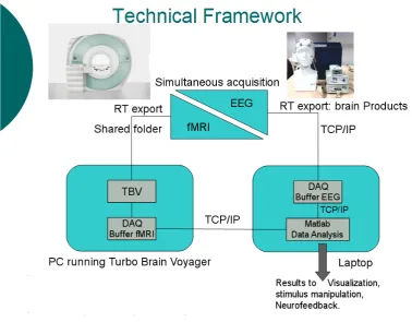
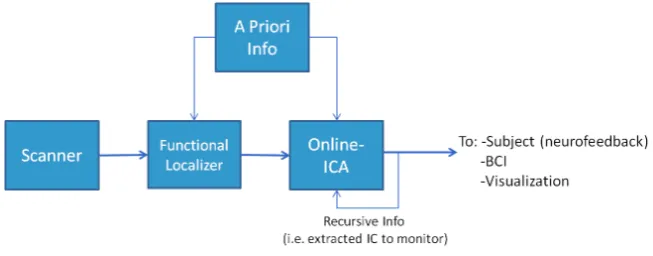
Figure 4.1: Experiment 1- Schematic Description of real-time EEG-fMRI
Hard-ware and SoftHard-ware Set-up- In this figure a scheme of the adopted set-up is reported. It
is highlighted the flux of data and information from both EEG and fMRI subsystems, along with the location of the software subsystems for data analysis and visualization. TBV is
the Turbo Brain Voyager software, DAQ is the data acquisition buffer used by matlab to mimicry multi-threading.
4.2
Data Acquisition
them jointly in the system and in a shared folder while they are acquired (i.e. volume by volume). This sequence has been slightly modified in order to export data corrected for inhomogeneities of the magnetic field using the Point Spread Function (Zaitsev et al., 2004). From the shared folder the data will be made available to external computers. It is worth noting that the new versions of Siemens control software makes it possible to easily export the data as an intrinsically implemented feature of the software by enabling an option.
4.3
Data Analysis
4.4
Stimulus Delivery
The stimulus delivery system has been implemented in Matlab exploiting an adapted version of Tools for NIfTI and ANALYZE image by Jimmy Shenhttp://www.mathworks. com/matlabcentral/fileexchange/8797. This tool has been used to deliver to the subject the spatial map of his brain activation detected by the data analysis techniques developed. Along with the spatial map, even the associated time course can be send to the subject in different formats, such as a bar whose magnitude is adapted to the level of activations or as an ongoing graph. This visual stimulus delivery is obtained by starting a parallel Matlab session on the same computer as the data analysis is ongoing. This new Matlab session is located on a secondary screen of the computer, which will be shown to the subject within the scanner.
4.5
Limitations
5
Experiment 2: Selecting ICA
algorithms and parameters for
real-time fMRI applications
5.1
Introduction
im-plemented in different ways depending on the characteristics of the algorithms. It can be as low invasive as a simple tailoring in the nature of the statistical distribution to be extracted, i.e. weighting more super-gaussian or sub-gaussian distributions, or as constrained as targeting a specific timecourse or spatial map. This approach is known as semi-blind decomposition, and its main property is to fuse the positive principles of data-driven algorithms with some kind ofa prioriknowledge on the problem of interest. The introduction of a priori knowledge can be done in several ways, e.g. by orienting the decomposition of data into sources with some specific properties. An example of a semi-blind approach is presented in (Lin et al., 2010), in which a spatial a priori constraint is introduced in the decomposition algorithm with the aim of extracting the source most congruent with a predefined spatial target. The motivation of considering priors include reduced computational time (asa priori information suggests short cuts in the decomposition to the algorithm), and improved quality of the sources obtained (given that the results are closer to what is expected). In general not all ICA imple-mentations foresee the possibility of introducing prior knowledge at spatial or temporal level. In this context, and given the noisy data of rt-fMRI experiments from the limited data available for analysis, it is of interest to extend the evaluation of real-time ICA strategies with the consideration of temporal and spatial priors.
fraction of the time series. The obtained results will be the base for the implementation of novel ICA-based method presented in the next chapter.
5.2
Materials and Methods
We refer the reader to the Appendix for details on the acquisition paradigm for the fMRI dataset adopted in the analysis performed in both Chapter 4 and 5.
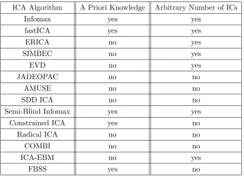
5.2.1 ICA Algorithms
A total of 14 different ICA algorithms were considered (see Table 5.1). The algorithms were available from the GIFT toolbox and most of them were discussed in a recent comparative study (Correa et al., 2005). The list includes algorithms already used in real-time fMRI experiments, like the fastICA algorithm (Esposito et al., 2003). These algorithms, which are public and were taken as in their original distributions, differ in their data reduction preprocessing steps (e.g., centering, whitening, dimensionality re-duction) and independence criteria for source separation (e.g., minimization of mutual information, maximization of non-Gaussianity) (Cichocki and Amari, 2002).
5.2.2 Parameters Analysed in the ICA Simulations
The main purpose of this real-time fMRI simulation study is to investigate a number of ICA algorithms to find the one that performs best across subjects using a trade-off of the following parameters:
1. Window length (i.e time length of data acquisition).
2. Model order (i.e. number of ICs).
3. Type of a priori information (none, spatial or temporal).
ICA Algorithm A Priori Knowledge Arbitrary Number of ICs
Infomax yes yes
fastICA yes yes
ERICA no yes
SIMBEC no yes
EVD no yes
JADEOPAC no no
AMUSE no no
SDD ICA no no
Semi-Blind Infomax yes yes
Constrained ICA yes no
Radical ICA no no
COMBI no no
ICA-EBM no yes
FBSS yes no
Table 5.1: ICA Algorithms - List of tested ICA algorithms and their possibility to
accept as parameters arbitrary a priori knowledge (both Spatial and Temporal) and a
varying number of ICs. Those algorithms which cannot accept an arbitrary number of ICs extract a numer of ICs equal to the time window length. These algorithms references
are contained in GIFT toolbox (GIFT:http://mialab.mrn.org/software/gift/index.
5.2.3 Window Length and Model Order
The amount of data that an ICA algorithm uses depends directly on the number of brain volumes available in the growing time window, which in turn defines a limit to the maximum number of ICs that may be computed. In this study we focus on a growing window approach because were interested in finding an optimal window length. As the time window length becomes longer there may be a more accurate representation of the averaged dynamic responses of the brain because more data is available. However, this may come at the cost related to both reduce temporal resolution of the dynamics characterized and increase the computation time. Conversely, with shorter windows the characterizations may be faster yet less accurate. For the simulation of each ICA algorithm the window length was varied between 3 and 35 brain volumes (the full time series consisted of 220 brain volumes, and 35 TRs was approximate to the known period of the visual-motor tasks). For each time window length the number of ICs was varied between 2 (minimum meaningful value of model order in BSS) and the actual window length. Moreover, since for computational reasons the model order must be less than or equal to the window length, the window length minimum value has been set to 3. Thus while increasing the window length all possible model orders between 2 and window length were evaluated to find the best performing pair of parameters (window length and model order). Not all the ICA algorithms considered permit an arbitrary selection of the number of desired ICs. Some of them (jade-opac, amuse, Radical ICA, combi, ICA-ebm, FBSS) allow the possibility to extract only a number of ICs that is fixed for each run and is equal to the number of available data points. In our case, this means that for these algorithms the spanned parameter space will be represented by a line identified by the points in the space with equal number of ICs and time window length.
5.2.4 Use of A Priori Information
other ones. To address this problem the concept of either spatial (Lin et al., 2010) or temporal (Esposito et al., 2003) a priori information has been explored in literature. Other ways to solve the problem of ICs ranking could be represented by exploitation of characteristic expected feature of the ICs of interest via a classifier (DeMartino et al., 2007).
In the context of real-time fMRI, a priori information may be available from a localizer scan that elicits aspects of activation that are then to be tracked dynamically in a subsequent experiment. The priors can make the mathematical computation of ICA easier, driving the algorithm initial conditions closer to the basin of attraction of the target IC. In this simulation study the temporal and spatial IC priors were determined from the ICA analysis of the full timeseries. This a priori information is incorporated into the ICA algorithms as an initial estimation of the weighted matrix or as a final constraint of the shapes of the target IC. Due to the intrinsic characteristics of the ICA algorithms, only a subset of them allows us to incorporate spatial and/or temporal a priori knowledge in the analysis (see Table 5.1).
5.2.5 Computation of template ICs for performance evaluations
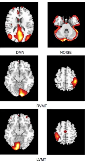
Four template ICs were identified on each subject by applying the Infomax ICA algo-rithm with 20 components to the full timeseries. The spatial maps and associated time courses of these networks were later used as reference and a priori knowledge options for the performance evaluation of different ICA implementations, in particular shorter time series to simulate real-time fMRI conditions.
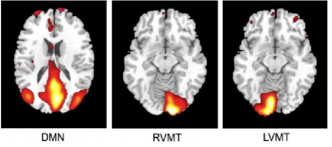
Figure 5.1: Experiment 2- Investigated brain activations - Spatial maps of ICs considered in the simulation obtained from Group ICA 20 ICs. For ease of visualization
only the relevant slices are reported here. First row depicts Default Mode Network (DMN) and residual motion artifact (Noise). Second and third rows depict the two task related
5.2.6 Evaluation of performance for different ICA implementations
The performance of each ICA algorigthm was assessed separately for each subject and network (RVMT, LVMT, DMN, NOISE) by systematically sampling the space of algorithm variables, finding for each variable set the target network ICs and comparing them with the corresponding template networks.
The ICA implementations for each subject and network were manipulated through the following variables:
• ICA algorithm: 14 algorithms listed in Table 5.1.
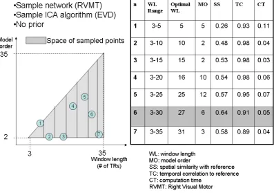
• Prior: all 14 algorithms were tested without priors. A subgroup of four algo-rithms (Fast ICA, Constrained ICA, ICA-EBM, FBSS) allowed the additional implementation of either spatial or temporal priors taken from the template ICs. • Window length (WL): for each algorithm the window length varied from 3 TRs to 35 TRs in a growing window scheme. The lower limit of 3 TRs was chosen as the minimum time course length for which an ICA can be computed. The upper limit of 35 TRs was chosen because it is approximate to the period of the cognitive task.
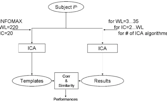
• Model Order (MO): for each WL the model order was varied between 2 and WL.
These parameters were manipulated according to an iterative automatic procedure (Soldati et al., 2012), as schematically shown in Figure 5.2. Overall this meant that for each subject (a total of 3), network (a total of 4) and ICA algorithm (a total of 22: 14 with no priors, 4 with spatial and 4 with temporal priors), a total of 561 ICA computations were made (33x34, given 33 WL values and for each WL-1 possible MO choices). At each iteration the extracted IC results were compared with the templates to estimate the performance of the iteration’s parameters.
The performance of each algorithm was characterized from the following three pa-rameters:
Figure 5.2: Experiment 2- Scheme of the procedure for testing ICA algorithms
- Diagram of adopted method for ICA algorithm comparison. For each subject data are exploited for creating templates using INFOMAX with model order (ICs) of 20 and window
length (WL) equal to the entire available time course. The ICA algorithms are then tested iteratively for each combination of IC and WL. Results of each computation are compared
with templates and evaluated in terms of spatial similarity and temporal correlation.
overlap) between the ICs extracted and the template IC for the corresponding network. The spatial similarity metric was computed as the absolute value of
Similarity= a∗b
norm(a)∗norm(b) (5.1)
where a and b are the vectors representing the spatial map (reshaped to 1D) of extracted and the template IC of interest respectively.
2. Temporal correlation with template network: The temporal correlation between the IC extracted and the template IC derived was computed, with its statistical significance (p-value <0.05).
Considering a fixed subject, brain network and ICA algorithm (with or without prior), the best performing ICA implementation (choice of WL and MO) was considered the one that gave the highest spatial similarity with a significant temporal correlation to the reference network and a computational time below the 200s threshold.
5.3
Results
This study evaluates the performance of different ICA algorithms in a real-time fMRI simulation that uses public fMRI data (Calhoun et al., 2003). The performance of various real-time ICA implementations is characterized on three task activated networks (motor, visual and default mode) and an IC representative of noise using three metrics: spatial and temporal similarity with the reference networks derived from the full fMRI time series as well as computational time (see Figure 5.3). The following variables were systematically manipulated in the various ICA implementations using a growing window approach: ICA algorithm, time window length, model order, use of spatial, temporal or no prior information. The goal was to find the implementations that would give the best compromise between highest similarity between the detected IC and the reference IC at minimal computation time. Figure 5.3 shows an example (mean over subjects) of the search for optimal combination of parameters in a sample network (right visual-motor cortex) when using one of the ICA algorithms (EVD). For illustration purposes, the growing explored space of window lengths (horizontal axis) and model orders (vertical axis) is sampled incrementally by seven areas covering different ranges of WL (MO is always from 2 to WL-1). For each of the sampled areas the optimal point is shown with its relevant information listed on the table in the figure. The figure shows how, for a fixed network and algorithm, the optimal performance can fluctuate depending on the amount of data available (window length) and the model order chosen. In particular, it can be seen that the optimal performance in different parts of the space is not given by the highest model order for a given window length.
Figure 5.3: Experiment 2- Example of results- Example of the path (which follows
the increment of the window length) resulting from iterative evaluation of the best per-formance (mean over subjects) for the EVD algorithm extracting the RVMT component.
Each point is associated to the similarity result and other parameters obtained in the grow-ing space spanngrow-ing a number of ICs and window lengths. Given the trade-off criteria, the
time was<35 TRs (200 s) and for which the significant temporal correlation with the reference time course was obtained (p value<0.05). The ICA algorithms that offered the best trade-off between high spatial/temporal correlation and low computation time were evd, jade-opac and amuse.
5.4
Discussion
The aim of the present study was to evaluate the performance of ICA algorithms in ill-posed conditions, i.e. with a small amount of data availability and constraints on computational time. The issue here was to understand if it is possible to adapt an ICA algorithm to a none-ideal environment, as presented in (Esposito et al., 2003). Moreover the analysis has been extended to investigate which ICA algorithm is more suitable to this kind of conditions from the perspectives of monitoring a brain activity of interest.
Testing ICA algorithms in ill-posed conditions must deal with several intrinsic ambi-guities and problems, both theoretical and practical, which must be taken into account. In particular, one issue is the comparison of algorithms using different model orders and information. Our goal was to explore the performance in terms of ability to reach the spatial and temporal network characteristics that can be derived from the full dataset in a standard offline analysis. Thus, we assumed as reference the optimal results obtained via group ICA with all time-points available, a model order of 20 and using the infomax algorithm, considering stochastic differences not critical. Another intrinsic issue is due to the fact that the differences in results between off-line and ill-posed conditions can be related not only to computation, but also to the extraction of dynamic behaviour with respect to the stationary one typically extracted by off-line ICA.
One issue that deserves special consideration in these simulations is that of circular-ity. The use of a validating reference template obtained from the same data used in the simulations does not introduce circularity issues since we are in principle just checking that the same information can be extracted in different ways, with only differences due to noise.
A practical issue to consider is that the high dimensionality of the space of param-eters results in a high computational load to run the simulations spanning the entire multidimensional parameter space. The best performance can be evaluated in a trade-off perspective, since different combination of parameters can give similar results. The consequence of this is that the optimality of performance is heavily connected to the practical application and conditions in which the ICA algorithm will be adopted.
that they offer (Tab. 5.1). In fact the tested ICA algorithms can be divided into three groups: those which accept setting of model order anda priori knowledge (i.e. infomax, fastICA, semi-blind infomax), those which do not accept neither setting of model order nor a priori knowledge (i.e. jade-opac, amuse, radical ICA, combi), and those which accept only one of the two (i.e. erica, simbec, evd, constrained ICA, ICA-ebm, FBSS). These constraints were intrinsic to the algorithms as in their public distributions. It was beyond the scope of this work to try to change any of the algorithms to eventually make them more flexible. The more flexible algorithms (i.e. those accepting full ma-nipulability of parameters) will not necessarily be better, since the most rigid could be the most adaptable for specific circumstances. Putting everything in a real-time fMRI experiment perspective, it is possible to distinguish the algorithms on the basis of the tasks and conditions they must face. Those algorithms which do not accept anya priori knowledge could work very well to define the target networks from the functional lo-calizer step that usually precedes a real-time fMRI acquisition, a step in whicha priori knowledge may not be necessary or even available. For this use it is possible to permit a higher computational load, since usually the localizer part of an experiment can have more time allocated to it. The algorithms that tended to be more suitable for this use are evd and amuse, which result in particularly fast computation, with evd performing slightly better. The jade-opac and fastICA algorithms also performed well but at the cost of a higher computational time (Tab. 5.2). The results show that the use of a priori knowledge can drastically improve computation time and spatial similarity to a target IC. This suggests that use of priors may be crucial in the dynamic analysis part of the real-time fMRI experiment, where any information from the localizer can be exploited to speed up the process and increase accuracy. From this point of view the flexibility of the ICA algorithm is essential. Thus among the algorithms which accept a priori knowledge, constrained ICA provides the most optimal solution, followed by fastICA (Tables 5.3 and 5.4).
to the on-line computation, since the scale of the resolution in monitoring the brain dynamics will be directly associated to that. This means that different algorithms can shown to be more or less adapt to be directly exported to a real-time set-up.
Another observation is related to the type of brain activity monitored (i.e. if it represents a resting state brain activity, a task related activity or physiological noise). Monitoring ICs with different origins conveys different information. Variability in the capability of ICA algorithms to correctly extract target ICs can be directly justified by the fact that the less variance of data is explained by the IC, the more difficult is to extract it by decreasing the amount of data available. For this reason ICs whose rank is low in a canonical ICA decomposition are critical to identify in the ill-posed conditions. Nonetheless, as the simulations show they can still be identified.
The periodicity of the ICs of interest affects the choice of optimal parameters. From simulations it can be seen that a significant performance may be reached even reduc-ing the data to a sreduc-ingle period of the task in the ICs of interest. The DMN deserves particular considerations due to the low frequency nature of its signal sources. Its iden-tification, despite being easily done by data driven algorithm, is dramatically harder in ill-posed conditions given that its periodicity is significatively longer, thus it results dif-ficult to observe enough of its dynamic given the real-time compatible vincula on time window length. Given these new dimensions (type of brain activity and periodicity) it is possible to see that different algorithms have different effectiveness in adequately identify brain activity coming from different kinds of sources. It can be seen that the same algorithm can outperform all the others in detecting task related activity, while suffering in dealing with non-structured noise or, viceversa, as for example it happens in the case of evd and jade-opac, or evd and combi with no a priori knowledge. The same reasoning holds for the use of a priori knowledge. Even if in this case not all algorithms permit the introduction ofa priori knowledge in performing the ICA decom-position, for those which accept this input the performance varies considering different target sources. Indeed fastICA and constrained ICA alternate best performance, with constrained ICA performing slightly better overall.
time of ICA decomposition: in general it grows linearly with the increase of the window length, and this can be easily justified by the fact that the more data are to be processed the more time it takes to do that. But as the data become more descriptive of the source to be extracted, the algorithm is able to extract it easier, thus reducing the computational time needed, independently by the data length.
One limitation of this study is that the adopted implementations of ICA algorithms are not directly optimized for ill-posed conditions. This opens the doors to further de-velopment oriented towards their methodological and algorithmic optimization, which would make them more efficient and flexible. Another element to be taken into account is the relatively small number of subjects adopted in the simulations. This constraint was driven by the necessity of having a dataset whose behaviour was well known in the ICA domain and which could confirm the stability and validity of obtained results. Nonetheless, this work demonstrates a methodology for evaluating different ICA im-plementations for the purpose of finding the ICA algorithms and analysis parameters for the optimal detection of a target brain network under ill-posed conditions. Further experiments are needed to evaluate the performance of ICA implementations on larger datasets and also other networks.
The results of this study can be used to evaluate ICA implementations for the dynamic analysis of fMRI data. In particular, in a potential real-time fMRI perspec-tive, the best performing ICA algorithm without the use ofa priori knowledge can be adopted to analyse the functional localizer data in a data-driven way. In this approach the target ICs to be then followed dynamically in the real-time experiment are defined without considering spatial or temporal constraints. The sources defined by the func-tional localizer can then be used in different algorithms that include a priori spatial, temporal or spatio-temporal knowledge for the dynamic monitoring of target ICs in a real time fMRI experiment, such as for neurofeedback.
5.5
Conclusions
6
Experiment 3: Evaluating novel
methods for real-time fMRI by
use of
a priori
conditions
6.1
Introduction
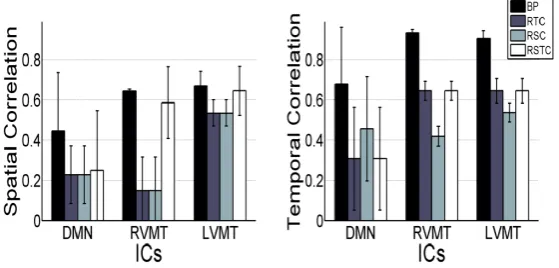
a reference optimal ICA. Moreover, the ICA algorithm (FastICA) is stochastic, which means that multiple repetitions of the analysis on the same dataset can give slightly different results, both in the spatial and temporal domains. This problem has been extensively discussed in literature, where one of the main proposed solution has been a method based on multiple ICA runs and clusterization of the obtained components, with the aim of reducing the issue of stochastic variability (Himberg et al., 2004). Such instabilities can be characterized by the standard deviation of the derived (STD) re-sults (spatial and/or temporal) when the analysis is repeated multiple times on the same dataset. The STD can be considered as a stability performance parameter of the algorithm, lower STD algorithms corresponding to more stable ones. This parameter may be particularly relevant if different ICA-based algorithms are to be considered and compared for real-time fMRI, where the analysis is repeated dynamically during data acquisition.
In this chapter we exploit results from the previous chapter and extend them by proposing and evaluating novel ICA-based algorithms that use different types ofa pri-ori knowledge for the dynamic monitoring of ongoing fMRI activity. The a priori information considered is either temporal, spatial, or both spatial and temporal. In addition, thea priori information is considered both in its static version when derived from the functional localizer, as well as dynamic when estimated recursively as the sliding-window progresses over the time course throughout the run. The different anal-ysis methods are tested by simulating a real-time fMRI experiment using an existing and public dataset. In