Folia Morphol. Vol. 60, No. 4, pp. 259–280 Copyright © 2001 Via Medica ISSN 0015–5659 www.fm.viamedica.pl O R I G I N A L A R T I C L E
Address for correspondence: Prof. Janusz Moryś, MD, PhD, Department of Anatomy and Neurobiology, Medical University of Gdańsk, ul. Dębinki 1, 80–210 Gdańsk, Poland, tel: +48 58 349 1401, fax: +48 58 349 1421, e-mail: [email protected]
The amygdaloid complex of the rabbit
— morphological and histochemical study
Hanna Jagalska-Majewska, Jerzy Dziewiątkowski, Sławomir Wójcik,
Anna Łuczyńska, Renata Kurlapska, Janusz Moryś
Department of Anatomy and Neurobiology, Medical University of Gdańsk, Poland
[Received; Revised 29 October 2001; Accepted 29 October 2001]
The aim of the present paper is to describe the morphology and topography of the nuclei of the amygdaloid complex in the rabbit. In the current study we also investigated the intensity of the enzymatic reaction for acetylcholinesterase (AChE) in the amygdaloid complex and the morphology of its neurones. Material con-sisted of 5 brains of adult New Zealand rabbit, stained either with cresyl violet or for AChE activity.
Although, as in other mammals, the rabbit amygdala consists of two main nu-clear groups (corticomedial and basolateral), it reveals a peculiar morphology pattern, forming a transition structure between those observed in the cat and rat. Especially characteristic is the arrangement of the basolateral complex. Within that the ventromedial division of the lateral nucleus seems to be the largest, while its dorsolateral division — the smallest. The arrangement of the cortico-medial complex in the rabbit is similar to both the cat and rat.
In the rabbit the highest acetylcholinesterase activity is found in the basolateral nucleus and the nucleus of the lateral olfactory tract. The lowest AChE staining is observed in the cortical and medial nuclei, amygdalohippocampal and ante-rior amygdaloid areas and intercalated masses.
key words: amygdala, rabbit, acetylcholinesterase and morphology
INTRODUCTION
The amygdaloid complex has attracted considerable attention over the last few decades and many of its organisational and structural features have been re-vealed. It belongs to the limbic system and is implicat-ed in memory and emotional behaviour [42]. It occu-pies a central position of the telencephalon, lying me-dially to the endopiriform nucleus. In the rabbit it extends in frontal sections from the level of the ante-rior commissure to the lateral geniculate body [15].
The morphology and subdivisions of the amygda-loid complex have been studied in many species: the rat [14, 43, 51, 53, 64], mouse [65], squirrel [66], guinea pig [66], hamster [62, 66, 70], cat [20, 29,
syn-Folia Morphol., 2001, Vol. 60, No. 4
aptic transmission. It is present in cholinergic and noncholinergic neurones, which are in the focus of interests of neurobiology, because cholinergic inner-vation of the cerebral cortex and amygdala plays a cri-tical role in the neuronal modulation of attention, arousal and memory [59]. Severe damage of cholin-ergic pathways is one of the earliest neuropathologi-cal features of Alzheimer’s disease [9, 10, 38, 59], espe-cially in the basolateral complex of the amygdala [5].
AChE activity of the amygdaloid complex was studied in man [16, 36, 58, 61], monkey [39], cat [48], guinea pig [21] and rat [3, 4, 47]. It is charac-teristic that the same nucleus in different species shows an approximately similar level of AChE activi-ty. Thus AChE is a valuable and comparable substrate in morphological studies.
According to Humphrey [23] and Johnston [25], the amygdala consists of two main nuclear groups: the corticomedial and basolateral. The corticome-dial complex, being phylogenetically older, is parti-tioned into: the central, medial nuclei, cortical amygdalohippocampal area, nucleus of the lateral olfactory tract, bed nucleus of the accessory olfac-tory tract and amygdalopiriform cortex. On the oth-er hand the phylogenetically youngoth-er basolatoth-eral complex is divided into the basal and lateral nuclei. There are other amygdaloid areas such as the ante-rior amygdaloid area and intercalated masses.
It is well known that the corticomedial (superfi-cial) division plays an important role in the conver-gence of sensory (especially olfactory), autonomic and endocrine inputs from the hypothalamus and brain stem, whereas the basolateral (deep) group receives direct sensory and limbic inputs, being an important structure in learning, memory, emotional and social behaviour [44]. The lateral and basolate-ral nuclei project to the centbasolate-ral nucleus [13, 26, 30], which then projects to the hypothalamic and brain stem target areas [56] modulating some aspects of emotions. There are a lot of data concerning the amygdala as a crucial structure in classical Pavlovian conditioned fear [7, 8, 69] and emotional learning in long term potentiation mechanism [32]. The amygdala also plays an important role in uncondi-tioned fear, anxiety, aggression and attention [13, 30]. Researchers started to indicate the lateralisa-tion of the amygdala’s involvement in emolateralisa-tionally influenced memory [6, 11]. It is responsible for re-cent but not remote memories [13] and is involved in memory consolidation [17].
There are many clinical aspects of studies on the amygdala. For example, there are some data
sug-gesting that developmental malformations of the amygdala may underlie autism [22]. The amygdala is susceptible to development of the temporal lobe epilepsy [60, 68]. Pitkänen et al. [54] have proved that some parts of the amygdala (e.g. the medial division of the lateral nucleus, basomedial nucleus, accessory basal nucleus, posterior cortical nucleus, anterior cortical nucleus and medial nucleus) in the rat are regions sensitive to status epilepticus-induced neuronal damage.
The above-mentioned involvement of the amygda-loid nucleus in physiological and pathological process-es taking part in a changeable external environment should be based on their morphological properties. Because of the lack of such a detailed description in one of the most common laboratory animals, the aim of the current study was to give a detailed description of the topography and morphology of the amygda-loid complex in the rabbit with the usage of classical techniques: cresyl violet and AChE staining.
MATERIAL AND METHODS
The material consisted of five brains of female New Zealand rabbits, aged 9–24 months and weighing 2650–4550 g. Care and treatment of animals were in accordance with the guidelines of the EC as well as of the Local Ethical Committee. Animals were deeply anaesthetised with a lethal dose of Thiopen-tal (80 mg/kg of body weight i.p.), then transcar-dially perfused with 250 ml of 0.9% solution of sa-line containing 10000 units of Heparin, followed by 1000 ml of Baker fluid (4% paraformaldehyde solu-tion in 0.1 M phosphate buffer with calcium chlo-ride (22,350-6 Aldrich), pH 7.4). The brains after immediate removal from skulls were fixed in Baker fluid for 3 hours, kept in 15% and next in 30% su-crose solution in 0.1 M phosphate buffer (pH 7.4, 4oC) until sunk. Frontal
40-mm-thick, serial sections of brains were cut on JUNG 1800 cryostat (Leica, Germany). The first set of sections was collected on glass slides and dried at room temperature and fi-nally stained with cresyl violet. The second group of sections was used for AChE histochemistry. The stain-ing procedure was based on Koelle method in the modification of Gerebtzoff and was carried out at a pH of 7.4 [18, 36, 37, 45].
For selective inhibition of unspecific cholinesteras-es serial sections were incubated at a temperature of 37o
Hanna Jagalska-Majewska et al., The rabbit amygdaloid complex
water and ethopropazine (E-2880 Sigma) [18, 45]. Sections were rinsed 2–3 times in distilled water. Then sections were postincubated in ammonium sulphide (17,250-2 Aldrich) and rinsed again. Sections were plated on glass slides, allowed to dry and covered; then analysed under light microscope (Labophot 2, Nikon, Japan). The set of low magnification images was saved as bitmaps to display topographic rela-tions between amygdaloid nuclei (light microscope (Leica DMLC, Germany) equipped with camera).
RESULTS
The corticomedial amygdaloid complex
In frontal sections the corticomedial amygdaloid com-plex appears at the level of the optic chiasm (Fig. 1) and extends throughout the whole amygdaloid
com-plex (Fig. 1–11). Its nuclei together with the anterior amygdaloid area build a rostral pole of the amygda-la (Fig. 1). This complex in AChE staining is hetero-geneous, although this differentiation is less visible then in the basolateral complex. Medial and cortical nuclei are poorly but homogeneously stained, while subdivisions of the central nucleus are characterised by poor to moderate intensity (Fig. 2b–9b).
The central nucleus (CE)
In the frontal sections the central nucleus appears in the plane where the medial and anterior cortical nuclei are well developed (Fig. 2). It is of an easily distinguishable ovoid shape. Its ventrolateral bound-ary is formed rostrally by the external capsule and caudally by the basolateral complex (Fig. 2–8).
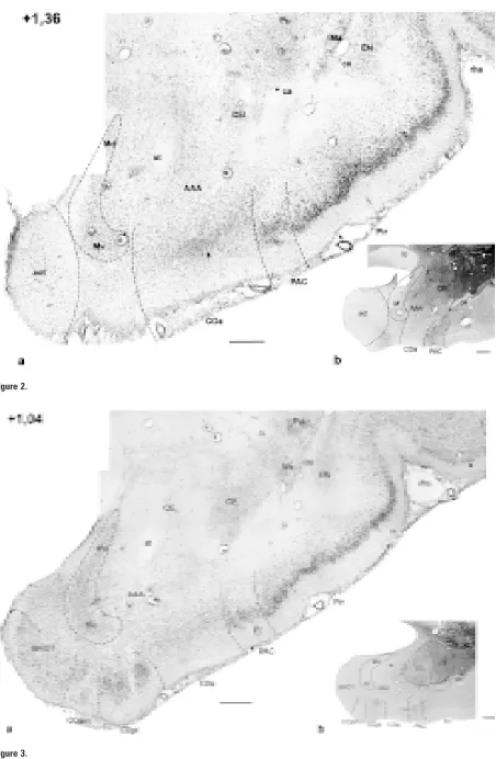
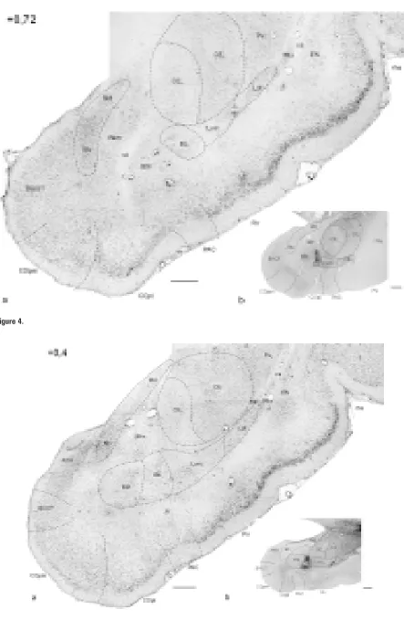
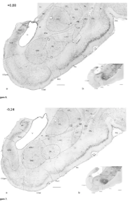
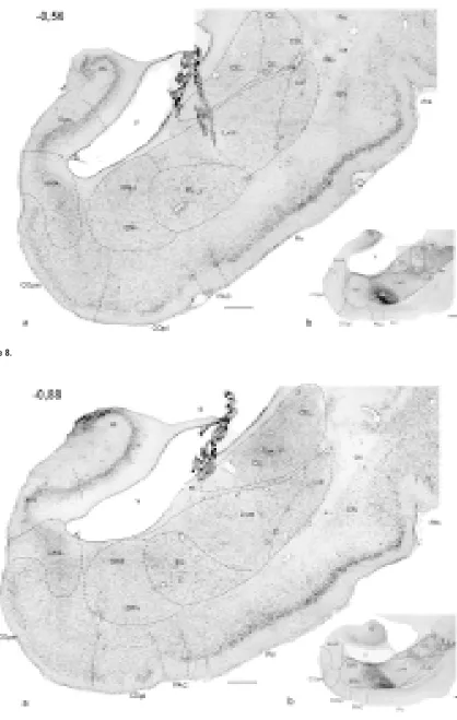
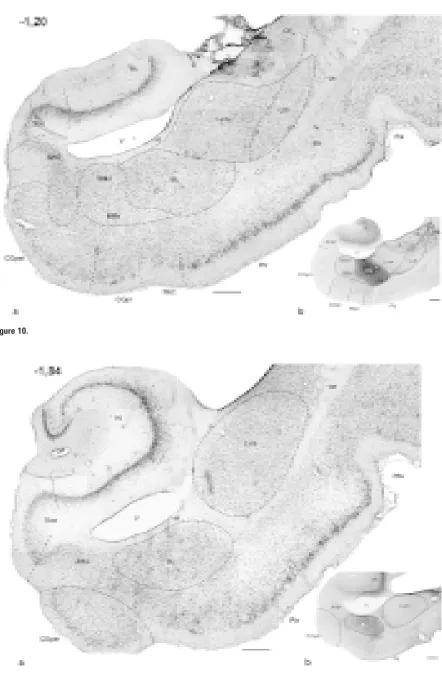
Me-Figure 1–11. Photomicrographs of frontal sections through the amygdaloid complex in the rabbit: a) cresyl violet; b) AChE histochemistry. The number in the upper left corner means the distance with reference to Bregma [mm].
AAA — anterior amygdaloid area, AHA — amygdalohippocampal area, aot – accessory olfactory tract, BAOT — bed nucleus of the ac-cessory olfactory tract, BL — basolateral nucleus, BM — basomedial nucleus (d, v — dorsal and ventral part, respectively), ca — anteri-or commissure, ce — external capsule, CE — central amygdaloid nucleus (CEm — medial, CEim — intermediate, CEc — capsular and CEl — lateral subdivisions), Coa — anterior cortical nucleus, COpm — posteromedial cortical nucleus, COpl — posterolateral cortical nucleus, EN — endopiriform nucleus, fi — fibres of hippocampus, Hi — hippocampus, IM — intercalated masses (IMa — anterior, IMl — longitudinal, IMm — medial), Ldl — dorsolateral division of the lateral nucleus, Lvm — ventromedial division of the lateral nucleus, M — medial amygdaloid nucleus (Md — dorsal, Mv — ventral subdivisions), NLOT — nucleus of lateral olfactory tract,
Folia Morphol., 2001, Vol. 60, No. 4
Hanna Jagalska-Majewska et al., The rabbit amygdaloid complex
Figure 4.
Folia Morphol., 2001, Vol. 60, No. 4
Figure 6.
Hanna Jagalska-Majewska et al., The rabbit amygdaloid complex
Folia Morphol., 2001, Vol. 60, No. 4
Figure 10.
Hanna Jagalska-Majewska et al., The rabbit amygdaloid complex
dially, the central nucleus is surrounded by fibres of the terminal stria and medial nucleus (Fig. 3–10). In rostral sections clear delineation of the central nu-cleus from the dorsolaterally located striatum is dif-ficult due to the similarity of their cellular structure (Fig. 2–4). The only difference is that neurones of the central nucleus are less dense. Nevertheless, in middle sections the dorsal aspect of the central nu-cleus is surrounded by fibres marking the boundary with the putamen (Fig. 6–8).
Figure 12. Photomicrographs of cells in the central, medial, anterior cortical nuclei and bed nucleus of the lateral olfactory tract in the rabbit: a) medial division of the central nucleus (CEm) with oval, pyra-midal-like and round neurones (arrows); b) lateral division of the central nucleus (CEl) with denser py-ramidal-like and oval neurones (arrows); c) the dorsal division of the medial nucleus (Md) with loosely packed large oval and round neurones (arrows); d) the ventral division of the medial nucleus (Mv) with round densely packed cells (arrows); e) the bed nucleus of the lateral olfactory tract, arrows indicate large oval and round neurones; f) the anterior cortical nucleus, oval and round neurones (arrows); cresyl violet; scale bar = 25 mm.
Folia Morphol., 2001, Vol. 60, No. 4
rostral sections and is characterised by low neuronal density (Fig. 12a) and low AChE activity (Fig. 3b–4b). The remaining divisions of the central nucleus are characterised by the homogeneity of the cellular
structure, however some cytoarchitectonic differenc-es are visible. The lateral division has smaller and more densely packed cells (Fig. 12b) and shows moderate heterogeneous AChE activity (Fig. 3b–7b).
Figure 13. Photomicrographs of cortical area in the rabbit: a, b, c — the posteromedial cortical nucle-us; a) layer I with loosely packed pyramidal-like neurones (arrows); b) layer II with densely packed oval, fusiform and pyramidal-like cells (arrows); c) layer III with oval, fusiform and pyramidal-like neu-rones (arrows); d) the chosen superficial cortical structures of the amygdaloid complex;
e, f, g — the periamygdaloid complex; e) layer I, only glial cells; f) layer II with densely packed oval and round neurones (arrows); g) layer III with loosely packed oval and round neurones
Hanna Jagalska-Majewska et al., The rabbit amygdaloid complex
It appears in the very rostral pole of the central nu-cleus and extends through the anterior and middle portions of the amygdala (Fig. 2–8).
The capsular and intermediate divisions appear from the middle portion of the amygdaloid complex (Fig. 6). The capsular division constitutes the ventro-lateral aspect of the central nucleus (Fig. 6–8). It is well visible in AChE preparations as it performs mo-derate enzymatic activity (Fig. 6b–8b), although higher than in the lateral division.
The intermediate division appears in the dorsal aspect of the central nucleus and is inserted between the medial and lateral ones (Fig. 6–8). It shows the lowest AChE activity of all divisions of the central nu-cleus (Fig. 6b–10b), which makes it easily distinguish-able from the putamen. In caudal sections only medial and intermediate divisions are present (Fig. 9–10).
The medial nucleus (M)
The medial nucleus is a group of cells that appears in the anterior aspect of the amygdaloid complex (Fig. 2). At rostral levels it neighbours the terminal stria, the anterior amygdaloid area — laterally, the bed nucleus of the accessory olfactory tract — medially and the anterior cortical nucleus — ventrolaterally (Fig. 2–3). At this level it has the outline of a bow, surrounding the anterior amygdaloid area from the ventral side (Fig. 2–3). Its vertical part inserts between the optic tract and terminal stria, while its horizontal one is directed to the anterior cortical nucleus (Fig. 2–3). More cau-dally the medial nucleus is inserted between the amygdalohippocampal area medially, the central nu-cleus, intercalated masses and basomedial nucleus — laterally (Fig. 5). Its most posterior portion is situated above the inferior horn of the lateral ventricle (Fig. 6). Cytoarchitectonical features enable the division of the medial nucleus into two fields: dorsal and ventral, but the boundary between them is not clear. The dorsal division is characterised by loosely-packed, bigger, oval and round, palely-stained neurones (Fig. 12c). The ventral division consists of neurones of simi-lar shape, but they are more densely packed, smaller and darkly stained (Fig. 12d).
The reaction for AChE is homogeneously low throughout the medial nucleus (Fig. 2b–6b).
The cortical nuclei
The cortical nuclei appear at the level of the optic chiasm and extend caudally throughout the whole amygdaloid complex lying superficially and ventrally to the anterior amygdaloid area and basolateral complex (Fig. 1–11).
On the basis of cytoarchitectonics as well as to-pographic relations, the following cortical nuclei can be distinguished: the anterior cortical nucleus (COa), posterior cortical nuclei (COp) (which splits into the posterolateral (COpl) and posteromedial cortical nucleus (COpm)).
In the cresyl violet staining cortical nuclei, ex-cept the anterior cortical nucleus, are composed of three cellular layers: layer I — containing small scat-tered cells, layer II — of densely-packed neurones and layer III — consisting of less densely packed cells (Fig. 13a–d). These nuclei are characterised by heterogeneous (from negative to moderate) AChE activity (Fig. 1b–11b).
The anterior cortical nucleus (COa)
The anterior cortical nucleus constitutes the most rostral aspect of the cortical nuclei, appearing at the level of the optic chiasm. At this level it borders the nucleus of the lateral olfactory tract medially and the periamygdaloid cortex — laterally (Fig. 1). More cau-dally the accessory olfactory tract and medial nucleus build the medial border of the anterior cortical nucle-us (Fig. 2). Further, the anterior cortical nuclenucle-us is re-placed by growing posterior cortical nuclei (Fig. 4)
The anterior cortical nucleus is characterised by its almost total lack of laminar structure. Darkly stained oval and round numerous cells are situated more deeply than some scattered neurones of the superficial part (Fig. 12f).
In AChE preparations the anterior cortical nucleus shows a negative staining, distinct from the dark stain-ing of the nucleus of the lateral olfactory tract (Fig. 1).
The posterior cortical nuclei (COp)
The posterior cortical nuclei appear just before the basolateral complex, and from the very beginning they constitute the posterolateral (COpl) and pos-teromedial cortical nuclei (COpm) (Fig. 3). They lie superficially and extend anteroposteriorly to the very caudal pole of the amygdaloid complex (Fig. 3–11).
Folia Morphol., 2001, Vol. 60, No. 4
The posteromedial cortical nucleus appears as a well-delineated mass of cells situated in the ven-tromedial edge of the hemisphere. It borders later-ally the posterolateral cortical nucleus (Fig. 3–10). Dorsomedially it abuts the bed nucleus of the acces-sory olfactory tract nucleus and amygdalohippocam-pal area (Fig. 3–5) and dorsolaterally — the basome-dial nucleus (Fig. 5–11). The posteromebasome-dial cortical nucleus together with the ventrolateral division of the lateral nucleus builds the most caudal aspect of the amygdala (Fig. 11).
The posterolateral cortical nucleus merges ros-trally with the anterior cortical nucleus (Fig. 3). It is
inserted between the periamygdaloid cortex lateral-ly and the posteromedial cortical nucleus — medial-ly (Fig. 4–10). Dorsalmedial-ly it borders the medial nucleus at rostral levels and the basal nucleus — at middle and caudal levels.
The low AChE activity of the posterolateral corti-cal nucleus is different from that in both the ante-rior and posteromedial cortical nuclei, where it is ne-gative (Fig. 3b–5b).
The periamygdaloid cortex (PAC)
The periamygdaloid cortex is a transition area between the piriform cortex and cortical nuclei (Fig. 1–11).
Hanna Jagalska-Majewska et al., The rabbit amygdaloid complex
Rostrally the periamygdaloid cortex borders the an-terior cortical nucleus medially (Fig. 1–3), whereas caudally — the periamygdaloid cortex abuts the posterolateral cortical nucleus (Fig. 4–10).
The periamygdaloid cortex possesses a charac-teristic laminar structure: layer I is wider and lay-ers II and III are not clearly separated. The latter consist of big darkly stained cells and differ signi-ficantly from the pale and small cells of the ante-rior cortical nucleus as well as from the distinct laminar structure of the posterolateral cortical nucleus (Fig. 13d–g).
The periamygdaloid cortex is characterised by differentiated AChE activity — its anterior portion shows laminar distribution of moderate activity, while its posterior one — negative (Fig. 1b–10b).
The nucleus of the lateral olfactory
tract (NLOT)
The nucleus of the lateral olfactory tract begins at the very rostral pole of the amygdaloid complex, at the level of the optic chiasm. At this level it is
locat-ed superficially in the mlocat-edioventral locat-edge of the hemi-sphere (Fig. 1), further it is replaced by the bed nucleus of the accessory olfactory tract (Fig. 2). It is surround-ed by the anterior amygdaloid area, and laterally neigh-bours the anterior cortical nucleus (Fig. 1).
The nucleus of the lateral olfactory tract consists of three layers: layer I — contains few scattered neurones (Fig. 14e), layer II — is characterised by large densely packed oval and round cells (Fig. 14d), layer III — con-sists of more loosely arranged, oval and round cells (Fig. 14c). These layers are also easily distinguishable in AChE preparations: the increase of the AChE activity is observed from superficial to deep layer (Fig. 1, 14a). Medially to the nucleus of the lateral olfactory tract the area of high AChE activity rich in fibres is found.
The bed nucleus of the accessory
olfactory tract (BAOT)
The bed nucleus of the accessory olfactory tract is a homogeneous group of loosely packed darkly stained cells, which appears ventromedially at the level where the optic tract begins. It is closely
Folia Morphol., 2001, Vol. 60, No. 4
nected with the accessory olfactory tract (aot) (Fig. 2). Cells of this nucleus are round and oval, they do not form layers (Fig. 3–5, 12e).
The bed nucleus of the accessory olfactory tract shows no AChE activity similarly to neighbouring structures: the posteromedial cortical nucleus — la-terally, the medial nucleus and further amygdalo-hippocampal area — dorsomedially (Fig. 3–5).
The amygdalohippocampal area (AHA)
The amygdalohippocampal area is a transitory area between the subicular complex and the amygdala. It appears in the medial aspect of the amygdala just before the ventral hippocampus (Fig. 5). Laterally it borders the medial, basomedial nuclei and inferior horn of the lateral ventricle and ventrally — the pos-teromedial cortical nucleus (Fig. 5–10). Caudally the amygdalohippocampal area fuses the periamygda-loid and piriform cortices.
The amygdalohippocampal area is a heteroge-neous group of neurones consisting of deeply si-tuated small oval and bigger round cells (Fig. 17d). In cresyl violet staining its limits are clearly distin-guishable, whereas low and homogeneous AChE ac-tivity does not allow their clear delineation (Fig. 5–11).
The basolateral complex
The basolateral complex — a large component of the amygdala — extends from the level just behind the optic chiasm almost to the joining of the ventral and dorsal hippocampi (Fig. 4–11). It is well developed at the level where the inferior horn of the lateral ventri-cle appears (Fig. 6). The nuventri-clei of the basolateral com-plex are differentiated cellular masses and are cha-racterised by variable AChE activity. AChE staining is especially useful for delineation of its nuclei, as we found big regional differences in the distribution of AChE enzyme in the basolateral complex.
The lateral amygdaloid nucleus
The lateral amygdaloid nucleus in the rabbit is well developed at the level of the beginning of the infe-rior horn of the lateral ventricle (Fig. 6). It extends throughout the amygdaloid complex lying medially to the external capsule separating it from the en-dopiriform nucleus, and ventrolaterally to the cen-tral nucleus (Fig. 4–11).
On the basis of the morphology and topogra-phic relations in the lateral amygdaloid nucleus we distinguished two main divisions: dorsolateral and ventromedial. The border between these divisions is clear in the anterior portion (Fig. 4–7), but
poste-riorly it is not so obvious (Fig. 8–10). This division is also supported by different intensity of AChE. In the dorsolateral division AChE activity is slightly higher than in the ventromedial one (Fig. 5b–8b); moreover the boundary between these divisions in the most rostral sections is clearly marked by a bundle of darkly stained fibres (Fig. 5b–6b).
The dorsolateral division of the lateral
nucleus (Ldl)
The dorsolateral division of the lateral nucleus begins the most rostrally among all nuclei of the basolateral complex. It borders laterally the external capsule and intercalated cells, but the border be-tween the intercalated masses and the dorsolateral division of the lateral nucleus is not sharp (Fig. 4–7). Medially it abuts the central nucleus (Fig. 4, 5) and more caudally the ventromedial division of the late-ral nucleus (Fig. 6–10). The dorsolatelate-ral division of the lateral nucleus prolongs ventrally to form some-thing like a shell of the nucleus (Fig. 6–10). Finally, in the most caudal sections, it is replaced by the growing ventromedial division of the lateral nucleus (Fig. 11).
The dorsolateral division is composed of darkly stained small pyramidal-like, oval and occasionally round neurones. These cells are densely packed. Axes of neu-ronal cell somata have no specific orientation (Fig. 15a). There are many glial cells among the neurones.
The enzymatic reaction for AChE in the dorsola-teral division is moderately intensive, higher than in the ventromedial division, especially in rostral sec-tions (compare Fig. 4b, 7b).
The ventromedial division of the lateral
nucleus (Lvm)
The ventromedial division of the lateral nucleus is the most caudally extended portion of the amygdala. It almost reaches the level of the fusion of the dorsal and ventral hippocampus. The ventromedial division of the lateral nucleus occupies the mediodorsal aspect of the basolateral complex. It borders the piriform cor-tex and dorsolateral division of the lateral nucleus — laterally, the central nucleus — dorsomedially, the basal nucleus — medially and ventrally (Fig. 4–11).
Hanna Jagalska-Majewska et al., The rabbit amygdaloid complex
In rostral sections neurones of the ventromedial part are large, palely stained and loosely packed (Fig. 15b), while caudally they become smaller and dens-er (Fig. 15c). These neurones are mainly pyramidal-like and occasionally fusiform; there are many glial cells among them (Fig. 15b, c).
The basal amygdaloid nucleus
The basal nucleus is located in the ventromedial as-pect of the basolateral complex (Fig. 4–11). Anteri-orly it reaches the anterior amygdaloid area (Fig. 6). It borders the central nucleus and intercalated masses dorsally, the terminal stria, inferior horn of the later-al ventricle and amygdlater-alohippocamplater-al area — me-dially, the bed nucleus of the lateral olfactory tract, posterior cortical nuclei and periamygdaloid cortex — ventrally (Fig. 5–11).
Two main cellular groups: basolateral and baso-medial are clearly visible in the basal nucleus. The AChE intensity in two parts of the basal nucleus is different and allows their clear delineation (Fig. 4b–11b).
The basolateral nucleus (BL)
The basolateral nucleus appears at the lateral aspect of the basal nucleus (Fig. 5–10). Fibres of the external capsule reach the lateral side of this nucleus.
Dorsally it borders the central nucleus (Fig. 4–6) and more caudally — the ventromedial division of the lateral nucleus (Fig. 7–11); its medioventral edge is surrounded by the basomedial nucleus. Further caudally the basolateral nucleus is located under the inferior horn of the lateral ventricle (Fig. 7–11). The ventromedial division of the lateral nucleus borders the basolateral nucleus dorsolaterally, whereas the piriform cortex and periamygdaloid cortex — ven-trally (Fig. 5–11).
Due to the presence of the largest neurones the basolateral nucleus is easily distinguishable. It pos-sesses scattered darkly stained pyramidal-like, mul-tipolar and oval neurones (Fig 16a), of smaller den-sity in comparison with the lateral amygdaloid nu-cleus. It also has the highest AChE activity (Fig. 4b–11b).
Folia Morphol., 2001, Vol. 60, No. 4
The basomedial nucleus (BM)
Among all nuclei of the basolateral complex, the baso-medial nucleus is located the most baso-medially and is com-posed of the smallest neurones. It is placed ventrolat-erally to the terminal stria and lateral ventricle and medially — to the basolateral nucleus (Fig. 5–10). From the ventral aspect the basomedial nucleus is sur-rounded by the amygdalohippocampal area, the bed nucleus of the lateral olfactory tract, the posterior cortical nuclei and neighbouring periamygdaloid cortex; in the very caudal part the delineation
be-tween the basomedial nucleus and above mentioned areas is not sharp (Fig. 4–10).
Neurones of the basomedial nucleus are pyra-midal-like, oval and fusiform (Fig. 16b, c). On the basis of their size and density we have divided the basomedial nucleus into dorsal and ventral divisions (Fig. 7–10). The ventral division has less dense and larger cells (Fig. 16c), while the dorsal one consists of smaller and more densely packed cells (Fig.16b). In the most caudal part only the dorsal division appears.
Hanna Jagalska-Majewska et al., The rabbit amygdaloid complex
The AChE activity is homogeneously low in the basomedial nucleus (Fig. 4b–10b) similarly to neigh-bouring structures (Fig. 8b–10b).
Other amygdaloid areas
The anterior amygdaloid area (AAA)
The anterior amygdaloid area builds a rostral ex-treme of the amygdala (Fig. 1). It is located dorsally to the anterior cortical nucleus and ventrally to the central nucleus (Fig. 2, 3). Caudally it is replaced by the basolateral complex (Fig. 4).This is the area of scattered cells and fibres. Its neurones are a heterogeneous population of mainly oval and round but also pyramidal-like and fusiform cells (Fig. 17b). AChE activity in this area is negative (Fig. 1b–3b).
Intercalated masses (IM)
Intercalated masses are very well developed in the rabbit. On the basis of topographic, cytoarchitec-tonic and histochemical properties, three large groups of intercalated masses can be distinguished. The first group — anterior (IMa) — precedes the lateral nucleus and constitutes a broad cellular band located along the external capsule from the level above the rhinal sulcus and infiltrating the dorsolat-eral division of the latdorsolat-eral nucleus (Fig. 1–7). It ap-pears as dense cell clusters consisting of two neu-ronal populations of darkly stained large or small cells (Fig. 17a). Large neurones are in the majority. The boundary between the anterior group of inter-calated masses and the dorsolateral division of the lateral nucleus is not clear.
The remaining groups of the intercalated mass-es — medial and longitudinal — differ only topo-graphically. The medial group of intercalated masses (IMm) is located medially between the central and basal nuclei (Fig. 4–6), whereas the longitudinal one (IMl) — among the fibres separating the central nucleus from the basolateral complex (Fig. 5–7). Neurones of these intercalated masses are mainly small, darkly stained and oval or round in shape (Fig. 17c, e).
The activity of AChE varies among groups of in-tercalated masses, although the difference is mini-mal: medial and longitudinal groups are negative while the anterior one is weakly stained.
DISCUSSION
In the literature we cannot find any detailed report concerning the morphology of the amygdala in the rabbit, available data on the subject are
fragmen-tary [19, 26, 45, 57, 66, 70]. Thus the aim of our study was an attempt at a precise delineation of nuclear boundaries and the settlement of the termi-nology in the amygdaloid complex.
According to Girgis et al. [19] the amygdaloid com-plex in the rabbit extends from +2.0 to –3.0 with refe-rence to Bregma — so the length of the amygdala equals approximately 5 mm. According to our observations it is closer to 4.3 mm, but there is a considerable variabil-ity between individuals within the species [19].
Our results may suggest that although general divisions of the amygdaloid nuclear complex in the rabbit are comparable to that in the cat and rat, they differ reasonably. The rabbit is taxonomically differ-ent from roddiffer-ents (it belongs to Lagomorpha) and consequently shows a peculiar aspect in the nuclear pattern. Besides that the pattern of the AChE acti-vity seems to be comparable (with some exceptions) within different species [3, 58].
We present in this paper a revised anatomical divi-sion of the amygdaloid complex in the rabbit based on both conventional Nissl cytoarchitectural studies and the differential distribution of AChE activity. In the majority of amygdaloid nuclei cresyl violet staining was adequate for designating their limits, for those cases the demar-cation made by AChE staining correlates strongly with Nissl cytoarchitectural boundaries. The boundaries of remaining nuclei (especially the central nucleus) were seen more clearly and additional subdivisions were more obvious in the histochemical preparations.
The highest enzymatic intensity is found in the basolateral nucleus and the nucleus of the lateral olfactory tract, low — in the basomedial nucleus, posterolateral cortical and medial nuclei; the pos-teromedial nucleus, amygdalohippocampal and an-terior amygdaloid areas are negative in AChE stain-ing. All these areas are characterised also by homo-geneous distribution of the enzymatic activity. Lateral and central nuclei as well as intercalated masses show negative to moderate and heterogeneous intensity of AChE staining. Differences in AChE activity in dif-ferent nuclei of the amygdaloid complex probably reflect differential innervation of these nuclei by cho-linergic neurones of the basal forebrain [35].
Folia Morphol., 2001, Vol. 60, No. 4
Our division of the lateral nucleus corresponds to the division of the lateral nucleus in the cat used by Krettek and Price [29], whereas the lateral nucle-us in the rat has more divisions [51, 53]. Krettek and Price [29] distinguished the lateral shell and the main body of this nucleus (further subdivided) in the cat, while in the rat – the dorsolateral, medial and ven-tromedial divisions are visible [51].
According to our studies the activity of AChE is higher in the dorsolateral part than in the ventro-medial one, although according to Nitecka [45] ac-tivity of AChE in the rabbit is exceptionally (in com-parison with other species) uniform throughout the whole lateral nucleus. Activity of this enzyme in the lateral nucleus in the human varies from relatively high to low depending on the topography [46] or is weak [58], whereas in the cat it is medium [48]. Ac-cording to de Olmos et al. [14] enzymatic activity in the rat varies through the lateral nucleus in a similar way as in the rabbit, which means that in the dorso-lateral division of the dorso-lateral nucleus is moderate and in the ventromedial division of the lateral nucleus is weak. Also in other species two areas of slightly dif-ferent activity, not very well distinguishable, were found in the lateral nucleus [35, 45].
The descriptions of the basal nucleus in the present paper are based on terminology used by Kapp et al. [26], Nitecka [45] and Young [70] in the rabbit. The same terms were used by Krettek and Price [29] in the cat and by Price et al. [55] in the rat. Totally different terminology of the basolateral amygdaloid complex (also of the other parts of the amygdala) was used by Japanese scientists (e.g. Uchida [66]). Uchida [66] named the basolateral nuclear com-plex as amygdala propria and subdivided it into lateral, intermediate and medial parts. Only the two latter terms can correspond to the basolateral nucleus.
In our data, the basolateral nucleus is a homoge-neous mass of large cells in cresyl violet staining. It agrees with the previous findings [26], whereas Young [70] has realised that the rabbit basolateral nucleus might be subdivided, but into lateral and medial parts. Posterior and anterior parts of the ba-solateral nucleus were delineated both in the rat [14, 35] and in the cat [29].
Also in the histochemical staining the basolate-ral nucleus shows uniformly strong positive reaction for AChE. This finding is supported in the rabbit [45], and in the rat [14, 45, 48]. According to de Olmos et al. [14] the rat basolateral nucleus appears as a sharp-ly delineated homogeneoussharp-ly densesharp-ly stained struc-ture throughout its whole extent.
In the cat and man two areas of different intensi-ty of AChE activiintensi-ty have been found [45, 46, 48, 58]. In the human the basolateral nucleus splits into supe-rior and infesupe-rior parts; the former shows stronger AChE reaction, while the latter — moderate or low [46]. Similarly — in the cat — this nucleus is also an area of the strongest but variable enzymatic activity [48].
The rabbit basomedial nucleus splits into two di-visions: dorsal and ventral, which are easily distinguish-able only in the middle part of the nucleus. Kapp et al. [26] and Young [70] did not partition this nucleus in the rabbit, however the topographic relations and the localisation described by them are similar to ours. In all studied species the basomedial nucleus shows much lower AChE activity than the basolate-ral nucleus [14, 46, 58]. In the rabbit AChE activity within this nucleus is homogeneously low, which is supported by others [45].
The localisation and extent of the central nucle-us in the rabbit are described in a similar way [26, 70]. Although Young [70] did not divide this nucleus in the rabbit, Kapp [26] has treated it as being built of only two groups of cells (medial and lateral), but he used cresyl violet as well as retrograde axonal transport methods. Nevertheless according to our results the central nucleus in the rabbit is composed in the same way as in the rat — we delineated four subdivisions, which closely correspond to those in the rat and are better visible in AChE preparations.
According to Pitkänen et al. [27,53] and Mc-Donald [34] in the rat there are four divisions (cap-sular, lateral, intermediate and medial). Their topo-graphical interrelations correspond to those observed in the rabbit, but their extent differs.
Two parts of the central nucleus (medial and la-teral) were distinguished in the cat by Hall [20] and Krettek and Price [29], and — on the basis of physio-logical properties — in the guinea-pig [33]. Also de Olmos [14] in the rat distinguished two main parts of the central nucleus, but he further subdivided them into the anterodorsal, anteroventral, postero-ventral, central and capsular subdivisions.
Hanna Jagalska-Majewska et al., The rabbit amygdaloid complex
The localisation of the medial nucleus in the rab-bit corresponds to its position in other species [26, 29, 53, 62], but its shape (bow-like) on coronal sec-tions in the rabbit is very characteristic. Previously the rabbit medial nucleus was treated in cresyl violet staining as a homogeneous mass of densely packed small neurones [15] (this was also supported by oth-er authors [26]) also charactoth-erised by a nearly com-plete lack of AChE activity [45]. According to our new data, the rabbit medial nucleus might be divid-ed into two parts: dorsal and ventral, but the bound-ary between them is not clear. In the cat two subdi-visions of the medial nucleus: anterior and postero-dorsal, were found [29], whereas in the rat Pitkänen et al. [53] distinguish its three main subdivisions: rostral, central, and caudal.
This area performs heterogeneous light to poor AChE activity in the rabbit, all studied species of ani-mals [14, 45, 48] and in man [58].
Cortical nuclei are divided in our study into the following distinct well differentiated areas, named the anterior, posteromedial and posterolateral nu-clei. This division closely corresponds to that used by Kapp et al. [26], although our previous data (Dziewiątkowski [15]) suggest the presence of ante-rior and posteante-rior parts. Young [70] and Girgis et al. [19] did not divide the cortical nuclei at all.
The morphology and topography of the cortical nuclei in the rabbit correspond to those observed in the rat and cat [29, 50, 53], although in the rabbit the posterior cortical nuclei appear medially to the anterior cortical nucleus, in contrast to the rat where they are located laterally to the anterior cortical nu-cleus [50].
Also the laminar pattern of the cortical nuclei in the rabbit is very similar to that in the cat and rat; the distribution of cells’ types constituting the lay-ers is close, too [20, 29].
In our investigations AChE activity in the cortical nuclei is heterogeneous and low. Similarly — both in the cat and rat there is almost negative enzymatic reaction for AChE [14, 48], but in the human the cortical nuclei have rather moderate intensity [58]. According to our data there is only a slight differ-ence of staining between three cortical nuclei, but it is very helpful in delineating boundaries.
According to our data the periamygdaloid cor-tex is an easily distinguishable structure lying medi-ally to the piriform cortex, although it was not de-scribed in previous findings of the amygdala in the rabbit [19, 26, 70]. In the cat [50, 53] a similar ho-mogeneous structure, of almost identical
topogra-phy but larger size, was delineated [29]. The peri-amygdaloid cortex was only divided by Kemppainen and Pitkanen [27] into sulcal and medial parts, al-though de Olmos [48] did not find them. However, such topographic areas might be delineated in our material, too.
In the corticomedial amygdaloid complex there are two superficial, cortical-like nuclei related to the olfactory system [12, 14]: the nucleus of the lateral olfactory tract and the bed nucleus of the accessory olfactory tract. In the rabbit they were not described. Within species the nucleus of the lateral olfacto-ry tract presents a veolfacto-ry close laminar arrangement as well as high AChE activity [3, 20, 50]. Also the morphology of the bed nucleus of the accessory ol-factory tract, although characterised by the lack of a layered structure, seems not to differ inter species [15, 29].
The anterior amygdaloid area is, among the re-maining amygdaloid nuclei, the one with poorly dis-tinguishable borders. Moreover, some authors con-sider that the term “anterior amygdaloid area” is proper only in a topographical sense, because tak-ing into account its connections it does not belong to the amygdala [31]. Its localisation in the rabbit corresponds to that in other species [14, 29, 46]: similarly to the rat it is characterised by the lack of any laminar cells’ arrangement present in the neigh-bouring cortical nuclei [14].
The AChE activity in the anterior amygdaloid area is negative. Also in man this area has little AChE ac-tivity [46, 58], while in the cat — it gives weakly positive reaction [48]. Our observations suggest the homogeneity of the anterior amygdaloid area, some authors suppose the presence of areas of various activity within this field [45]. Characteristically in the rat the anterior amygdaloid area shows a light to moderate “patchy” staining reaction [14].
Folia Morphol., 2001, Vol. 60, No. 4
also darkly stained but bigger and less densely packed neurones, which are slightly similar to the neighbour-ing basal nucleus cells [14]. In our study we also found two populations of cells but the boundary between them is not clear and we were not able to delineate two different areas in the amygdalohip-pocampal area.
The AChE activity of the amygdalohippocampal transition area shows a slight interspecies variabili-ty; it is moderate in the rat [14], negative in the cat [48] and rabbit.
In the comparison with other species, intercalat-ed masses in the adult rabbit differ significantly. In the rabbit we found three main groups of interca-lated masses: anterior, medial and longitudinal; their morphology and topography were never described. The localisation of intercalated masses is similar in studied species [14, 40, 49].
According to Pare and Smith [49] in the cat neu-rones of intercalated masses are mainly GABAergic small cells, but there is also a small population of larger ovoid neurones which are immunonegative. Also in the rat intercalated masses consist of two cell types: most of them are small and round, a minority (about 5%) — are large [40]. A similar distribution of cell types characterises, according to our data, medial and longitudinal groups of intercalated masses. The ante-rior group is mainly build of large neurones and small cells are in the minority, moreover this group appears very rostrally, and its cells reach the level of the rhinal sulcus Such an anterior and dorsal location of this group of intercalated masses was not observed in other species [40, 49, 63].
In contrast to the strong AChE reaction of interca-lated masses in man [58] but similarly to the weak enzymatic activity in the rat [40], the intensity of AChE reaction in the rabbit varies from negative (medial and longitudinal groups) to weak (anterior group).
To summarise our findings we can conclude that, although the general scheme of the topography as well as division of the amygdaloid complex in the rabbit resembles those in the rat and cat, it reveals a peculiar morphology pattern. Especially the ar-rangement of the nuclei of the basolateral complex is characteristic in the rabbit. The basolateral nucle-us changes its position on coronal sections into the ventral direction and is replaced from above by the ventrolateral division of the lateral nucleus. In the cat this nucleus changes its position into more dor-sal and the ventrolateral division of the lateral nu-cleus covers it from the lateral side. Within the
baso-lateral complex the ventromedial division of the la-teral nucleus occupies the biggest area, while its dor-solateral division is the smallest.
The similar arrangement of the corticomedial complex in the rabbit, rat and cat supports their phylogenetic older origin. Within the corticomedial complex the central nucleus is the largest and the most differentiated area.
Differentiation of the amygdaloid nuclei — easy for large nuclear complexes, or specific nuclei in cre-syl violet method — should be revised by immuno-histochemical (e.g. calcium binding proteins) or his-tochemical methods (e.g. AChE activity) for their detailed description. Although each species is cha-racterised by its own pattern of reactivity within the nuclear subdivisions, the general scheme is very si-milar, reflecting their physiological properties.
ACKNOWLEDGEMENTS
The authors wish to thank Ms Sylwia Scisłowska, MA, for the preparation of illustrations. This paper is sup-ported by funds from the Committee of Scientific Research, CSR Grant ST-11.
REFERENCES
1. Aggleton JP (1985) A description of intra-amygdaloid connections in old world monkey. Exp Brain Res, 57: 390–399.
2. Aggleton JP (1986) A description of the amygdalo-hip-pocampal interconnections in the macaque monkey. Exp Brain Res, 64: 515–526.
3. Ben-Ari Y, Zigmond RE, Shute CCD, Lewis PR (1977) Regional distribution of choline acetyltransferase and acetylcholinesterase within the amygdaloid complex and stria terminalis system. Brain Res, 120: 435–445. 4. Berdel B, Morys J, Maciejewska B, Narkiewicz O (1996) Acetylcholinesterase activity as a marker of matura-tion of the basolateral complex of the amygdaloid body in the rat. Int J Dev Neurosci, 14: 543–549.
5. Brashear HR, Godec MS, Carlsen J (1988) The distribu-tion of neuritic plaques and acetylcholinesterase stain-ing in the amygdala in Alzheimer’s disease. Neurolo-gy, 38: 1694–1699.
6. Cahill L, Haier RJ, White NS, Fallon J, Kilpatrick L, Lawrence C, Potkin SG, Alkire MT (2001) Sex-related difference in amygdala activity during emotionally influenced memo-ry storage. Neurobiol Learn Mem, 75: 1–9.
7. Cahill L, Vazdarjanova A, Setlow B (2000) The basola-teral amygdala complex is involved with, but is not necessary for, rapid acquisition of Pavlovian ’fear con-ditioning’. Eur J Neurosci, 12: 3044–3050.
8. Cahill L, Weinberger NM (1999) Is the amygdala a lo-cus of “conditional fear”? Some questions and cave-ats. Neuron, 23: 227–228.
Hanna Jagalska-Majewska et al., The rabbit amygdaloid complex
a novel high-affinity inhibitor of acetylcholinesterase that is of interest for treatment of Alzheimer’s disease. Mol Pharmacol, 57: 409–417.
10. Carlsen J (1989) New perspectives on the functional anatomical organization of the basolateral amygdala. Acta Neurol Scand Suppl, 122: 1–27.
11. Coleman-Mesches K, McGaugh JL (1995) Muscimol in-jected into the right or left amygdaloid complex dif-ferentially affects retention performance following aversively motivated training. Brain Res, 676: 183–188. 12. Crosby EC, Humphrey T (1941) Studies of the verte-brate telencephalon. II. The nuclear pattern of the an-terior olfactory nucleus, tuberculum olfactorium and the amygdaloid complex in adult man. J Comp Neu-rol, 74: 309–352.
13. Davis M (2000) The role of the amygdala in conditioned and unconditioned fear and anxiety. In: Aggleton JP (ed.). The amygdala, Vol. 2. Oxford University Press, Oxford, UK, pp. 213–287.
14. de Olmos J, Alheid GF, Beltramino CA (1985) Amygda-la. In: Paxinos G (ed.). The rat nervous system. Aca-demic Press, Sydney, pp. 223–334.
15. Dziewiatkowski J, Berdel B, Kowianski P, Kubasik-Ju-raniec J, Bobek-Billewicz B, Morys J (1998) The amygda-loid body of the rabbit — a morphometric study using image analyser. Folia Morphol (Warszawa), 57: 93–103. 16. Emre M, Heckers S, Mash DC, Geula C, Mesulam M-M (1993) Cholinergic innervation of the amygdaloid complex in the human brain and its alterations in old age and Alzheimer’s disease. J Comp Neurol, 336: 117–134.
17. Ferry B, Roozendaal B, McGaugh JL (1999) Role of nore-pinephrine in demiating stress hormone regulation of long-term memory storage: A critical involvement of the amygdala. Biol Psychiatry, 46: 1140–1152. 18. Gerebtzoff MA (1953) Recherches histochemiques sur
les acetylcholine et choline esterases. Acta Anat, 19: 366–379.
19. Girgis M, Shih-Chang W (1981) Stereotaxic atlas of the rabbit brain, Warren H. Green, Inc., St Louis, Mi. pp. 1–70.
20. Hall E (1972) The amygdala of the cat: A Golgi study. Z Zellforsch, 134: 439–458.
21. Hall E, Geneser-Jensen FA (1971) Distribution of acety-cholinesterase and monoamine oxidase in the amygda-la of the guinea pig. Z Zellforsch, 120: 204–221. 22. Howard MA, Cowell PE, Boucher J, Broks P, Mayes A,
Farrant A, Roberts N (2000) Convergent neuroanato-mical and behavioural evidence of an amygdala hy-pothesis of autism. Neuroreport, 11: 2931–2935. 23. Humphrey T (1968) The development of the human
amygdala during early embryonic life. J Comp Neurol, 132: 135–166.
24. Humphrey T (1972) The development of the human amygdaloid complex. In: Eleftherion BE (ed.). The neu-robiology of the amygdala. Plenum Press, New York, pp. 21–77.
25. Johnston JB (1923) Further contributions to the study of the evolution of the forebrain. J Comp Neurol, 35: 337–481.
26. Kapp BS, Schwaber JS, Driscoll PA (1985) Frontal cor-tex projections to the amygdaloid central nucleus in the rabbit. Neuroscience, 15: 327–346.
27. Kemppainen S, Pitkänen A (2000) Distribution of par-valbumin, calretinin, and calbindin-D28k immunoreac-tivity in the rat amygdaloid complex and colocaliza-tion with gamma-aminobutyric acid. J Comp Neurol, 426: 441–467.
28. Kosmal A, Malinowska M, Woznicka A (1997) Diversi-ty of connections of the temporal neocortex with amygdaloid nucli in the dog (Canis familiaris). Acta Neurobiol Exp, 57: 289–314.
29. Krettek JE, Price JL (1978) A description of the amygda-loid complex in the rat and cat with observations on intra-amygdaloid axonal connections. J Comp Neurol, 178: 255–280.
30. LeDoux JE (2000) Emotion circuits in the brain. Ann Rev Neurosci, 23: 155–184.
31. Magnus O, Lammers HJ (1956) The amygdaloid-nu-clear complex. Folia Psychiat Neurol Neurochir Neerl, 59: 555–582.
32. Maren S (1999) Long-term potentiation in the amygda-la — a mechanism for emotional learning and memo-ry. Trends Neurosci, 22: 561–567.
33. Martina M, Royer S, Paré D (1999) Physiological pro-perties of central medial and central lateral amygdala neurons. J Neurophysiol, 82: 1843–1854.
34. McDonald AJ (1982) Cytoarchitecture of the central amygdaloid nucleus of the rat. J Comp Neurol, 208: 401–418.
35. McDonald AJ (1984) Neuronal organization of the lat-eral and basolatlat-eral amygdaloid nuclei in the rat. J Comp Neurol, 222: 589–606.
36. Mesulam M-M, Geula C (1988) Nucleus basalis (Ch4) and cortical cholinergic innervation in the human brain: observations based on the distribution of acetylcho-linesterase and choline acetyltransferase. J Comp Neu-rol, 275: 216–240.
37. Mesulam M-M, Geula C (1991) Acetylcholinesterase-rich neurons of the human cerebral cortex: cytoarchi-tectonic and ontogenetic patterns of distribution. J Comp Neurol, 306: 193–220.
38. Mesulam M-M, Geula C, Moran MA (1987) Anatomy of cholinesterase inhibition in Alzheimer’s disease: Ef-fect of physostigmine and tetrahydroaminoacridine on plaques and tangles. Ann Neurol, 22: 683–691. 39. Mesulam M-M, Mufsom EJ, Levey AI, Wainer BH (1984)
Atlas of cholinergic neurons in the forebrain and upper brainstem of the macaque based on monoclonal choline acetyltransferase immunohistochemistry and acetylcho-linesterase histochemistry. Neuroscience, 12: 669–686. 40. Millhouse OE (1986) The intercalated cells of the
amygdala. J Comp Neurol, 247: 246–271.
41. Millhouse OE, DeOlmos J (1983) Neuronal configura-tions in lateral and basolateral amygdala. Neuroscience, 10: 1269–1300.
42. Morys J (1996) The limbic system and emotions. Post Psychiat Neurol, 5: 1–13.
de-Folia Morphol., 2001, Vol. 60, No. 4
velopment, morphology and functions. Folia Morphol (Warszawa), 58: 29–46.
44. Murray EA (1991) Contributions of the amygdalar com-plex to behavior in macaque monkeys. Prog Brain Res, 87: 167–180.
45. Nitecka L (1975) Comparative anatomic aspects of lo-calization of acetylcholinesterase activity in the amygda-loid body. Folia Morphol (Warszawa), 34: 167–185. 46. Nitecka L, Narkiewicz O (1976) Localization of
acetyl-cholinesterase activity in the amygdaloid body of man. Acta Neurobiol Exp, 36: 333–352.
47. Nitecka L, Narkiewicz O, Zawistowska H (1971) Acetyl-cholinesterase activity in the nuclei of the amygdaloid complex in the rat. Acta Neurobiol Exp, 31: 383–388. 48. Nitecka L, Zawistowska H, Bialowas J (1973) Nuclei of the amygdaloid body in cats - structure and acetyl-cholinesterase activity. Folia Morphol (Warszawa), 23: 40–49.
49. Pare D, Smith Y (1993) The intercalated cell masses project to the central and medial nuclei of the amygda-la in cats. Neuroscience, 57: 1077–1090.
50. Paxinos G, Watson C (1986) The rat brain in stereo-taxic coordinates. Second Edition, Academic Press Inc., San Diego, CA, pp. 1–262.
51. Pikkarainen M, Rönkkö S, Savander V, Insausti R, Pit-känen A (1999) Projections from the lateral, basal, and accessory basal nuclei of the amygdala to the hippo-campal formation in rat. J Comp Neurol, 403: 229–260. 52. Pitkänen A, Amaral DG (1993) Distribution of calbin-din-D(28K) immunoreactivity in the monkey temporal lobe: The amygdaloid complex. J Comp Neurol, 331: 199–224.
53. Pitkänen A, Jolkkonen E, Kemppainen S (2000) Ana-tomic heterogeneity of the rat amygdaloid complex. Folia Morphol, 59: 1–23.
54. Pitkänen A, Tuunanen J, Kälviäinen R, Partanen K, Salmen-perä T (1998) Amygdala damage in experimental and human temporal lobe epilepsy. Epilepsy Res, 32: 233–253. 55. Price JL, Russchen FT, Amaral DG (1987) Integrated sys-tems of the CNS. The limbic region. II. The amygdaloid complex. In: Björklund A, Hokfelt T, Swanson LW (eds.). Handbook of chemical neuroanatomy. Elsevier, Am-sterdam, pp. 279–388.
56. Salomé N, Viltart O, Leman S, Sequeira H (2001) Acti-vation of ventrolateral medullary neurons projecting to spinal autonomic areas after chemical stimulation of the central nucleus of amygdala: a neuroanatomi-cal study in the rat. Brain Res, 890: 287–295. 57. Shek JW, Wen GY, Wisniewski HM (1986) Atlas of the
rabbit brain and spinal cord. Karger, Basel, pp. 1–235.
58. Sims KS, Williams RS (1990) The human amygdaloid complex: a cytologic and histochemical atlas using Nissl, myelin, acetylcholinesterase and nicotinamide adenine dinucleotide phosphate diaphorase staining. Neuroscience, 36: 449–472.
59. Smiley JF, Mesulam MM (1898) Cholinergic neurons of the nucleus basalis of Meynert receive cholinergic, cat-echolaminergic and GABAergic synapses: An electron microscopic investigation in the monkey. Neuroscience, 88: 241–255.
60. Stoop R, Pralong E (2000) Functional connections and epileptic spread between hippocampus, entorhinal cor-tex and amygdala in a modified horizontal slice prepara-tion of the rat brain. Eur J Neurosci, 12: 3651–3663. 61. Svendsen CN, Bird ED (1985) Acetylcholinesterase
stain-ing of the human amygdala. Neurosci Lett, 54: 313–318. 62. ten Donkelaar HJ, Lammers GJ, Gribnau AAM (1979) Neurogenesis in the amygdaloid nuclear complex in a rodent (the Chinese hamster). Brain Res, 165: 348–353.
63. Tombol T, Szafranska-Kosmal A (1972) A Golgi study of the amygdaloid complex in the cat. Acta Neurobiol Exp, 32: 825–843.
64. Turner BH (1981) The cortical sequence and terminal distribution of sensory related afferents to the amygda-loid complex of the rat and monkey. In: Ben-Ari Y (ed.). The amygdaloid complex. Elsevier/North-Holland Bio-medical Press, Amsterdam, pp. 51–62.
65. Uchida Y (1950) A contribution to the comparative anatomy of the amygdaloid nuclei in mammals, espe-cially in rodents. Part I: rat and mouse. Folia Psychiat Neurol Jap, 4: 25–42.
66. Uchida Y (1950) A contribution to the comparative anatomy of the amygdaloid nuclei in mammals, espe-cially in rodents. Part II: guinea pig, rabbit and squir-rel. Folia Psychiat Neurol Jap, 4: 91–107.
67. Urban I, Richard P (1972) A stereotaxic atlas of the New Zealand rabbit’s brain, Charles C. Thomas Pub-lisher. Springfield pp. 1–86.
68. Van Elst LT, Woermann FG, Lemieux L, Trimble MR (1999) Amygdala enlargement in dysthymia — A vol-umetric study of patients with temporal lobe epilepsy. Biol Psychiatry, 46: 1614–1623.
![Figure 1–11. Photomicrographs of frontal sections through the amygdaloid complex in the rabbit: a) cresyl violet; b) AChE histochemistry.The number in the upper left corner means the distance with reference to Bregma [mm].AAA — anterior amygdaloid area, AHA — amygdalohippocampal area, aot – accessory olfactory tract, BAOT — bed nucleus of the ac-cessory olfactory tract, BL — basolateral nucleus, BM — basomedial nucleus (d, v — dorsal and ventral part, respectively), ca — anteri-or commissure, ce — external capsule, CE — central amygdaloid nucleus (CEm — medial, CEim — intermediate, CEc — capsular andCEl — lateral subdivisions), Coa — anterior cortical nucleus, COpm — posteromedial cortical nucleus, COpl — posterolateral corticalnucleus, EN — endopiriform nucleus, fi — fibres of hippocampus, Hi — hippocampus, IM — intercalated masses (IMa — anterior,IMl — longitudinal, IMm — medial), Ldl — dorsolateral division of the lateral nucleus, Lvm — ventromedial division of the lateral nucleus,M — medial amygdaloid nucleus (Md — dorsal, Mv — ventral subdivisions), NLOT — nucleus of lateral olfactory tract,PAC — periamygdaloid cortex, Pir — piriform cortex, Pu — putamen, rhs — rhinal sulcus, Suc — subicular complex,st — terminal stria, to — optic tract, V — lateral ventricle; scale bar = 50 mm.](https://thumb-us.123doks.com/thumbv2/123dok_us/9956504.1983692/3.581.64.520.323.632/photomicrographs-histochemistry-amygdalohippocampal-intermediate-posteromedial-posterolateral-corticalnucleus-periamygdaloid.webp)