Journal of Patient-Centered
Journal of Patient-Centered
Research and Reviews
Research and Reviews
Volume 2
Issue 4 -- Integrative Medicine Article 2
11-20-2015
In Vitro Growth Suppression of Renal Carcinoma Cells by
In Vitro Growth Suppression of Renal Carcinoma Cells by
Curcumin
Curcumin
Santhi D. Konduri
Madhavi Latha Yadav Bangaru Phu Thanh Do
Shenglin Chen Jeffrey Woodliff Sanjay Kansra
Follow this and additional works at: https://aurora.org/jpcrr
Part of the Cells Commons, Life Sciences Commons, Neoplasms Commons, and the Oncology Commons
Recommended Citation Recommended Citation
Konduri SD, Bangaru ML, Do P, Chen S, Woodliff J, Kansra S. In vitro growth suppression of renal carcinoma cells by curcumin. J Patient Cent Res Rev. 2015;2:156-164. doi: 10.17294/2330-0698.1197
Santhi D. Konduri, PhD,1* Madhavi Latha Yadav Bangaru, PhD,2* Phu Thanh Do, PhD,3*
Shenglin Chen, PhD,2 Jeffrey Woodliff, PhD,3 Sanjay Kansra, PhD1,2,3,4 1Aurora Research Institute, Aurora Health Care, Milwaukee, WI
2Department of Medicine, Medical College of Wisconsin, Milwaukee, WI
3Department of Pediatrics, Medical College of Wisconsin, Milwaukee, WI 4Department of Pharmacology, Medical College of Wisconsin, Milwaukee, WI
In Vitro Growth Suppression of Renal Carcinoma Cells by
Curcumin
The incidence of malignant clear cell renal carcinoma (ccRCC) has risen in the last two decades, particularly in African Americans, Hispanics and women. The standard cytokine-based therapy with interferon-α (IFN-α) or interleukin-2 has not been effective at suppressing ccRCC growth (5-year survival less than 10%), and toxicity is common.1 Although the use
of targeted therapies in the clinical management of
ccRCC has provided benefits in terms of prolonging
progression-free survival, eventually patients develop resistance and disease recurs.2-4 Therefore, more
effective and less toxic therapeutic agents are needed.
At the molecular level, disruption of several signaling pathways has been shown to contribute to the development and progression of ccRCC. Mutations in the tumor suppressor von Hippel-Lindau gene (VHL) are common in ccRCC.5,6 The protein product of VHL,
pVHL, functions to suppress transcriptional activity of hypoxia-inducible factor (HIF)-1α and HIF-2α. HIFs regulate the expression of genes involved in cell survival and angiogenesis (transforming growth * Drs. Konduri, Bangaru and Do contributed equally to this
report.
Correspondence: Sanjay Kansra, PhD, Aurora Research Institute, 960 N. 12th Street, Suite #4120, Milwaukee, WI, 53233, T: 414-219-5393, F: 414-219-5381,
Email: sanjay.kansra@aurora.org
Purpose Malignant clear cell renal carcinoma (ccRCC) is an aggressive tumor highly resistant to chemotherapy
and radiation. Current therapeutic approaches to management of ccRCC have not significantly improved patient survival, therefore novel therapies are needed. Activated NFκB and STAT3 expression is
associated with ccRCC pathogenesis. The dietary polyphenol curcumin is a well-documented antitumor
agent and a known inhibitor of NFκB and STAT3 activation. Given the lack of effective therapies that block ccRCC progression, our objective was to examine whether curcumin could suppress the growth and migration of ccRCC cells, and whether this suppression was mediated via inhibition of NFκB and
STAT3 activity.
Methods Human ccRCC cell lines (769-p, 786-o, Caki-1, ACHN and A-498 cells) were exposed to curcumin to
assess the impact of curcumin on ccRCC cell viability. Colony formation assay was used to assess the effect of curcumin on ccRCC cell renewal capability. Effect of curcumin on apoptosis was determined
by annexin V binding and mitochondrial membrane depolarization assays. The anti-migratory effect of curcumin on ccRCC cells was assessed using the wound healing assay. Effect of curcumin on NFκB
and STAT3 phosphorylation in 769-p cells was determined by western blot analysis.
Results In ccRCC cells, curcumin decreased cell proliferation and cell viability, abolished clonogenic property, induced apoptosis and blocked cellular migration. The growth suppressive and pro-apoptotic effects of
curcumin were accompanied by decreased phosphorylation of NFκB and STAT3.
Conclusions The ability of curcumin to induce apoptosis and inhibit migration of ccRCC cells justifies additional mechanistic and preclinical studies that examine the effect of curcumin or other NFκB and STAT3
inhibitors as potential suppressors of ccRCC tumorigenesis. (J Patient-Centered Res Rev. 2015;2:156-164.)
factor α, platelet-derived growth factor and vascular endothelial growth factor).7 Thus, the consequence of
an inactive pVHL is increased growth factor expression and growth factor receptor-mediated signaling. The increase in receptor-mediated signaling leads to the activation of transcription factors, including NFκB and
STAT3, which have well-defined roles in promoting
tumor growth.
Increased NFκB activity stimulates the expression of gene proteins (bcl-2, MMP-2 and MMP-9) critical to tumor survival and invasion. Indeed, increased NFκB activity has been correlated with ccRCC progression, and upregulation of the prosurvival protein bcl-2 in human ccRCC is a common occurrence.8,9 The highly metastatic
and invasive nature of ccRCC has been attributed to increased expression of matrix metalloproteinases (e.g. MMP-2 and MMP-9) that promote the disruption of extracellular matrices. Expression of bcl-2, MMP-2 and MMP-9 is regulated by NFκB.
Constitutively activated STAT3 is associated with tumor progression, angiogenesis and development of chemotherapy/radiotherapy resistance.10-12 These
effects of STAT3 are believed to be mediated through upregulation of cyclins, vascular endothelial growth factor and bcl-2.13 Increased STAT3 expression has
been detected in human ccRCC, and STAT3 was shown to promote interleukin-6–mediated proliferation of ccRCC cells.14,15
NFκB and STAT3 are activated by HIF-induced growth factors and play critical roles in promoting ccRCC tumorigenesis. To the best of our knowledge, successful pharmacological targeting aimed at suppressing HIF-mediated transcription in ccRCC has not been achieved. For this study we investigated whether the dietary polyphenol curcumin, a known suppressor of NFκB and STAT3 transcriptional activity,16,17 would be
effective at suppressing ccRCC tumor cell growth.
MATERIALS AND METHODS
Chemicals and Reagents
Curcumin was purchased from LTK Laboratories (St. Paul, MN) and Sigma-Aldrich (St. Louis, MO). 786-o, Caki-1, ACHN and A-498 cells were purchased from American Type Culture Collection (ATCC) (Manassas, VA). 769-p cells used in this study were either
provided by Dr. A. Sorokin (Medical College of Wisconsin, Milwaukee, WI) or purchased from ATCC. Phospho-p65 NFκB (S536), total-p65 NFκB, phospho-STAT3 (S727) and total-STAT3 antibodies were purchased from Cell Signaling Technology (Danvers, MA).
Cell Culture and Assessment of Cell Viability 769-p, 786-o, Caki-1, ACHN and A-498 cells were maintained in complete growth medium containing 10% fetal bovine serum (FBS). Medium was replaced every 2–3 days and subculturing done as required. To assess cell viability, equal number of cells were seeded in a 96-well plate in complete growth medium. After an overnight incubation at 37° C, cells were exposed to vehicle or curcumin in medium containing 0.5% FBS. In experiments that evaluated the dose-dependency of curcumin on ccRCC cell viability or on DNA synthesis, indicated cell lines were exposed to curcumin (0–200 µM) for 72 hours. In experiments that examined the time-course of curcumin-induced suppression of ccRCC cell viability, 769-p cells were exposed to curcumin (0, 5 or 50 µM) for 1, 2 or 3 days. After treatment, cell viability was quantitated by using either the colorimetric MTT assay or the Cell Titer-Glo®
Luminescent Cell Viability Assay (Promega, Madison, WI) as previously described.18,19 DNA synthesis was
measured by determining bromodeoxyuridine (BrdU) incorporation into DNA as previously described.20
Colony Formation Assays
Colony formation ability of 769-p cells was determined as previously described.18 To briefly summarize, 769-p
cells growing in log phase were seeded at a density of 3,000 cells/well in a 6-well plate in complete growth medium. Cells were allowed to adhere for an overnight period, following which the medium was replaced with complete medium containing curcumin (0, 5, 20, 50, 100 or 200 µM). Cells were cultured for approximately 2 weeks, with a medium change (containing fresh vehicle or curcumin) performed every 4–5 days. Crystal
violet was used to stain and visualize the colonies.
Annexin V Staining Assays
769-p cells were treated with curcumin (0, 5 or 50 µM) for 24 hours. After treatment, cells were washed with phosphate-buffered saline and stained with annexin
V-FITC and propidium iodide. Subsequent flow
Western Blotting
769-p cells were treated with curcumin (0, 5, 20 or 50 µM) for 30 minutes. After treatments, protein content was determined in the cellular lysates and an equal amount of protein subjected to electrophoresis, followed by western blotting with the indicated antibodies as previously described.18 The pixel intensities of the
western blot bands (p-p65 NFκB, t-p65 NFκB, p-STAT3 and t-STAT3) were quantitated using the intensity plot and area calculation functions of the ImageJ program available from the National Institutes of Health (http:// rsbweb.nih.gov/ij/). After normalization to the band
intensities of total proteins (t-p65 NFκB and t-STAT3), p-p65 NFκB and p-STAT3 protein expressions were calculated. The control (vehicle-treated sample) was assigned an arbitrary value of 1, and fold change in the curcumin-treated groups was calculated.
Mitochondrial Membrane Depolarization Assays 769-p cells were treated with curcumin (0, 5 or 50 µM) for 24 hours. Cells were washed, stained with JC-1 dye,
and then analyzed by flow cytometry, according to the
manufacturer’s protocol (Cell Technology, Fremont, CA) as previously described.18
Wound Healing Assay
Wound healing assay for migration was performed as previously described.21 Briefly, 769-p cells were grown
to monolayer confluency in 6-well plates. A sterile p200 pipette tip was used to inflict a scratch wound. Cellular
debris was removed by washing, and subsequently phase
contrast microscopy was used to capture an image of the wound. Impact of 24-hour curcumin treatment (0, 5 or 50 µM) on wound closure in 769-p cells was assessed.
RESULTS
Curcumin Inhibits ccRCC Cell Proliferation and Viability
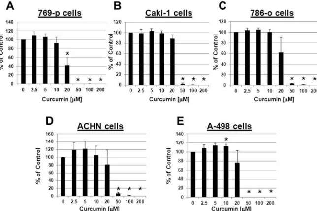
We initially investigated whether curcumin was effective in suppressing the growth of ccRCC cell lines. To address this, we evaluated the concentration-dependent effect of curcumin on cell viability in a panel of human ccRCC cell lines. 769-p, 786-o, Caki-1, ACHN and A-498 cells were treated with curcumin and cell viability assessed.
Our data demonstrated that in all five human ccRCC cell lines, curcumin was able to significantly (P<0.05)
suppress cell viability (Figure 1), with maximal (>90%) growth suppression in the concentration range of 50–200 µM. In 769-p cells significant (P<0.05) growth
suppression was also detected at a lower concentration (20 µM). Although of a small magnitude (~13%), we
did detect a significant (P<0.05) growth stimulation
in A-498 cells in response to a lower concentration (10 µM) of curcumin.
It has been reported that genetic or epigenetic variations in VHL can be detected in approximately 90% of human RCC cases.22 Given that 769-p cells do not express VHL
mRNA, we used this VHL-deficient system for our
subsequent studies that examined the mechanism by which curcumin suppressed ccRCC cell growth.23
Figure 1. Curcumin inhibits ccRCC cell proliferation and viability. 769-p cells (A), Caki-1 cells (B), 786-o cells (C), ACHN cells (D) and A-498 cells (E)
We next examined the kinetics of curcumin-induced inhibition of 769-p cell proliferation. Our data showed that compared to vehicle treatment, 5 µM curcumin had
no significant effect on 769-p cell proliferation at either
the early time points or later time points (Figure 2A). On the other hand 50 µM curcumin had a significant
inhibitory effect on cell proliferation as early as 1 day, and this inhibitory effect persisted up to 3 days.
To confirm that the growth inhibition detected in the cell
viability assays was due to decreased DNA synthesis, we examined the effect of curcumin on BrdU incorporation
in 769-p cells. A significant (P<0.05) inhibition of cell
proliferation was detected with 20 µM curcumin, and maximal suppression of cell proliferation was detected with 100–200 µM curcumin (Figure 2B).
Curcumin undergoes degradation in serum-depleted cell culture media, resulting in the formation of
trans-6-(4′-hydroxy-3′-methoxyphenyl)-2,4-dioxo-5-hexenal, ferulic acid, feruloyl methane and vanillin.24
Both ferulic acid and vanillin have been reported to mediate the antioxidant activity of curcumin.25-28
To address the question of whether the growth suppressive effects of curcumin were mediated by the degradation products of curcumin, 769-p cells were treated with the indicated concentration of ferulic acid and vanillin, and cell proliferation determined after 2 days. Our data showed that 50 µM ferulic acid had no
significant growth suppression of 769-p cells (Figure
2C). Although 50 µM vanillin did suppress 769-p cell
proliferation (<10%), it was approximately ninefold
less effective than curcumin (50 µM) at suppressing growth of 769-p cells.
Curcumin Blocks Colony Formation Ability, Induces Apoptosis and Blocks Migration of ccRCC Cells
Next, we questioned whether curcumin had any effect on the colony formation ability of 769-p cells. 769-p cells were seeded in medium containing 10% FBS and allowed to adhere for 24 hours. The medium was then replaced with fresh medium containing 10% FBS together with the indicated concentration of curcumin, and colony formation was monitored over the ensuing 2 weeks. We observed that 769-p cells have a robust ability to form colonies and that this is abolished by curcumin in a concentration-dependent manner, with a substantial decrease in the number of 769-p cell colonies detected in presence of 5 µM curcumin (Figure 3A). At higher concentrations of curcumin (20–200 µM) the ability of 769-p cells to form colonies was completely ablated. Therefore, we questioned whether curcumin would induce apoptosis in ccRCC cells. To test this, 769-p cells were treated with the indicated concentrations of curcumin for 24 hours, washed, and incubated with JC-1
dye (a mitochondria-specific dual fluorescence probe)
per manufacturer instructions (Cell Technologies). Figure 2. Curcumin inhibits ccRCC cell proliferation
in a time-dependent manner. A: 769-p cells were treated with vehicle control, or 5 or 50 μM curcumin for the indicated times, and cell viability was assessed. Data are presented as optical density and are the mean (± SEM) of at least three independent determinations. Data are representative of two separate experiments yielding similar results. Asterisks (*) designate a significant difference (P<0.05) from day 1 control.
In vehicle- and 5 µM curcumin-treated cells, 93.31% and 92.01% of the cells were detected with intact mitochondrial membranes, respectively. However, in 50 µM curcumin-treated cells, only 29.86% of cells presented intact mitochondrial membranes while 69.64% of the cell population presented with disruptions in the mitochondrial membrane (Figure 3B), indicating
increased mitochondrial membrane depolarization.
To examine whether the increase in mitochondrial
membrane depolarization would lead to increased
cell death, 769-p cells were treated with the indicated concentrations of curcumin for 24 hours, and following
trypsinization, annexin V-FITC binding and propidium iodide staining were analyzed by flow cytometry. Our
results showed that in vehicle- and 5 µM curcumin-treated cells, 1.74% and 2.29% of cells, respectively, underwent apoptosis (Figure 3C). However, treatment with 50 µM curcumin resulted in 75.49% of cells undergoing cell death.
Since ccRCC tumors are highly metastatic, we questioned whether curcumin would block migration of ccRCC
cells. To test this, 769-p cells were grown to monolayer
confluence and a scratch wound was inflicted. Cells were
incubated overnight in FBS-containing medium together with either vehicle or curcumin. Our data showed that in both vehicle- and 5 µM curcumin-treated cells, wound closure was complete (Figure 3D). However, 50 µM
curcumin significantly inhibited the migration of 769-p
cells, as evidenced by lack of wound closure.
Curcumin Decreases Phosphorylation of NFĸB and STAT3 in ccRCC Cells
Given the important role of NFκB and STAT3 in ccRCC pathogenesis, and since curcumin has been shown to suppress NFκB and STAT3 transcriptional activity, we questioned whether the ability of curcumin to induce apoptosis in ccRCC cells is accompanied with decreased NFκB and STAT3 activation. Because transcriptional activity of NFκB and STAT3 is positively regulated by phosphorylation, the impact of curcumin on the phosphorylation status of p65 NFκB and STAT3 was examined by western blotting with antibodies that
specifically detect either the phosphorylated or total
form of p65 NFκB and STAT3. Our data demonstrated Figure 3. Curcumin blocks colony formation ability, induces apoptosis and blocks migration of ccRCC cells.
A: 769-p cells were cultured in medium containing (0–200 μM) curcumin, and colony formation was assessed after approximately 2 weeks by crystal violet staining. Data shown are from a single experiment that was representative of three similar experiments yielding similar results. B: 769-p cells were treated with indicated concentration of curcumin for 24 hours, and cells were washed and labeled with JC-1 dye. Decreased red and increased green fluorescence intensities were measured by flow cytometry. Quantitative changes in percentage of gated cells are presented as the mean (± SEM) of three independent determinations. Data are from a single experiment that was representative of two independent experiments. Asterisks (*) designate a significant difference (P<0.05) from control values. C: 769-p cells were treated with indicated concentrations of curcumin for 24 hours, subjected to annexin V-FITC and propidium iodide staining, and analyzed by flow cytometry. Live/dead cell ratios were calculated. Data are presented as
that curcumin significantly (P<0.05) decreased
phosphorylation of p65 NFκB (S536) by 27.18% and 48.71% at concentrations of 20 and 50 µM, respectively (Figure 4A, 4B). In addition, 20 and 50 µM curcumin
significantly (P<0.05) decreased STAT3 (S727)
phosphorylation in 769-p cells by 31.14% and 48.76%, respectively (Figure 4C, 4D). The observed decrease in the phosphorylated forms of p65 NFκB and STAT3 were not due to an effect of curcumin on the expression levels of p65 NFκB and STAT3 (Figure 4A, 4C).
DISCUSSION
Both in the United States and worldwide, the incidence of ccRCC and ccRCC-related mortality has increased over the past decade. The National Cancer Institute reports that approximately 54,000 new cases of renal cancer are diagnosed and approximately 13,000 deaths by renal cancer occur annually. Although ccRCC is highly resistant to chemotherapy/radiotherapy, a better understanding of the biology of ccRCC has led to the
identification of several signaling pathways currently
being examined as potential targets. It is now well-established that constitutively active STAT3 and NFκB participate in ccRCC pathogenesis.9,14 Thus, suppression
of STAT3 and NFκB provides a novel therapeutic approach to suppressing ccRCC cell proliferation. Given the lack of effective therapies for ccRCC, and the well-documented ability of the dietary polyphenol curcumin to suppress STAT3- and NFκB-mediated signaling, we examined whether ccRCC tumor suppression could be achieved with curcumin. It is also of interest to note that the age-adjusted incidence of kidney cancer in the United States is the highest in the world and approximately
six times greater than rates in Asian countries where curcumin is regularly consumed through diet. Our in vitro study reveals that curcumin is effective at inducing apoptosis and suppressing migration of ccRCC cells.
In vitro characterization of the growth suppressive effects
of curcumin in ccRCC cells revealed that curcumin, in a concentration- and time-dependent manner, suppressed 769-p cell viability and colony formation ability, induced
apoptosis and blocked migration. We confirmed that the
growth suppressive effect of curcumin was not cell
type-specific, as in addition to the 769-p cells,
curcumin-induced growth suppression was detected in a panel of human ccRCC cell lines, including Caki-1, 786-o, ACHN and A-498 cells. We also examined the ability of curcumin to suppress the growth of RENCA (mouse renal carcinoma cells) and observed a similar growth suppressive pattern (data not shown). Of particular
significance is the ability of curcumin to suppress the
growth of IFN-α–resistant 786-o cells.29 Our future
studies will examine whether curcumin sensitizes
IFN-α–resistant ccRCC cells to IFN-α–induced growth suppression.
We next questioned whether curcumin would decrease viability of normal renal proximal epithelial tubule cells. We observed that curcumin failed to induce apoptosis in cultured normal rat renal tubular epithelial cells (data not shown). Our observations are consistent with both in vitro and in vivo studies demonstrating a protective effect of curcumin on renal tubular epithelial cells.30,31
Taken together, our data clearly showed that curcumin could be an effective growth suppressor of ccRCC cells.
In most cases metastasis has already occurred when ccRCC is diagnosed. Therefore, the ability of curcumin to block cell migration of ccRCC cells was examined. Our wound healing assay data showed that curcumin was effective at blocking serum-induced wound closure.
Since degraded curcumin is known to yield dietary ferulates, especially in buffered medium and serum-free medium, we questioned whether the growth suppressive effects of curcumin we observed were due to the generation of ferulic acid and vanillin. However, because 50 µM ferulic acid had no significant growth
suppressive effect on 769-p cell viability and vanillin’s modest growth suppressive effect on 769-p cell viability was approximately nine times less than curcumin at an equimolar concentration, we concluded that the growth suppressive effect of curcumin on ccRCC cell viability is not mediated by its degradation products, suggesting a direct effect of free curcumin. Our conclusion would be consistent with a response to a query raised from a recent article that described the antitumor effects of curcumin in pancreatic cancer patients.32 In their rebuttal
the authors demonstrated that the suppressive effect of curcumin on tumor necrosis factor-α–activated NFκB in human myeloid cells is not mediated by dietary ferulates, as both ferulic acid and vanillin failed to block NFκB activation by this protein.
Phosphorylation of the p65 subunit of NFκB regulates the DNA binding and transcriptional activity of NFκB.33 Likewise, in melanocytic cells, constitutively
phosphorylated STAT3 has been shown to accumulate in the nucleus and promote cell survival.34 In examining
the impact of curcumin on NFκB- and STAT3-mediated transcription in ccRCC cells, we found there is a robust expression of constitutively phosphorylated p65 NFκB and STAT3 in 769-p cells. Further, when 769-p cells are exposed to curcumin, a rapid decrease in the phosphorylation of transcription factors p65 NFκB and STAT3 was observed. Although our data clearly showed that curcumin decreases STAT3 and NFκB phosphorylation, the mechanism of this suppression in ccRCC cells remains to be determined. One of the key kinases that phosphorylate p65 NFκB is IKKα, which phosphorylates p65 NFκB on serine 536.35 In multiple
myeloma cells, curcumin inhibited constitutively activated IKK.36 It is possible the observed decrease
in p65 NFκB phosphorylation in ccRCC cells could be due to curcumin-induced suppression of IKKα
activity. Likewise, the inhibitory effect of curcumin on STAT3 phosphorylation could involve suppressing receptor-associated Janus kinase(s) or the nonreceptor
tyrosine kinase Src. Interestingly, Yonezawa et al.
demonstrated the suppression of RCC cell growth by Src kinase inhibitor PP1 and that this was accompanied with decreased STAT3 activation.37 Our future studies
will examine the role of IKKα and Src in mediating the growth suppressive effects of curcumin in ccRCC cells.
Recent studies suggested the potential of targeting NFκB and STAT3 signaling axes in ccRCC as novel therapeutic approaches.38,39 To this end, the
proteasome inhibitor bortezomib induced apoptosis in
ccRCC cells in a NFκB-dependent manner; however, just the suppression of NFκB was not sufficient to
induce apoptosis in ccRCC cells, demonstrating the requirement of engaging additional signaling targets in inducing apoptosis in ccRCC cells.40 It is reasonable
to conclude that the ability of curcumin to target two essential signaling arms in ccRCC, STAT3 and NFκB, might be the underlying basis of its pro-apoptotic effect.
Although curcumin is an effective and selective (i.e. targets tumor cells but not normal cells41,42)
antitumor agent, its use in a clinical setting has been limited, primarily due to its well-documented poor
pharmacokinetic profile. To overcome this limitation,
novel approaches to enhancing the bioavailability of curcumin are now the focus of several studies. These include coadministration of curcumin and piperine (an inhibitor of glucuronidation), which resulted in a 2,000% increase in bioavailability of curcumin.43 Another
approach has led to the generation of nanoparticles of curcumin. Nanoparticle curcumin was effective at suppressing the growth of MCF-7 breast cancer cells,44
and at inhibiting NFκB activity and inducing apoptosis in human pancreatic tumor cell lines.45 Liposomal curcumin
also was shown to be an effective anticancer agent in a pancreatic cancer xenograft model.46,47 Additionally,
complexing curcumin with micelles or phospholipids has proved to improve its bioavailability.48,49 Finally,
synthetic curcumin analogs (e.g. EF-24, HO-3867) were reported to have increased bioavailability when compared to curcumin and are more effective than curcumin at suppressing tumor growth.50,51 Interestingly,
Given the excellent safety profile of curcumin, and the
recent efforts aimed at enhancing its bioavailability, we
anticipate that clinical trials examining the efficacy of
curcumin or its synthetic analogs in suppressing ccRCC tumorigenesis will be conducted.
CONCLUSIONS
We report a robust growth suppressive and pro-apoptotic in vitro effect of curcumin on ccRCC cells.
Although in vitro findings do not necessarily translate
into clinical care, if the growth suppressive effects of curcumin on ccRCC are validated in clinical trials, this therapeutic approach could complement existing therapies.
Patient-Friendly Recap
• Curcumin, a commonly used food additive in
Southeast Asia, is known to inhibit the growth of several types of cancer cells in experimental settings.
• The authors studied whether curcumin could
block growth and migration of malignant kidney cancer cells.
• The authors found that curcumin decreased the
viability of kidney cancer cells as well as their ability to migrate.
• Given its few side effects, curcumin (or its
synthetic analogs) could be considered for clinical trials aimed at deveoping novel therapies for kidney cancer.
Conflicts of Interest None.
References
1. Motzer RJ, Bander NH, Nanus DM. Renal-cell carcinoma. N Engl J Med. 1996;335:865-75. CrossRef
2. Gore ME, Szczylik C, Porta C, et al. Safety and efficacy of
sunitinib for metastatic renal-cell carcinoma: an expanded-access trial. Lancet Oncol. 2009;10:757-63. CrossRef
3. Escudier B, Eisen T, Stadler WM, et al. Sorafenib in advanced clear-cell renal-cell carcinoma. N Engl J Med. 2007;356: 125-34. CrossRef
4. Hudes GR, Carducci MA, Choueiri TK, et al. NCCN Task
Force report: optimizing treatment of advanced renal cell
carcinoma with molecular targeted therapy. J Natl Compr Canc Netw. 2011;9 Suppl 1:S1-29.
5. Kim WY, Kaelin WG. Role of VHL gene mutation in human cancer. J Clin Oncol. 2004;22:4991-5004. CrossRef
6. Ashida S, Furihata M, Tanimura M, et al. Molecular detection of von Hippel-Lindau gene mutations in urine and lymph node samples in patients with renal cell carcinoma: potential biomarkers for early diagnosis and postoperative metastatic status. J Urol. 2003;169:2089-93. CrossRef
7. Maxwell PH, Dachs GU, Gleadle JM, et al. Hypoxia-inducible factor-1 modulates gene expression in solid tumors and
influences both angiogenesis and tumor growth. Proc Natl Acad Sci U S A. 1997;94:8104-9. CrossRef
8. Tomita Y, Bilim V, Kawasaki T, et al. Frequent expression of Bcl-2 in renal-cell carcinomas carrying wild-type p53. Int J Cancer. 1996;66:322-5.
9. Oya M, Takayanagi A, Horiguchi A, et al. Increased nuclear factor-kappa B activation is related to the tumor development of renal cell carcinoma. Carcinogenesis. 2003;24:377-84.
CrossRef
10. Aggarwal BB, Kunnumakkara AB, Harikumar KB, et al. Signal
transducer and activator of transcription-3, inflammation, and
cancer: how intimate is the relationship? Ann N Y Acad Sci.
2009;1171:59-76. CrossRef
11. Al Zaid Siddiqui K, Turkson J. STAT3 as a target for inducing
apoptosis in solid and hematological tumors. Cell Res.
2008;18:254-67. CrossRef
12. Huang S. Regulation of metastases by signal transducer and activator of transcription 3 signaling pathway: clinical implications. Clin Cancer Res. 2007;13:1362-6. CrossRef
13. Niu G, Wright KL, Huang M, et al. Constitutive Stat3 activity up-regulates VEGF expression and tumor angiogenesis.
Oncogene. 2002;21:2000-8. CrossRef
14. Horiguchi A, Oya M, Shimada T, Uchida A, Marumo K, Murai M. Activation of signal transducer and activator of transcription 3 in renal cell carcinoma: a study of incidence and its association with pathological features and clinical outcome. J Urol. 2002;168:762-5. CrossRef
15. Guo C, Yang G, Khun K, et al. Activation of Stat3 in renal tumors. Am J Transl Res. 2009;1:283-90.
16. Singh S, Aggarwal BB. Activation of transcription factor NF-kappa B is suppressed by curcumin (diferuloylmethane) [corrected]. J Biol Chem. 1995;270:24995-5000. CrossRef
17. Bharti AC, Donato N, Aggarwal BB. Curcumin
(diferuloylmethane) inhibits constitutive and IL-6-inducible STAT3 phosphorylation in human multiple myeloma cells.
J Immunol. 2003;171:3863-71. CrossRef
18. Bangaru ML, Chen S, Woodliff J, Kansra S. Curcumin (diferuloylmethane) induces apoptosis and blocks migration of human medulloblastoma cells. Anticancer Res. 2010;30: 499-504.
19. Bobustuc GC, Smith JS, Maddipatla S, et al. MGMT inhibition restores ERalpha functional sensitivity to antiestrogen therapy.
Mol Med. 2012;18:913-29. CrossRef
20. Kansra S, Yamagata S, Sneade L, Foster L, Ben Jonathan N. Differential effects of estrogen receptor antagonists on pituitary lactotroph proliferation and prolactin release. Mol Cell Endocrinol. 2005;239:27-36. CrossRef
21. Kansra S, Stoll SW, Johnson JL, Elder JT. Src family kinase inhibitors block amphiregulin-mediated autocrine ErbB signaling in normal human keratinocytes. Mol Pharmacol.
2005;67:1145-57. CrossRef
22. Nickerson ML, Jaeger E, Shi Y, et al. Improved identification
Murai M. Renal cancer cells lacking hypoxia inducible factor (HIF)-1alpha expression maintain vascular endothelial growth
factor expression through HIF-2alpha. Carcinogenesis.
2007;28:529-36. CrossRef
24. Wang YJ, Pan MH, Cheng AL, et al. Stability of curcumin
in buffer solutions and characterization of its degradation
products. J Pharm Biomed Anal. 1997;15:1867-76. CrossRef
25. Srinivasan M, Sudheer AR, Menon VP. Ferulic acid: therapeutic potential through its antioxidant property. J Clin Biochem Nutr. 2007;40:92-100. CrossRef
26. Sudheer AR, Muthukumaran S, Kalpana C, Srinivasan M, Menon VP. Protective effect of ferulic acid on nicotine-induced DNA damage and cellular changes in cultured rat peripheral blood lymphocytes: a comparison with N-acetylcysteine.
Toxicol In Vitro. 2007;21:576-85. CrossRef
27. Makni M, Chtourou Y, Fetoui H, Garoui EM, Boudawara T,
Zeghal N. Evaluation of the antioxidant, anti-inflammatory and
hepatoprotective properties of vanillin in carbon tetrachloride-treated rats. Eur J Pharmacol. 2011;668:133-9. CrossRef 28. Kanski J, Aksenova M, Stoyanova A, Butterfield DA. Ferulic
acid antioxidant protection against hydroxyl and peroxyl radical oxidation in synaptosomal and neuronal cell culture systems in vitro: structure-activity studies. J Nutr Biochem.
2002;13:273-81. CrossRef
29. Tomita S, Ishibashi K, Hashimoto K, et al. Suppression of SOCS3 increases susceptibility of renal cell carcinoma to interferon-alpha. Cancer Sci. 2011;102:57-63. CrossRef
30. Tong QS, Zheng LD, Lu P, et al. Apoptosis-inducing effects of curcumin derivatives in human bladder cancer cells.
Anticancer Drugs. 2006;17:279-87. CrossRef
31. Chiu J, Khan ZA, Farhangkhoee H, Chakrabarti S. Curcumin prevents diabetes-associated abnormalities in the kidneys by inhibiting p300 and nuclear factor-kappaB. Nutrition.
2009;25:964-72. CrossRef
32. Dhillon N, Aggarwal BB, Newman RA, et al. Phase II trial of curcumin in patients with advanced pancreatic cancer. Clin Cancer Res. 2008;14:4491-9. CrossRef
33. Karin M, Ben-Neriah Y. Phosphorylation meets ubiquitination:
the control of NF-[kappa]B activity. Annu Rev Immunol.
2000;18:621-63. CrossRef
34. Sakaguchi M, Oka M, Iwasaki T, Fukami Y, Nishigori C. Role and regulation of STAT3 phosphorylation at Ser727
in melanocytes and melanoma cells. J Invest Dermatol.
2012;132:1877-85. CrossRef
35. Sakurai H, Chiba H, Miyoshi H, Sugita T, Toriumi W. IkappaB kinases phosphorylate NF-kappaB p65 subunit on serine 536 in the transactivation domain. J Biol Chem. 1999;274: 30353-6. CrossRef
36. Bharti AC, Donato N, Singh S, Aggarwal BB. Curcumin (diferuloylmethane) down-regulates the constitutive activation of nuclear factor-kappa B and IkappaBalpha kinase in human multiple myeloma cells, leading to suppression of proliferation and induction of apoptosis. Blood. 2003;101:1053-62.
CrossRef
37. Yonezawa Y, Nagashima Y, Sato H, et al. Contribution of
the Src family of kinases to the appearance of malignant phenotypes in renal cancer cells. Mol Carcinog. 2005;43: 188-97. CrossRef
38. Sourbier C, Danilin S, Lindner V, et al. Targeting the nuclear factor-kappaB rescue pathway has promising future in human renal cell carcinoma therapy. Cancer Res. 2007;67:11668-76.
CrossRef
39. Horiguchi A, Asano T, Kuroda K, et al. STAT3 inhibitor WP1066 as a novel therapeutic agent for renal cell carcinoma.
Br J Cancer. 2010;102:1592-9. CrossRef
40. An J, Sun Y, Fisher M, Rettig MB. Maximal apoptosis of
renal cell carcinoma by the proteasome inhibitor bortezomib
is nuclear factor-kappaB dependent. Mol Cancer Ther.
2004;3:727-36.
41. Ohori H, Yamakoshi H, Tomizawa M, et al. Synthesis and
biological analysis of new curcumin analogues bearing an enhanced potential for the medicinal treatment of cancer. Mol Cancer Ther. 2006;5:2563-71. CrossRef
42. Syng-Ai C, Kumari AL, Khar A. Effect of curcumin on normal and tumor cells: role of glutathione and bcl-2. Mol Cancer Ther. 2004;3:1101-8.
43. Shoba G, Joy D, Joseph T, Majeed M, Rajendran R, Srinivas
PS. Influence of piperine on the pharmacokinetics of curcumin
in animals and human volunteers. Planta Med. 1998;64:353-6.
CrossRef
44. Kumar SS, Mahesh A, Mahadevan S, Mandal AB. Synthesis
and characterization of curcumin loaded polymer/lipid based
nanoparticles and evaluation of their antitumor effects on MCF-7 cells. Biochim Biophys Acta. 2014;1840:1913-22.
CrossRef
45. Bisht S, Feldmann G, Soni S, et al. Polymeric nanoparticle-encapsulated curcumin (“nanocurcumin”): a novel strategy for human cancer therapy. J Nanobiotechnology. 2007;5:3.
CrossRef
46. Li L, Braiteh FS, Kurzrock R. Liposome-encapsulated
curcumin: in vitro and in vivo effects on proliferation, apoptosis, signaling, and angiogenesis. Cancer. 2005;104:1322-31.
CrossRef
47. Li L, Ahmed B, Mehta K, Kurzrock R. Liposomal curcumin
with and without oxaliplatin: effects on cell growth, apoptosis, and angiogenesis in colorectal cancer. Mol Cancer Ther.
2007;6:1276-82. CrossRef
48. Ma Z, Shayeganpour A, Brocks DR, Lavasanifar A, Samuel J. High-performance liquid chromatography analysis of curcumin in rat plasma: application to pharmacokinetics
of polymeric micellar formulation of curcumin. Biomed
Chromatogr. 2007;21:546-52. CrossRef
49. Liu A, Lou H, Zhao L, Fan P. Validated LC/MS/MS assay for curcumin and tetrahydrocurcumin in rat plasma and application to pharmacokinetic study of phospholipid complex of curcumin. J Pharm Biomed Anal. 2006;40:720-7. CrossRef
50. Adams BK, Cai J, Armstrong J, et al. EF24, a novel synthetic curcumin analog, induces apoptosis in cancer cells via a redox-dependent mechanism. Anticancer Drugs. 2005;16:263-75.
CrossRef
51. Dayton A, Selvendiran K, Kuppusamy ML, et al. Cellular
uptake, retention and bioabsorption of HO-3867, a fluorinated
curcumin analog with potential antitumor properties. Cancer Biol Ther. 2010;10:1027-32. CrossRef