Officially Associated with
“The Egyptian Society of Oral Implantology”
An Official Publication of
“Ivano-Frankivsk National Medical University, Ukraine”
Review Article
COLOUR MATCHING: A REVIEW OF CONVENTIONAL AND
CONTEMPORARY DENTAL COLOUR MATCHING SYSTEMS
Mukut Seal, * Pratim Talukdar, ** Virag Srivastav, ***
Kartik Pendharkar
†* MDS, Department of Endodontics, Private Practitioner, Guwahati, Assam, India **MDS, Department of Prosthodontics, Private Practitioner, Guwahati, Assam, India
*** Senior Lecturer, Department of Prosthodontics, BBD College of Dental Sciences, Lucknow, Uttar Pradesh, India † MDS, Department of Endodontics, Private Practitioner, Mumbai, Maharashtra, India
________________________________________________________________________
ABSTRACT
Precise color communication is integral to the development of esthetic harmony and overall restorative success. While traditional shade taking procedures have enabled some degree of shade information transfer, computerized shade analysis device allow for standardized, repeatable shade determination for increased accuracy by placing technology in the role of observer while eliminating the influence of negative visual illusion effect to deliver exact and reproducible information that will allow the dentist and lab technician to produce accurately matched restoration.
KEYWORDS: Colour; colour matching; shade guides; contemporary devices
INTRODUCTION
The study of colour is an integral part of esthetic dentistry. If the colour of a restoration is off-even slightly, the mistake can be glaringly evident; it looks fake. Unfortunately colour is tricky. Slight variances in shade play with our eyes, our minds and our dentistry. Cosmetic and esthetic dental outcomes are essential to meet patients‟ high expectations and positively influence their self-esteem.[1,2] Shape and color determine the aesthetics of both natural and restored teeth.[3-6] Attractive restorative and prosthodontic outcomes begin with a consistent buccal shape and silhouette that reflect light.[7] The perceived color of natural teeth depends on the illuminating light source, critical to translucency, opalescence and fluorescence.[8,9] Determining a precise shade is dependent on clinical skill, shade guide system and lighting conditions. This paper reviews some of the significant factors in the process of tooth
shade matching and benefits of modern light correcting techniques.
ESSENTIALS OF COLOUR CONCEPT
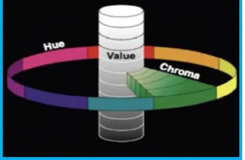
According to the Glossary of Prosthodontic terms,[10] color can be defined as „the quality of an object or substance with respect to light reflected or transmitted by it. Color is usually determined visually by measurement of hue, saturation, and luminous reflectance of the reflected light‟.[12,13] For indirect cases, the clinician must convey the primary tooth shade characteristics of hue, chroma, and value to the technician who, in turn, produces restorations that match to the remaining dental structure.[14,15] Hue refers to the varying wavelengths of observed radiant energy (red, yellow, green, blue, purple, etc). Approximately 80 percent of natural teeth fall into the A hue range.[16,17] Chroma describes the strength or saturation of the hue.[10] Value, lightness or brightness, distinguishes a color‟s relative darkness,[11] Value is often the most important dimension of shade.[12-20] The greater the total amount of light reflected the higher the value). The scale ranges from low of 0 (i.e., pure black) to high of 10 for pure white. Chroma has been found to increase with age in both enamel and dentin, while hue and value do not based on in vitro testing.[1] Natural teeth exhibit translucency, fluorescence and opalescence, all of which information is necessary for successful shade matching. Translucency is the gradient between transparent and opaque.[8] Fluorescence is the absorption of short wavelength light with the spontaneous emission of longer wavelength light.[21] Opalescence makes a material appear one color with reflected light and another color with transmitted light.[22]
PERCEPTION OF COLOUR
Shade selection involves the perception of colour,
Conventional and contemporary dental colour matching Seal M, Talukdar P, Srivastav V, Pendharkar K
which depends on three entities: 1. Light source (illuminant); 2. Object; and
3. Detector (ocular or instrumental).
The visual system of the eye is only capable of detecting wavelengths from 380 (violet) to 780nm (red). Isaac Newton showed that light had no colour, as it is only when it interacts with an object that colour is produced (Fig. 1).[23]
ILLUMINANT
The colour of an object can change depending on the illuminant, e.g. tungsten light may cast a yellow colour compared to daylight. The property of light source to influence colour of objects is called “colour rendition”. There are three main illuminants within any dental practice: natural, incandescent and fluorescent. Natural sunlight is
itself variable with light appearing blue at noon when the sun has less atmosphere to penetrate, and red/orange during the morning and evening. Incandescent lighting is predominantly red/yellow and lacking in blue while fluorescent lighting is high in blue tones and low in red. There are special lights (Fig. 2) that are colour corrected to emit light with a more uniform distribution of colour that can be utilised. Initial shade selection should be initially made with these lights then the shade should be matched under different lights to avoid metamerism (the phenomenon that occurs when shades appear to match under one lighting condition.
OBJECT
Colour possess three dimensions: value, hue and chroma. A high value object often reflects most
Fig. 1: Isaac Newton‟s experiment of splitting light by a prism into the colours of the spectrum - red, orange, yellow, green, blue, indigo and violet
Fig. 2: Use of Optilume TrueShade – which consists of LED lighting to provide a colour
corrected light output of 5500K
Fig. 3: The Munsell color system, showing: a circle of hues, and levels of value and chroma
Fig. 4: VITA classical shade guide
Table 1
the light falling on its surface and appears bright. The converse is true with a dark object absorbing most of the light and appearing dull or of low value. Hue is wavelength of light, and dependent on the spectral reflectance from an object. Chroma is the concentration of colour or colour intensity (Fig. 3).
DETECTOR (SENSATION)
The third part of stimulus for colour is the spectral response of the detector, or eye. The difficulty of shade selection is that clinicians must be able to interpret a multi-layered structure of varying thickness, opacities and optical surface characteristics. This can affect the way that the eye perceives colour. The basic hue of the tooth is determined by the colour of the underlying dentine, while value is a quality of the enamel overlay. Muia in 1993 stated, “The dentine imparts all the colour. Enamel is like a fiberoptic structure conducting light through its rods”. Chroma is the saturation of colour in the dentine, but is influenced by the value and thickness of the enamel. Teeth are often termed “polychromatic” and have the variation in hue, value and chroma within the teeth and give three dimensional depth and characteristics. A young dentition is characterised by opaque, high value enamel, which blocks underlying dentine. As teeth age, the enamel becomes more translucent and dull (low value) revealing the underlying dentine. This layering can make reading of tooth colour difficult since the value of enamel and surface
lustre often complicate colour evaluation of the underlying dentine.[23]
PROTOCOLS FOR SHADE SELECTION[1] Remove bright color from the working field.
If the patient is wearing bright clothing, it is prudent to cover the patient with neutral color bib (grey). Any dark color lipstick should be removed, because it could affect shade matching.
Always clean the tooth by using prophylaxis paste prior to shade selection.
Its important not to view the shade comparison for more than 7 s to avoid eye fatigues.
Clinician should be at a distance of 28‑33 cm from the patient during shade selection.
Always determine shade when the teeth are most hydrated, because enamel dehydration reduces its translucency by 82%, misleading the clinician.
Shade comparison should always be done in between 10 am and 2 pm, because at this time color temperature is around 5500 K and then under color corrected light to ensure the accuracy of the match.
During the shade comparison always place shade tabs either above or below the tooth to be match, never place shade tab adjacent to the tooth to avoid binocular effect.
Always, value is analyzed first, followed by chroma and then hue.
Conventional and contemporary dental colour matching Seal M, Talukdar P, Srivastav V, Pendharkar K
immediately after bleaching, patient should be recalled after 2‑3 weeks for shade comparison.
Always during shade selection teeth should be divided in 3 regions. Gingival area (gives accurate determination of dentinal chroma), Body area and Incisal area (enamel is thickest here and varies from translucent to transparent).
SHADE GUIDES (CONVENTIONAL)
Tooth shade matching is most frequently performed visually using dental shade guide. The first shade guide was introduced in 1956 by Vita Zahnfabrik. The most popular shade guide are ‑ VITA Classical, Chroma scope, Vitapan 3D ‑
Master shade guide. VITA classical shade guide ‑ It consist of 16 tabs, arranged into four groups based on the hue and within the group according to increasing Chroma. Hue is categorized by letter i.e., A = Orange, B = Yellow, C = Yellow/Gray and D = Orange/Gray. Chroma and Value are categorized by numbers i.e., 1 = least chromatic and highest value, = most chromatic and lowest value (Fig. 4).[1] Another popular shade guide is the chromascope. It uses only numbering system to identify the shade. Chromascope is arranged in groups based on the hue (100 = white, 200 = yellow, 300 = orange, 400 = gray, 500 = brown) and within the groups according to increasing chroma from 10 to 40.[1] Another choice of shade guide is the Vita System 3D-Master (Fig. 5). It consists of 11 fired porcelain tooth shaped samples built up with cervical, dentinal and incisal powders and composed of feldspar nepheline and high temperature ceramic pigments. The 11 sets consist of 26 samples ranging from lightest to darkest value, from lowest to highest intensity and from yellow to red. Vita Value, Chroma and Hue correspond similarly to Munsell value, hue and chroma representing the three dimensions of color. The tabs are grouped into 5 categories, sequentially numbered with increasing value (1-5). All tabs within the value group have the same brightness. In each of the groups the chroma increases from top to bottom. All the groups except 1 and 5 have 3 letters: L, M, R, which allows the hue to be chosen. L (light) is yellow, M (medium) is yellow-red, and R is a red hue. Documenting of this shade is with a number/letter/number system.
The first number indicates the value group (1-5), letter is the hue (L, M, R) and the chroma (1-3). E.g. 3M2 is the 3rd value group, M hue sub-group, and 2 chroma levels. The shades of the lightness level 3 cover 50% of the natural tooth shades. The shades of the lightness levels 2 and 4 cover an additional 46%. VITA has also created a bleached tab, labeled the “0” (zero) group, to allow dentists to create bleached restoration shades. One of the other advantages of the Vita System 3D-Master is the repeatability of shade selections with the system (Table 1). It was concluded that use of this system compared to the classic guide improved intrarater repeatability among general practitioners. A number of related factors in selecting shades must also be understood to achieve a successful result. These factors include, translucency, contour, surface texture, and lustre. Selecting the basic shade or color is only the first step.[23]
Types of Technology Shade System (Contemporary Devices)
RGB DEVICES
Devices that acquired red, green and blue image information to create a color image, such as most consumer video or digital still cameras, are commonly referred to as RGB devices. Digital cameras and other RGB devices represent the most basic approach to electronic shade taking and still require a certain degree of subjective verification by the human eye. Various approaches have been used to translate this data into useful dental information. The information accuracy of RGB devices is questionable sine they are not measurement instrument; rather they infer the color properties of a captured image. These systems are useful for providing lab technicians with a referential starting point, but should not be relied upon solely to determine the shade of a tooth. The shade scan system from Cynovad is an example of an RGB device.[24]
DIGITAL CAMERAS AND IMAGING SYSTEMS
taking, still requiring a certain degree of subjective shade selection with the human eye.[25] Various approaches have been used to translate this data into useful dental color information.
ClearMatch (Smart Technology, Hood River, OR) is a software system that uses high-resolution digital images and compares shades over the entire tooth with known reference shades.[26-30] Similar to the software associated with color measuring devices, ClearMatch contains the color database of industry-standard shade guides.[26]
SPECTROPHOTOMETERS
Spectrophotometers are amongst the most accurate, useful and flexible instruments for overall color matching and color matching in dentistry.[24] They measure the amount of light energy reflected from an object at 1–25 nm intervals along the visible spectrum.[27,28] A spectrophotometer contains a source of optical radiation, a means of dispersing light, an optical system for measuring, a detector and a means of converting light obtained to a signal that can be analyzed. The data obtained from spectrophotometers must be manipulated and translated into a form useful for dental professionals. The measurements obtained by the instruments are frequently keyed to dental shade guides and converted to shade tab equivivalent.[29] Compared with observations by the human eye, or conventional techniques, it was found that spectrophotometers offered a 33% increase in accuracy and a more objective match in 93.3% of cases.[24]
Crystaleye (Olympus, Tokyo, Japan) combines the benefits of a traditional spectrophotometer with digital photography. Through the development of optical and image processing technology, this product allows the practitioner to match tooth shade and color more accurately and simply compared with the traditional spectrophotometer.[27] The significant benefit of this system is that „virtual shade tabs‟ in the computers database can be cross-referenced and superimposed visually onto the natural tooth image to be matched giving the technician the ability to visualize the correct shade tabs. The digital image produced by the Crystaleye uses a 7-band LED light source, which results in a more precise depiction of color than the conventional systems used with digital cameras. Moreover, the image produced by the Crystaleye is taken from
inside the oral cavity and consequently is devoid of the external light that can cause discrepancies.
Vita Easyshade Compact (Vita Zahnfabrik, Bad Sackingen, Germany) is cordless, small, portable, cost efficient, battery operated, contact-type spectrophotometer that provides enough shade information to help aid in the color analysis process. Different measurement modes are possible with Easyshade Compact: tooth single mode, tooth area mode (Fig. 6), (cervical, middle and incisal shades), restoration color verification (includes lightness, chroma and hue comparison) and shade tab mode (practice/training mode).[30]
Shade-X (X-Rite, Grandville, MI) is also compact and cordless „„spot‟‟ measurement‟‟ spectrophotometer with 3-mm probe diameter, and keyed to the majority of popular shade guides. Shade-X has two databases to match the color of the dentin (more opaque) and the incisal tooth regions (more translucent).[31]
SpectroShade Micro (MHT Optic Research, Niederhasli, Switzerland) is an imaging spectrophotometer. It uses a digital camera/LED spectrophotometer combination. It has an internal computer with the analytical software. The tooth positioning guidance system, shown on the LCD touch screen, is used during color measurement. Images and spectral data can be saved on the internal memory and transferred to a computer.[32]
COLORIMETERS
Colorimeters measure tristimulus values and filter light in red, green and blue areas of the visible spectrum. Colorimeters are not registering spectral reflectance and can be less accurate than spectrophotometers (aging of the filters can additionally affect accuracy).[33]
Conventional and contemporary dental colour matching Seal M, Talukdar P, Srivastav V, Pendharkar K
cameras. Combination of visual color determination (Vitapan 3D‑Master shade guide and Linear guide) with digital cameras and electronic devices (spectrophotometers) increase chances for successful shade matching.[36] The VITA easy shade had the best combination of accuracy and reliability. Reliability percentages were 99.0, for shade vision, 96.9, for Spectro Shade, 96.4 for VITA Easyshade and 87.4 for Shade Scan. Accuracy percentages were 92.6 (highest) for the VITA Easyshade.[33]
CONCLUSION
Accurate shade selection that allows restorations to match the natural dentition positively influences the patient‟s appearance and esthetic self-esteem. Patients are demanding contemporary esthetic dentistry, which has prompted the industry to continuously raise the bar with regard to esthetic detail. Many factors can influence the perception of color; by taking advantage of today‟s shade-matching technology, the subjectivity of color assessment can be minimized and accurate diagnosis of a restoration‟s shade is more easily communicated. Besides the clinical applications, dental color measuring instruments and systems are increasingly used in research. Although we all are health care providers first, we are also artist. With a good working knowledge of colour, your artistry will become as natural as your dentistry.
CONFLICT OF INTEREST & SOURCE OF FUNDING
The author declares that there is no source of funding and there is no conflict of interest among all authors.
BIBLIOGRAPHY
1. Chu SJ. Fundamentals of Color: Shade Matching and Communication in Esthetic Dentistry. Quintessence Publishing Co. Inc 2004.
2. Freedman G. Buyer‟s guide to cosmetic imaging systems. Cosmetic imaging creates projection of restorative treatment. Dent Today 2009;28(7):134-8.
3. Glick K. Cosmetic dentistry is still dentistry. J Can Dent Assoc 2000;66(2):88-9.
4. Jeannin T, Ubassy G. Anterior prosthetic restoration. Cah Prothese 1984;12(46):93-100.
5. Ubassy G. Fabrication and natural stratification of dental ceramics 1. Rev Fr Prothes Dent 1990;14:61-70.
6. Terry DA, Geller W, Tric O. Anatomical form defines color: function, form, and aesthetics. Pract Proced Aesthet Dent 2002;14(1):59-67.
7. Glick KL. Color management of cosmetic restorations. Curr Opin Cosmet Dent 1995:36-40.
8. Villarroel M, Fahl N, De Sousa AM, De Oliveira OB Jr. Direct esthetic restorations based on translucency and opacity of composite resins. J Esthet Restor Dent 2011;23(2):73-87.
9. Meyenberg KH. Dental esthetics: a European perspective. J Esthet Dent 1994;6(6):274-81.
10. The glossary of prosthodontic terms. J Prosthet Dent 2005;94(1):10-92.
11. Burkinshaw SM. Color in relation to dentistry. Fundamentals of color science. Br Dent J 2004;196(1):33-41.
12. Jaint N, Verma P, Mittal S, Singh AK, Munjal S. Gender based alteration in color perception. Indian J Physiol Pharmacol 2010;54(4):366-70.
13. Fondriest J. Shade matching in restorative dentistry: the science and strategies. Int J Periodontics Restorative Dent 2003;23(5):467-79.
14. Winkler S, Boberick KG, Weitz KS, Datikashvili I, Wood R. Shade matching by dental students. J Oral Implantol 2006;32(5):256-8.
15. Baltzer A, Kaufmann-Jinoian V. Shading of ceramic crowns using digital tooth shade matching devices. Int J Comput Dent 2005;8(2):129-52.
16. Touati B. Excellence with simplicity in aesthetic dentistry. Pract Periodontics Aesthet Dent 1997;9(7):806-8.
17. Hall NR, Kafalias MC. Composite color matching: the development and evaluation of a restorative color matching system. Aust Prosthodont J 1991;5:47-52.
19. Miller LL. Shade matching. J Esthet Dent 1993;5(4):143-53.
20. Sorensen JA, Torres TJ. Improved color matching of metal-ceramic restorations. Part I: A systematic method for shade determination. J Prosthet Dent 1987;58(2):133-9.
21. McLaren EA. Luminescent veneers. J Esthet Dent 1997;9(1):3-12.
22. Sundar V, Amber PL. Opals in nature. J Dent Technol 1999;16(8):15-7.
23. Christopher CK. Shade selection. Aust Dent Pract 2007;10:116-9.
24. Paul S, Peter A, Pietrobon N, Hammerle CH. Visual and spectrophotometric shade analysis of human teeth. J Dent Res 2002;81:578-92.
25. Blaes J. Today‟s technology improves the shade-matching problems of yesterday. Journal of Indiana Dental Association 2002-2003;81:17-9.
26. http://www.clearmatch.com/index.htm [accessed 06.04.10].
27. Khurana R, Tredwin CJ, Weisbloom M, Moles DR. A clonical evaluation of the individual repeatability of three commercially available color measuring devices. British Dental Journal 2007;203:675-80.
28. Kielbassa AM, Beheim-Schwarzbach NJ, Neumann K, Zantner C. In vitro comparison of visual and computeraided pre-and post-tooth shade determination using various home bleaching procedures. Journal of Prosthetic Dentistry 2009;101:92-100. 29. Lagouvardos PE, Fougia AG,
Diamantopoulou SA, Polyzois GL. Repeatability and interdevice reliability of two portable color selection devices in matching and measuring tooth color. Journal of Prosthetic Dentistry 2009;101:40-5. 30.
http://www.vita-zahnfabrik.com/resourcesvita/shop/en/ en_3055212.pdf [accessed 06.04.10]. 31. http://www.xrite.com/product_overview.asp
x?ID=812 [accessed 06.06.10].
32. Ristic I, Paravina RD. Color measuring instruments. Acta Stomatologica Naissi 2009;25:925-32.
33. Kim-Pusateri S, Brewer J, Davis EL, Wee AG. Reliability and accuracy of four dental
shade-matching devices. Journal of Prosthetic Dentistry 2009;101:93-9.
34. http://www.xrite.com/product_overview.asp x?ID=339 [accessed 06.04.10].
35. Miller L. Organizing color in dentistry. Journal of the American Dental Association 1987;115:26-40.