ISSN Online: 2164-3016 ISSN Print: 2164-3008
DOI: 10.4236/ojo.2019.95012 May 27, 2019 123 Open Journal of Orthopedics
Mallet Toe of the Hallux Treated by Bridging
Suture Technique Using Suture Anchors:
A Case Report and Literature Review
Akira Ando1* , Tsutomu Kobayashi1, Masashi Koide1, Michimasa Matsuda1,
Yoshihiro Hagiwara2, Eiji Itoi2
1Department of Orthopaedic Surgery, Matsuda Hospital, Sendai, Japan
2Department of Orthopaedic Surgery, Tohoku University School of Medicine, Sendai, Japan
Abstract
An avulsion fracture of the extensor hallucis longus at the distal end of the great toe is called “mallet toe” of the hallux. It is a rare injury and the treat-ment options are conservative treattreat-ment using a splint, percutaneous or open Kirschner wire fixation similar to that in the mallet finger, or suture anchor fixation. We present a case treated by the bridging technique using two suture anchors. A 57-year-old Japanese man injured his left great toe after a fall while walking barefoot on the bed. His great toe was forced into a hyperplantarflexion position. Plain radiography and computed tomography showed a small bone fragment at the base of the dorsal distal phalanx, sug-gesting an avulsion fracture of the extensor hallucis longus. He was treated by bridging suture technique with two suture anchors. At first, two suture anc-hors were inserted to the fracture bed of the distal phalanx, and then the bone fragment and extensor hallucis longus tendon were secured with two hori-zontal mattress sutures. Finally, bridging sutures were performed using the remaining sutures and the sutures used for mattress suturing. He obtained bony union and symmetric range of motion of the interphalangeal joint. This technique allowed us to fix the small bone fragment rigidly and mobilize the interphalangeal joint earlier to preserve the range of motion. It would be a valuable procedure when the bone fragment is small.
Keywords
Mallet Toe, Hallux, Avulsion Fracture, Extensor Hallucis Longus, Suture Anchor
How to cite this paper: Ando, A., Ko-bayashi, T., Koide, M., Matsuda, M., Hagi-wara, Y. and Itoi, E. (2019) Mallet Toe of the Hallux Treated by Bridging Suture Technique Using Suture Anchors: A Case Report and Literature Review. Open Jour-nal of Orthopedics, 9, 123-129.
https://doi.org/10.4236/ojo.2019.95012
Received: April 29, 2019 Accepted: May 24, 2019 Published: May 27, 2019
Copyright © 2019 by author(s) and Scientific Research Publishing Inc. This work is licensed under the Creative Commons Attribution International License (CC BY 4.0).
DOI: 10.4236/ojo.2019.95012 124 Open Journal of Orthopedics
1. Introduction
An avulsion fracture of the extensor hallucis longus at the distal phalanx of the great toe is called “mallet toe” of the hallux [1]. The underlying mechanism of injury is the plantarflexion of distal phalanx to the extended interphalangeal joint similar to that in the mallet injury of the finger [2] [3]. It is a rare injury and its treatment has been reported only in six case reports [1] [4] [5] [6] [7] [8]. These treatments included conservative therapy with a splint to prevent plantar-flexion of the hallux [1] [4], Kirschner wire fixation similar to that in bony mal-let finger [5] [6] [7], or open reduction and internal fixation with a suture anc-hor [8]. It is difficult to immobilize the interphalangeal joint in extension with a splint unlike in the mallet injury of the finger [5]. Kirschner wire fixation tends to be less invasive but temporary interphalangeal joint fixation is inevitable, though joint stiffness, osteoarthritis, and infection due to temporary fixation may be rare [5] [6] [7]. One case report described a technique using a suture anchor without transarticular immobilization of the interphalangeal joint [8]. We present a case treated by the suture bridging technique using two suture anchors. This technique allowed us to fix the small bone fragment rigidly and mobilize the interphalangeal joint earlier than the technique with Kirschner wires.
2. Case Report
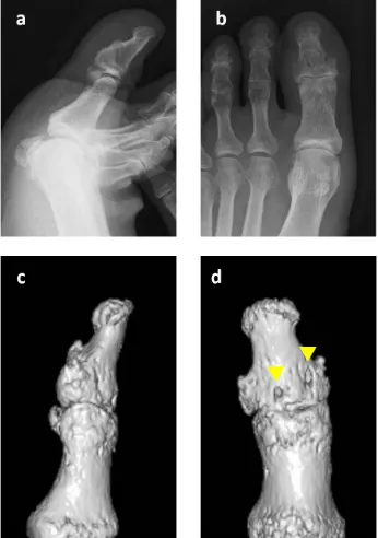
A 57-year-old male banker presented with pain and swelling in his left great toe after a fall, while walking barefoot on the bed. His great toe was forced into a hyperplantarflexion position. On physical examination, he had swollen great toe and a bony prominence at the dorsal aspect of the interphalangeal joint (Figure 1(a)). His hallux was in a flexed position and he could not actively extend the great toe (Figure 1(b)). Plain radiography showed a small bone fragment at the base of the dorsal distal phalanx (Figure 2(a) and Figure 2(b)). The joint was congruent and subluxation was not observed. Computed tomography revealed the size and location of the fragment suggesting an avulsion fracture of the ex-tensor hallucis longus (Figure 2(c) and Figure 2(d)).
DOI: 10.4236/ojo.2019.95012 125 Open Journal of Orthopedics No complications were observed during surgery and the great toe was immo-bilized with buddy taping to the second toe to prevent plantarflexion of the in-terphalangeal joint for two weeks. Heel weight bearing was allowed the day after surgery and passive range of motion exercises were cautiously initiated once the wound was completely healed. At four weeks, partial toe weight bearing was al-lowed and full toe weight bearing was initiated at two months after the surgery. Three months after the surgery, bony union was confirmed both with plain ra-diography and computed tomography (Figure 4), and symmetric range of mo-tion of the interphalangeal joint was obtained. At one year, the patient did not complain of any pain during walking and running. Written informed consent was obtained from the patient for publication of this case report and accompa-nying images.
Figure 1. Macroscopic findings of the great toe. A bony prominence (arrow) at the dorsal aspect of the interphalangeal joint was observed (a), the great toe was in the flexed position active extension was not possible (b).
[image:3.595.290.458.410.657.2]DOI: 10.4236/ojo.2019.95012 126 Open Journal of Orthopedics
[image:4.595.265.484.68.239.2]Figure 3. Surgical technique applied to the great toe. Intraoperative photographs after exposure of the fracture site (a), insertion of the two suture anchors (b) and fixation of the bone fragment with the bridging technique (c). Schematic illustration of the surgery (d).
Figure 4. Lateral (a) and anteroposterior (b) plain radiographs and lateral (c) and dorsal (d) aspect of the computed tomography at three months after the surgery. Bone union was achieved and two drill holes were observed (arrowheads).
3. Discussions
[image:4.595.287.460.311.557.2]DOI: 10.4236/ojo.2019.95012 127 Open Journal of Orthopedics
Table 1. Summary of previously reported and present cases of mallet toe of the hallux.
Age (Sex)
Mechanism of injury
Size of bony
fragment Treatment Outcomes
Rapoff 36 (M) Caught on the edge of the step 45% of JS
Rigid-soled sandal for 10 weeks to prevent plantarflexion
Returned at 16 weeks and 10% ROM restriction
Hennessy 45 (M) Fall during descending stairs
Small Dorsal thermoplastic extension splint for 8 weeks
50% ROM restriction and minor flexion deformity
Nakamura 51 (M)
Stubbed on the threshold of
sliding door Small
IP joint fixation with a K-wire for 5 weeks after failed conservative treatment
Slight limitation in flexion at 1 year
Wada 49 (M) Caught on a carpet barefoot
More than 50% of JS
Extension block method with two K-wire for 4 weeks
Returned at 8 weeks and no description about ROM
Martin 16 (M) Kicking a ball barefoot 40% of JS
Open reduction and fixation with two K-wire and temporal IP joint fixation for 5 weeks after failed closed reduction
Full activity except for sports at 3.5 months and symmetric ROM
Hong 1) 39 (F) 2) 46 (M)
1) Fall at the slope 2) Kicking an opponent
1) 40% of JS 2) 30% of JS
One suture anchor fixation with two drill holes into the fragment
Returned at 2 and 3 months and no and slight ROM limitation
Ando 57 (M) Fall from the bed barefoot Small
Two suture anchor fixation with suture bridging technique
Returned at 2 months and no ROM limitation
M, male; F, female; JS, joint surface; K-wire, Kirschner wire; IP, interphalangeal; ROM, range of motion.
Treatment options for the injury are conservative treatment [1] [4], percuta-neous or open Kirschner wire fixation [5] [6] [7], or suture anchor fixation [8]. Rapoff et al. reported a case treated with a rigid soled sandal for ten weeks [4], and Hennesy et al. reported using a dorsal thermoplastic extension splint for eight weeks [1]. Although these studies reported acceptable results, restriction in range of motion, flexion deformity, and extensor lag of the hallux persisted after these treatments [1] [4].
DOI: 10.4236/ojo.2019.95012 128 Open Journal of Orthopedics Hong et al. reported two cases treated with a suture anchor for reattaching the avulsed fragment without transarticular immobilization of the interphalangeal joint [8]. They reported that a suture anchor was inserted into the fracture bed of the distal phalanx and two drill holes were made into the avulsed fragment. Thereafter, the fragment was secured with sutures [8]. This technique is applica-ble when the avulsed fragment is large enough for two drill holes. However, it is difficult when the fragment is small for drilling holes directly into it, like in our case. The reported size of the fragment was divided into two groups, 30% to 50% of the joint surface [4] [6] [7] [8] or too small to measure as in our case [1] [5]. In the present case, two suture anchors were inserted into the fracture bed of the distal phalanx, and two mattress sutures were made just proximal to the avulsed fragment in the interphalangeal joint extension. Finally, the fixation was rein-forced with bridging sutures. This technique provided sufficient fixation of the avulsed fragment to allow early postoperative mobilization of the interphalan-geal joint and good clinical results were obtained without limitation of the in-terphalangeal joint range of motion. This technique can be a valuable procedure when the bone fragment is small.
4. Conclusion
Bridging suture technique with two suture anchors allowed us to fix the small bone fragment rigidly and mobilize the interphalangeal joint earlier to preserve the range of motion.
Conflicts of Interest
The authors declare no conflicts of interest regarding the publication of this pa-per.
References
[1] Hennessy, M.S. and Saxby, T.S. (2001) Traumatic ‘Mallet Toe’ of the Hallux: A Case Report. FootandAnkleInternational, 22, 977-978.
https://doi.org/10.1177/107110070102201209
[2] Bendre, A.A., Hartigan, B.J. and Kalainov, D.M. (2015) Mallet Finger. TheJournal oftheAmericanAcademyofOrthopaedicSurgeons, 13, 336-344.
https://doi.org/10.5435/00124635-200509000-00007
[3] Leinberry, C. (2009) Mallet Finger Injuries. The Journal of Hand Surgery, 34, 1715-1717. https://doi.org/10.1016/j.jhsa.2009.06.018
[4] Rapoff, A.J. and Heiner, J.P. (1999) Avulsion Fracture of the Great Toe: A Case Re-port. FootandAnkleInternational, 20, 337-339.
https://doi.org/10.1177/107110079902000514
[5] Nakamura, S. (2007) Temporary Kirschner Wire Fixation for a Mallet Toe of the Hallux. JournalofOrthopaedicScience, 12, 190-192.
https://doi.org/10.1007/s00776-006-1106-x
DOI: 10.4236/ojo.2019.95012 129 Open Journal of Orthopedics
[7] Martin, E.A., Barske, H.L. and DiGiovanni, B.F. (2013) Open Surgical Treatment of an Acute, Unstable Bony Mallet Injury of the Hallux. FootandAnkleInternational, 34, 295-298. https://doi.org/10.1177/1071100712465850
[8] Hong, C.C. and Tan, K.J. (2013) Suture Anchor Fixation of Unstable Bony Mallet Injuries of the Hallux. FootandAnkleInternational, 34, 1737-1741.