ISSN Online: 2327-509X ISSN Print: 2327-5081
DOI: 10.4236/jbm.2019.75006 May 17, 2019 27 Journal of Biosciences and Medicines
N-Methylcantharidinimidum Induces
Apoptosis in Human Hepatocellular
Carcinoma Cell HepG2
Li Tang, Yi Shen
*Department of Pathophysiology, Laboratory of Stem Cell and Tissue Engineering, Chongqing Medical University, Chongqing, China
Abstract
Hepatocellular carcinoma (HCC) is one of the most common malignant tumors and the prognosis of patients was poor. Cantharidin is an effective anti-tumor component of Chinese traditional medicine. N-methylcantharidinimidum is a novel cantharidin derivative with better curative effect and less side effect. In this study, the effects of N-methylcantharidinimidum on proliferation, apoptosis and cell cycle of human hepatocellular carcinoma cell line HepG2 were examined. The results demonstrated that N-methylcantharidinimidum significantly induced apoptotic cell death in a dose and time-dependent man-ner and arrested cell cycle in HepG2 cells. N-methylcantharidinimidum also suppressed cyclin D1. This study suggests that N-methylcantharidinimidum may serves as a potential chemopreventive agent for live cancer.
Keywords
N-Methylcantharidinimidum, HepG2, Apoptosis, HCC
1. Introduction
Liver cancer is the third leading cause of cancer-related death worldwide. Al-though young patients have an improved prognosis, older patients have an in-creased mortality rate after being diagnosed with liver cancer. Cantharides is a kind of arthropod insect and cantharidin is a sesquiterpenoid derivative ex-tracted from cantharides. Cantharidin is an effective anti-tumor component of Chinese traditional medicine [1]. It has been shown to effectively inhibit cell proliferation in numerous cancer cell lines including colorectal, hepatic, blad-der, pancreatic and breast cancers [2] [3] [4]. The clinical application of can-How to cite this paper: Tang, L. and Shen,
Y. (2019) N-Methylcantharidinimidum Induces Apoptosis in Human Hepatocellu-lar Carcinoma Cell HepG2. Journal of Biosciences and Medicines, 7, 27-33.
https://doi.org/10.4236/jbm.2019.75006
DOI: 10.4236/jbm.2019.75006 28 Journal of Biosciences and Medicines tharidin is restricted due to its severe irritant to the digestive and urinary sys-tems. N-methylcantharidinimidum is a novel cantharidin derivative. In xeno-graft model, the survival time of liver cancer mice was prolonged by N-methylcantharidinimidum at the concentration of 16 - 18.5 mg/ml. The clini-cal trial was used to treat primary liver cancer, and its curative effect was better than cantharidin and did not have apparent toxic side effect on digestive and urinary systems. In this study, HepG2 cells were used to observe the anti-tumor effect and its mechanism of N-methylcantharidinimidum.
2 Materials and Methods
2.1. Cell Culture
N-methylcantharidinimidum was a generous gift from Chongqing Institute of Chinese Materia Medica. Hepatocellular carcinoma cell HepG2 was obtained from the American Type Culture Collection (Rockville, MD, USA). Cells were cultured with complete medium MEM/EBSS (HyClone, UT, USA) containing 10% fetal bovine serum (Gibco BRL, MD, USA), 2 mM glutamine, 100 μg/ml streptomycin, and 100 U/ml penicillin. Cells were maintained at 37˚C in a hu-midified atmosphere of 5% CO2.
2.2. Cell Proliferation Assay
The effect of N-methylcantharidinimidum on cell proliferation was assessed by MTT assay. HepG2 cells were seeded at a density of 1 × 10⁴ cells per well in triplicate in 96-well plates and cultured for 24 h to allow the cells for attach-ment. N-methylcantharidinimidum was dissolved in DMSO and diluted in MEM/EBSS. The cells were treated with 5, 6, 7 and 8 mg/ml of
N-methylcantharidinimidum for 24, 48 and 72 h. After treatment, 20 μl of 5 mg/ml MTT solution was added to each well and incubated for 4 h at 37˚C. The supernatant was removed and 200 μl of DMSO was added to each well. The ab-sorbance was quantified using enzyme-linked immunosorbent assay (ELISA) reader at 570 nm.
2.3. Morphological Analysis
According to the results of MTT assay, HepG2 cells (5 × 10⁴/ml) were inoculated in 24-well plates with presetted slides, the cells were treated with 5, 6 and 7 mg/ml of N-methylcantharidinimidum for 1 to 6 days. Then the slide were re-moved and stained with Wright’s stain, and observed under light microscope.
2.4. Flow Cytometric Analysis
DOI: 10.4236/jbm.2019.75006 29 Journal of Biosciences and Medicines mg/ml, 5 ml) was added and followed by incubation at room temperature in dark for 30 min. The apoptosis rate was immediately measured by FACSCalibur (BD Biosciences, USA). To evaluate the effect of N-methylcantharidinimidum on the cell cycle, the harvested cells were washed twice with ice-cold PBS, fixed with ice-cold 70% ethanol and maintained overnight at −20˚C. DNA was stained with 100 μg/ml propidium iodide (PI) solution. The cell cycle distribution was analyzed by flow cytometry.
2.5. TUNEL Assay
In situ cell death, detection kit-POD (Boehringer Mannheim, German) was used to detect the late-stage apoptosis. Cells were seeded in 96-well plates and treated with 6 mg/ml of N-methylcantharidinimidum. Cells were fixed with 4% para-formaldehyde. Fixed cells were penetrated with 0.5% Triton X-100 for 2 min on the ice and then incubated with TUNEL reaction mixture for 1 h at 37˚C. All the slides were stained by DAB coupling and counterstained with hematoxylin. The cells were then examined using a light microscope.
2.6. Western Blot Analysis
HepG2 cells were treated with 6 mg/ml N-methylcantharidinimidum for 3 d. Total protein of cells was resolved by SDS-PAGE and transferred onto a PVDF membrane. After blocking with 5% non-fat milk in tris-buffered saline with 0.1% Tween 20 (TBST), membranes were incubated with respective primary an-tibodies directed against CyclinD1 (1:1000) and β-actin (1:3000) at 4˚C over-night. Membranes were washed and then incubated with HRP-conjugated rabbit anti-IgG (1:5000) for 1 h at room temperature. Protein bands were assessed by enhanced chemiluminescence system (ECL, KeyGEN, China).
2.7. Statistical Analysis
All statistical analyses were evaluated using SPSS 17.0 software. The significance of difference between the groups was analyzed with two-way ANOVA test or two-tailed unpaired Student’s t-test. P-values < 0.05 were considered as statisti-cally significant.
3. Results
3.1. N-Methylcantharidinimidum Inhibits Cell Proliferation
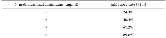
The effect of N-methylcantharidinimidum on HepG2 cells was investigated by MTT assay. The results showed that N-methylcantharidinimidum inhibited the proliferation of HepG2 cells in dose- and time-dependent manners (Table 1). The cytostatic does of 6 mg/ml was taken for further study of
N-methylcantharidinimidum on HepG2 cells.
3.2. N-Methylcantharidinimidum Induces Cell Apoptosis
DOI: 10.4236/jbm.2019.75006 30 Journal of Biosciences and Medicines Table 1. Inhibitory Effect of N-methylcantharidinimidum on the growth of HepG2 cell.
N-methylcantharidinimidum (mg/ml) Inhibitory rate (72 h)
5 24.3%
6 40.4%
7 67.2%
8 89.6%
After treated with N-methylcantharidinimidum, vacuoles appeared in the toplasm. Some cells were shrunken, with condensed nuclear chromatin and cy-toplasm. Condensed nuclei were frequently fragmented. Apoptosis was induced after 3 days of treatment, and the number of apoptotic cells increased gradually with the extension of treat time.
2) Apoptosis was detected by flow cytometric analysis. 6 mg/ml
N-methylcantharidinimidum promoted HepG2 cells apoptosis after an exposure of 3, 4 and 5 days (22.17%, 23.87% and 33.79% of cells, respectively). The result also showed that N-methylcantharidinimidum induced cell cycle arrest at G2/M phase.
3) In situ determination of apoptosis by TUNEL. The TUNEL assay was used to detect DNA fragmentation characteristic for apoptosis, seen as nuclei stained dark brown. A number of TUNEL positive cells substantially increased after 6 mg/ml N-methylcantharidinimidum treated for 4 days, dark brown granules were seen in the nucleus, indicated that DNA strand breaks had occurred.
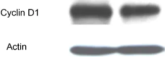
3.3. N-Methylcantharidinimidum Decreased the Expression of
Cyclin D1
The protein level of cyclin D1 was determined by immunoblotting. As shown in Figure 1, expression of cyclin D1 decreased in HepG2 cell after treated with 6 mg/ml N-methylcantharidinimidum for 3 days. β-actin served as an internal control.
4. Discussion
[image:4.595.204.539.101.179.2]DOI: 10.4236/jbm.2019.75006 31 Journal of Biosciences and Medicines Figure 1. Expression of cell cycle-controlling
protein Cyclin D1 in HepG2 cell.
compounds inhibit cell proliferation in numerous cancer cell lines such as he-patic, bladder, pancreatic, colorectal, leukemic, oral, and breast cancers [9] [10] [11]. In order to improve pharmacokinetic properties, increase efficacy and re-duce toxic side effects, some new cantharidin derivatives were synthesized and be studied. Cantharidin showed inhibitory effects on murine ascites hepatoma and ascites reticulum cell sarcoma [12]. Cantharidin sodium and Shenmai injec-tion combined with chemotherapy significantly reduced the incidence of side effects in postoperative breast cancer patients in clinical trial [13]. Norcanthari-din, a demethylated analogue of canthariNorcanthari-din, induced cell apoptosis in human oral cancer cells through a mitochondria-mediated pathway [14]. Here, we inves-tigated the anti-tumor effect and its mechanism of N-methylcantharidinimidum, a novel cantharidin derivative, in hepatocellular carcinoma cell line HepG2.
In this study, N-methylcantharidinimidum was shown to exert strong cyto-toxicity against human hepatocellular carcinoma cell line HepG2, inhibiting cell proliferation in vitro through inducing apoptosis. The result of the cell prolifera-tion assay showed that N-methylcantharidinimidum exerted a potent cytotoxic effect on HepG2 in a dose and time-dependent manner. The inhibition of proli-feration in HepG2 cells was a result of apoptosis induction and cell cycle arrest. Apoptosis is an important homeostatic mechanism that is characterized by unique morphological and biochemical features and is used to maintain the ap-propriate numbers of cells in the body. In current study, we demonstrated that N-methylcantharidinimidum treated cells express an apoptotic reaction. Cyclin D1 is an oncoprotein that plays a key role in the development of tumor. High expression of cyclin D1 is considered to be indicative of a poor prognosis and related to an unfavorable therapeutic outcome. Overexpression of cyclin D1 protein leads to increased cell proliferation, which gives neoplastic cells a growth advantage and may also favor the occurrence of additional genetic lesions with potential oncogenic effects. Cyclin D1 is an important regulator of G1 phase pro-gression in many different cell types. In this study, N-methylcantharidinimidum treatment decreased the level of cyclin D1 in HepG2 cells, which is correlated with the cell cycle analysis showing G2/M phase arrest.
DOI: 10.4236/jbm.2019.75006 32 Journal of Biosciences and Medicines
Conflicts of Interest
The authors declare no conflicts of interest regarding the publication of this paper.
References
[1] Wang, G., Dong, J. and Deng, L. (2018) Overview of Cantharidin and Its Analogues. Curr Med Chem., 25, 2034-2044.
https://doi.org/10.2174/0929867324666170414165253
[2] Kok, S.H., Hong, C.Y., Kuo, M.Y., et al. (2003) Comparisons of Norcantharidin Cy-totoxic Effects on Oral Cancer Cells and Normal Buccal Keratinocytes. Oral Oncol., 39, 19-26. https://doi.org/10.1016/S1368-8375(01)00129-4
[3] Chen, Y.N., Chen, J.C., Yin, S.C., et al. (2002) Effector Mechanisms of Norcanthari-din-Induced Mitotic Arrest and Apoptosis in Human Hepatoma Cells. Int J Cancer., 100, 158-165. https://doi.org/10.1002/ijc.10479
[4] Xiao, W., Dai, B., Zhu, Y., et al. (2015) Norcantharidin Induces Autophagy-Related Prostate Cancer Cell Death through Beclin-1 Upregulation by miR-129-5p Suppres-sion. Tumour Biol., 37, 15643-15648. https://doi.org/10.1007/s13277-015-4488-6
[5] Ling, N., Zhou, X., Ji, Y., et al. (2017) Immuno-Modulatory and Cellular Antioxi-dant Activities of κ-Selenocarrageenan in Combination with Epirubicin in H22 He-patoma-Bearing Mice. Biomedicine & Pharmacotherapy, 91, 132-137.
https://doi.org/10.1016/j.biopha.2017.04.064
[6] Chen, F., Sun, Y., Zheng, S.L., et al. (2017) Antitumor and Immunomodulatory Ef-fects of Ginsenoside Rh2 and Its Octyl Ester Derivative in H22 Tumor-Bearing Mice. Journal of Functional Foods, 32, 382-390.
https://doi.org/10.1016/j.jff.2017.03.013
[7] Sarkar, F.H. and Li, Y. (2006) Using Chemopreventive Agents to Enhance the Effi-cacy of Cancer Therapy. Cancer Research, 66, 3347-3350.
https://doi.org/10.1158/0008-5472.CAN-05-4526
[8] Wang, J., Liu, W., Chen, Z., et al. (2017) Physicochemical Characterization of the Oolong Tea Polysaccharides with High Molecular Weight and Their Synergistic Ef-fects in Combination with Polyphenols on Hepatocellular Carcinoma. Biomedicine & Pharmacotherapy, 90, 160-170. https://doi.org/10.1016/j.biopha.2017.03.059
[9] Zhang, C., Chen, Z., Zhou, X., et al. (2014) Cantharidin Induces G2/M Phase Arrest and Apoptosis in Human Gastric Cancer SGC-7901 and BGC-823 Cells. Oncol Lett., 8, 2721-2726. https://doi.org/10.3892/ol.2014.2611
[10] Liu, B., Gao, H.C., Xu, J.W., et al. (2012) Apoptosis of Colorectal Cancer UTC116 Cells Induced by Cantharidinate. Asian Pac J Cancer Prev., 13, 3705-3708.
https://doi.org/10.7314/APJCP.2012.13.8.3705
[11] Li, W., Xie, L., Chen, Z., et al. (2010) Cantharidin, a Potent and Selective PP2A In-hibitor, Induces an Oxidative Stress-Independent Growth Inhibition of Pancreatic Cancer Cells through G2/M Cell-Cycle Arrest and Apoptosis. Cancer Sci., 101, 1226-1233. https://doi.org/10.1111/j.1349-7006.2010.01523.x
[12] Wang, G.S. (1989) Medical Uses of Mylabris in Ancient China and Recent Studies. J Ethnopharmacol., 26, 147-162. https://doi.org/10.1016/0378-8741(89)90062-7
DOI: 10.4236/jbm.2019.75006 33 Journal of Biosciences and Medicines
https://doi.org/10.7314/APJCP.2014.15.14.5597