Synergistic Anti-Tumor Effect of Cisplatin When
Combined with an Anti-Src Kinase Integrin-Based
Peptide
Michael Agrez1, Madhu Garg2,3, Douglas Dorahy3, Stephen Ackland2,3
1
Division of Surgery, John Hunter Hospital, Newcastle, New South Wales, Australia; 2Department of Medical Oncology, Calvary Mater Hospital, Waratah, New South Wales, Australia; 3Hunter Medical Research Institute, John Hunter Hospital, Newcastle, New South Wales, Australia.
Email: michael.agrez@hnehealth.nsw.gov.au
Received May 31st, 2011; revised June 20th, 2011; accepted June 27th, 2011.
ABSTRACT
Background: It is known that activeSrc kinasepromotes survival of ovarian cancer cell lines and inhibition of c-Src
has been shown to restore sensitivity of drug-resistant human ovarian cancer cells to cisplatin. In this study we
exam-ined the effects of a 10 mer peptide on proliferation of human colon and ovarian cancer cells when used alone and in
combination with cisplatin. Materials and Methods: A 10 mer peptide, RSKAKNPLYR, derived from a 15 mer ERK2
binding sequence present on the cytoplasmic domain of the β6 integrin subunit was tested for its effect on proliferation
of HT29 colon cancer cells under serum-free conditions by means of the MTT assay. Cell proliferation studies to
exam-ine the effects of cisplatin combexam-ined with peptide were conducted in serum-containing medium using the 10 mer peptide
fused to a hydrophobic signal peptide sequence. Drug combination studies were performed on HT29 cells and a
cis-platin-resistant cell line (ADDP) derived from an ovarian cancer cell line A2780. The effects of peptides on Src kinase
activity were assessed in a cell-free in vitro kinase assay. Results: The 10 mer peptide was as effective as the 15 mer
parent compound at inhibiting proliferation of HT29 cells. Exposure of HT29 and ADDP cells to a combination of
cis-platin and the fusion peptide resulted in synergistic inhibition of cell growth. Both the 10 mer peptide alone and when
fused to the signal peptide sequence inhibited Src kinase activity. Conclusion: Our findings raise the possibility of
com-bination therapy comprising peptide and cisplatin for cisplatin-resistant ovarian cancers and other cancers that are high expressors of c-Src.
Keywords: Cisplatin, Peptides, Cancer Cell Lines, MTT Assay, Synergy, C-Src
1. Introduction
Tumor chemotherapy with platinum-based compounds has met with mixed success. Cis-diamminedichloropla- tinum (II)(cisplatin) is a long established anticancer drug with activity in a variety of solid tumor types including head and neck cancer, ovarian cancer and non-small cell lung cancer. However, major disadvantages of cisplatin include relapse in most tumors after an initial response [1] and the observed resistance to cisplatin as seen with co-lon cancer cells [2].
Inhibition of Src tyrosine kinase has been shown to enhance the cytotoxicity of cisplatin and restore sensitiv-ity in drug-resistant cells [3]. Moreover, in colon cancer decreased Src expression in the cell line HT29 (that has high constitutive Src expression and activity) by means
[5].
C-Src activation has been documented in upwards of 50% of tumors derived from the colon, liver, lung, breast and pancreas [6,7]. C-Src activity increases with pro-gressive stages of disease in colon cancer and is thought to be predictive of poor clinical prognosis suggesting that c-Src activation confers growth and/or survival advan-tages to metastatic colon tumor cells [8-10]. It has been postulated that c-Src activation may contribute to colon tumor progression and metastasis by activating Akt- mediated survival pathways that decrease sensitivity of detached cells to anoikis [4]. C-Src has also been shown to be over-expressed and activated in a high proportion of ovarian cancers [11] and inhibition of c-Src sensitizes ovarian cancer cells to chemotherapeutic agents such as paclitaxel and cisplatin [3].
A functional interaction between activation of c-Src and integrins is well-recognised. Integrins comprise a family of cell adhesion receptors composed of alpha/beta heterodimeric subunits that provide a functional and structural bridge between the extracellular matrix and intracellular signaling molecules [12]. For example, it has been suggested that binding of the c-terminal portion
of the cytoplasmic tail of the β3 integrin subunit to the
c-Src SH3 domain can disrupt the auto-inhibitory inter-actions between the SH3 and SH2 domains of Src [13].
Expression of the αvβ6 integrin in ovarian cancers
may contribute to the invasive potential of ovarian
can-cers [14] and expression of the αvβ6 integrin in colon
cancer has been identified as an independent prognostic indicator for worse outcome in patients suffering from this disease [15]. We have previously reported that a se-quence of 15 amino acids, RSKAKWQTGTNPLYR,
located within the cytoplasmic tail of the β6 integrin
subunit binds to extracellular signal-regulated kinase 2 (ERK2) and proposed that this contributes to tumor growth [16]. Moreover, a non-naturally occurring peptide, RSKAKNPLYR, derived from this binding sequence also binds with equal affinity to ERK2 [17]. In the pre-sent study we examined the effect of the 10 mer peptide
on in vitro growth of human colon and ovarian cell lines
in the absence and presence of cisplatin.
2. Materials & Methods
2.1. Cell Lines and Culture Conditions
The human colon cancer cell line HT29, an ovarian can-cer cell line, A2780, and its cisplatin resistant subline,
ADDP, were used for the in vitro studies. The
cis-platin-resistant cell subline, ADDP, was developed from long term exposure of the ovarian cancer cell line, A2780, to cisplatin [18] and both cell lines were obtained with kind permission from Dr G.J. Peters, Amsterdam,
Neth-erlands [19]. Cell lines were cultured at 37˚C, under air
containing 5% CO2 and passaged regularly for optimal
growth. Cells were maintained in DMEM medium con-taining 10% fetal bovine serum. All culture medium preparations were further supplemented with penicillin/ streptomycin (100 µg/ml), and glutamine (2 mM).
2.2. Peptides
All peptides were synthesized by Auspep, Melbourne, Australia. For cell growth assays performed in the presence of serum, the 10 mer RSKAKNPLYR, was linked at its amino terminus to a modification of the hy-drophobic signal peptide from the Kaposi fibroblast growth factor, AAVALLPAVLLALLAP [20] that lacked the c-terminal proline (AAVALLPAVLLALLA) and the fused peptide was designated IK2 (AAVALPAVLL- ALLARSKAKNPLYR).
2.3. In Vitro Growth Inhibition Assay
Cells in logarithmic growth were transferred to 96-well plates in 100 µl medium at a density of 2500 cells per well for serum-containing experiments and at a density of 4000 cells per well for serum free medium (SFM) ex-periments. For serum-containing experiments, 100 µl medium with or without the test agent (50 µl single agent with 50 µl medium or 50 µl of both agents for combina-tion studies) were added to each well in triplicate 24 hours after plating, while for SFM experiments the pre-viously added serum-containing medium was removed and 200 µl SFM medium with or without the test agent added to each of triplicate wells. Drug exposure experi-ments were carried out on cell lines using varying con-centrations of agents: peptides (50 nM - 100 µM), cis-platin (0.5 µM - 100 µM) as single agents, and for com-bination studies a fixed concentration of IK2 (30 µM) was added to varying concentrations of cisplatin (in trip-licate) for 48 hr in serum-containing medium. For single agent peptide studies, cells were exposed to peptides for 72 - 96 hours in serum-free culture medium.
to be expected based on percentage inhibition of cell growth in the presence of peptide alone multiplied by the inhibition of cell proliferation for cisplatin alone at each concentration of cisplatin for both HT29 and ADDP cells (termed “additive” in the graphed data shown). Growth inhibition in compound combination studies that ex-ceeded the calculated additive effect was indicative of a synergistic effect.
2.4. C-Src Kinase Activity Assay
In vitro c-Src kinase activity assays were performed by
Upstate Kinase Profiling, Dundee, Scotland, according to the manufacturer’s instructions. In brief, in a final reac-tion volume of 25 µL, c-Src (h) (5 mU - 10 mU) was incubated with 8 mM MOPS pH 7.0, 0.2 mM EDTA, 250 µM KVEKIGEGTYGVVYK (Cdc2 peptide), 10
mM MgAcetate and [ԃ-33P-ATP] (specific activity
ap-proximately 500 cpm/pmol). The reaction was initiated by the addition of the MgATP mix. After incubation for 40 minutes at room temperature, the reaction volume was stopped by the addition of 5 µL of a 3% phosphoric acid solution. 10 µL of the reaction volume was then spotted onto a P30 filtermat and washed three times for 5 min-utes in 75 mM phosphoric acid and once in methanol prior to drying and scintillation counting.
3. Results
The 10 mer peptide, RSKAKNPLYR, was as effective as the 15 mer parent compound RSKAKWQTGTNPLYR at
inhibiting in vitro proliferation of HT29 cells cultured
under serum-free conditions as shown in Figure 1, Panel
A. Cleavage of amino acids from the amino terminus of RSKAKNPLYR in the presence of serum enzymes (un-published data) accounts for its lack of effect in the presence of serum (see Figure 1, Panel B).
To enhance both metabolic stability and transmembrane transport, the active peptide was fused to the hydrophobic signal peptide sequence, AAVALLPAVLLALLA, de-rived from Kaposi’s fibroblast growth factor [19]. As
shown in Figure 1, Panel B, the anti-proliferative effect
of RSKAKNPLYR on HT29 cells cultured in serum- containing medium was restored when the 10 mer was fused with the signal peptide sequence (AAVALL-PAVLLALLARSKAKNPLYR, designated IK2) and was not accounted for by effects of the signal peptide itself which was ineffective at inhibiting cell growth (Figure 1, Panel B).
Exposure of HT29 and ADDP cells to a combination of cisplatin and IK2 (30 µM) resulted in synergistic inhi-bition of cell growth (thicker graphed lines for observed
inhibition of cell growth shown in Figure 1, Panels C
and D, respectively). The observed values for growth inhibition at each cisplatin concentration in combined
studies exceeded expected additive values at concentra-tions of cisplatin above 10 µM for HT29 cells and 5 µM
for ADDP cells (Figure 1, Panels C and D, respectively).
The selection of a fixed concentration of 30 µM of IK2 in combination studies was based upon the approximate
mean GI50 values for HT29 and ADDP cells in the
pres-ence of peptide alone as shown in Table 1.
The GI50 values for cells cultured under serum-
containing and serum-free conditions in the presence of
compounds alone and in combination are shown in Table
1. For ADDP cells, GI50 values for the combination of
compounds exceeded the values for the calculated addi-tive effect of cisplatin and IK2 alone (Table 1). However,
for HT29 cells, the GI50 values for observed and additive
effects for combination of compounds overlapped at lower concentrations of cisplatin and synergism was seen only at concentrations above 10 µM of cisplatin. Notably,
as seen in Figure 1, Panels B, C and D, the surviving
fraction of cells exposed to IK2, IK2 plus cisplatin and cisplatin alone, respectively, goes below zero at higher concentrations indicating actual cell killing and not just
growth arrest or inhibition. In cell-free in vitro kinase
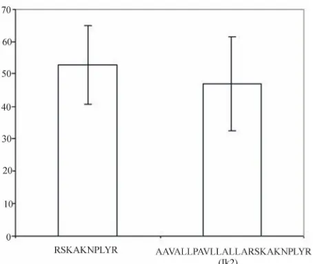
assays both the active peptide, RSKAKNPLYR, and the fusion peptide, IK2, inhibited Src kinase activity to the same degree (Figure 2).
4. Discussion
The c-Src oncogene is over-expressed and activated in a high proportion of ovarian cancers [11]. Accordingly, targeted therapy with the Src inhibitor dasatinib has been shown to interact in a synergistic manner with plati-num-based chemotherapy in ovarian cancer cells [21]. A 15 mer peptide, RSKAKWQTGTNPLYR, representing
the ERK2 binding sequence located within the β6
in-tegrin tail has previously been reported to also inhibit c-Src activity [22]. In the present report we show that a deletion variant of this peptide, RSKAKNPLYR, lacking
the middle five amino acids (i.e., WQTGT) inhibits both
c-Src activity as well as cancer cell growth in vitro.
Moreover, the combination of this 10 mer peptide with cisplatin results in synergistic inhibition of proliferation of colon and ovarian cancer cells.
0 0.1 0.2 0.3 0.4 0.5 0.6 0.7 0.8 0.9 1 1.1
0.01 0.10 1.00 10.00 100.00
Conc ( µM )
S u rv iv in g Fr a c ti o n RSKAKWQTGTNPLYR RSKAKNPLYR
HT29 Cells exposed to peptides in serum-free medium
HT29 Cells exposed to peptides in serum-containing medium
-0.2 -0.1 0 0.1 0.2 0.3 0.4 0.5 0.6 0.7 0.8 0.9 1 1.1
0.10 1.00 10.00 100.00
Conc ( µM )
S ur v iv ing Fr a c ti on RSKAKNPLYR AAVALLPAVLLALLA AAVALLPAVLLALLARSKAKNPLYR (IK2) A
(a )
B (b)
HT29 cells exposed to cisplatin ± IK2 peptide
-0.2 -0.1 0 0.1 0.2 0.3 0.4 0.5 0.6 0.7 0.8 0.9 1 1.1
0.10 1.00 10.00 100.00
Conc ( µM )
S ur v iv ing Fr a c ti on IK2 Cisplatin (CP) observed CP+ 30µM IK2 additive CP+ 30µM IK2 30µM IK2 C -0.2 -0.1 0 0.1 0.2 0.3 0.4 0.5 0.6 0.7 0.8 0.9 1 1.1
0.01 0.10 1.00 10.00 100.00
Conc ( µM )
S u rv iv ing Fr a c ti on
ADDP - IK2 ADDP-Cisplatin (CP) ADDP-observed CP+ 30µM IK2 ADDP-additive CP+ 30µM IK2 ADDP-30µM IK2
A2780-Cisplatin (CP)
Ovarian cancer cell lines exposed to cisplatin ± IK2 peptide
D
(c) (d)
[image:4.595.63.533.88.405.2]Figure 1. (a) Growth response curves for HT29 colon cancer cells cultured in the presence of peptides under serum-free con-ditions for 72 hours. (b) Growth response curves for HT29 cells cultured in the presence of peptides in serum-containing me-dium for 72 - 96 hours. (c) Growth response curves for HT29 cells cultured for 48 hours in the presence of either cisplatin or peptide alone and in combination using a fixed concentration of peptide, 30 µM. (d) Growth response curves for ovarian can-cer cells cultured for 48 hours in the presence of either cisplatin alone (A2780 cell line that is cisplatin-sensitive and the ADDP cell line that is cisplatin resistant), peptide alone (ADDP cells) and in combination using a fixed concentration of pep-tide, 30 µM (ADDP cells). Values are means ± SEM.
Table 1. GI50 values (Mean ± SEM) for cells cultured under serum-free and serum-containing medium exposed to single and
combination of compounds.
Mean ± SEM (µM)
COMPOUNDS HT29 ADDP A2780
RSKAKNPLYR 5.06 ± 1.01
Serum- free
medium RSKAKWQTGTNPLYR 6.33 ± 2.18
AAVALLPAVLLALLA-RSKAKNPLYR
(IK2) 36.67 ± 6.01 35.67 ± 4.70
Cisplatin (CP) 16.33 ± 2.96 34.50 ± 4.38 1.40 ± 0.61
Observed CP + 30 µM IK2 6.83 ± 3.65
Serum- cotaining
medium
Additive CP + 30 µM IK2 28.33 ± 8.82
of mammalian cells [23]. This is consistent with our own unpublished observation that IK2 (signal peptide fused to RSKAKNPLYR) is 10-fold more effective at inhibiting proliferation of HT29 cells under serum-free culture
conditions, i.e., in the absence of peptide-degrading
en-zymes, than the 10 mer sequence alone.
[image:4.595.87.512.527.634.2]Figure 2. Percentage inhibition of c-Src kinase activity by peptides as determined by in vitro kinase assay. Values rep-resent the mean percentage inhibition for three separate assays ± SEM.
there are no published data on comparative levels of c-Src expression or activity between the cisplatin- sensitive A2780 cell line and the cisplatin-resistant sub-line ADDP. Cisplatin-sensitive A2780 cells are known to be high expressors of Src [21] and whether Src deregula-tion is further enhanced in A2780 cells with acquired resistance to cisplatin such as the ADDP subline is not known.
The cause of clinical resistance to platinum com-pounds remains unknown and is likely to reflect a mul-ti-factorial problem that is not exclusively related to in-creased c-Src activity or expression. For example, cis-platin-resistant ovarian cancer cells have been shown to be more effective at repairing cisplatin-DNA lesions and effluxing cisplatin or preventing influx of the drug [24,25]. Resistance to cisplatin has also been linked to expression of Stat1 (signal transducer and activator of transcription) in A2780 cells and to activation of the phosphatidylinositol 3-kinase (PI3K)/Akt signaling pathway in the ovarian cancer cell line OVCAR-3 [26,27]. Cell survival signals are mediated via PI3K/Akt and the tumor suppressor phosphatase and tensin ho-molog (PTEN) negatively regulates survival mediated by the PI3K/Akt pathway [28]. Interestingly, levels of PTEN in an A2780 subline with acquired resistance to cisplatin have been found to be lower than in the parental A2780 cell line [28]. Furthermore, the cellular response to cisplatin involves activation of multiple signal trans-duction pathways, including the mitogen-activated pro-tein (MAP) kinase pathway [29]. For example, inhibition of cisplatin-induced ERK activation using the MAP/ERK kinase synthetic inhibitor PD98059 has been shown to
enhance sensitivity to cisplatin in A2780 cisplatin- sensitive and resistant cells [30].
We acknowledge several limitations of our study. Firstly, we have not shown that peptide-enhanced sensi-tivity to cisplatin in the A2780 cisplatin-resistant subline, ADDP, is a result of c-Src inhibition in these cells. Sec-ondly, we cannot exclude the possibility that the 10 mer peptide derived from the ERK2 binding sequence on the
cytoplasmic domain of the β6 integrin subunit acts by
inhibiting MAP kinase activation in ADDP cells in re-sponse to cisplatin. Moreover, whether or not the 10 mer peptide inhibits upstream kinases involved in MAP
kinase signaling remains to be determined by further in
vitro kinase assays. Thirdly, we do not know whether the
β6 integrin cytoplasmic domain binds directly to c-Src.
The 15 amino acid sequence on β6 that has been reported
to inhibit c-Src activity shares 53% - 60% homology with
the β3 and β5 integrin cytoplasmic domains across that
region [22]. Arias-Salgado and colleagues have recently shown in direct binding assays that the interaction be-tween the GST-c-Src SH3 domain and the cytoplasmic
tail of the β3 integrin subunit required the four
car-boxyl-terminal residues [13]. Interestingly, this short amino acid sequence does not overlap with the
homolo-gous sequence on the cytoplasmic tail of the β6 integrin
subunit that inhibits c-Src activity.
Upon activation, c-Src is auto-phosphorylated at tyro-sine residue 419 and phosphorylation of tyrotyro-sine 527 leads to an intra-molecular interaction between the SH2 and SH3 domains of c-Src that inhibits catalytic activity [31]. It has been suggested that binding of the c-terminal portion of the cytoplasmic tail of the β3 integrin subunit to the c-Src SH3 domain can disrupt this auto-inhibitory interaction between the SH3 and SH2 domains of Src [13]. Possible mechanisms whereby integrin-derived peptides may interfere with this process include competi-tion between peptide and integrin cytoplasmic domains for binding to c-Src or peptides themselves acting as in-tra-molecular ligands that engage both SH2 and SH3 domains, thereby, reducing Src activity [31].
c-Src.
REFERENCES
[1] K. J. Scanlon, M. Kashai-Sabet, T. Tone and T. Funato, “Cisplatin Resistance in Human Cancers,” Pharmacology
and Therapeutics, Vol. 52, No. 3, 1991, pp. 385-406.
doi:10.1016/0163-7258(91)90033-I
[2] S. Huerta, D. M. Harris, A. Jazirehi, B. Bonavida, D. Elashoff, E. H. Livingston, et al., “Gene Expression Pro-file of Metastatic Colon Cancer Cells Resistant to Cis-platin-Induced Apoptosis,” International Journal of
On-cology, Vol. 22, No. 3, 2003, pp. 663-670.
[3] T. Chen, Y. Pengetnze and C. C. Taylor, “Src Inhibition Enhances Paclitaxel Cytotoxicity in Ovarian Cancer Cells by Caspase-9-independent Activation of Caspase-3,”
Molecular Cancer Therapeutics, Vol. 4, No. 2, 2005, pp.
217-224.
[4] T. C. Windham, N. U. Parikh, D. R. Siwak, J. M. Summy, D. J. McConkey, A. J. Kraker, et al., “Src Activation Regulates Anoikis in Human Colon Tumor Cell Lines,”
Oncogene, Vol. 21, No. 51, 2002, pp. 7797-7807.
doi:10.1038/sj.onc.1205989
[5] T. G. Bivona, I. P. De Castro, I. M. Ahearn, T. M. Grana, V. K. Chin, P. J. Lockyer, et al., “Phospholipase Cgamma Activates Ras on the Golgi Apparatus by Means of RasGRP1,” Nature,Vol.424, 2003, pp. 694-698. doi:10.1038/nature01806
[6] S. C. Dehm and K. Bonham, “SRC Gene Expression in Human Cancer: The Role of Transcriptional Activation,”
Biochemistry and Cell Biology, Vol. 82, No. 2, 2004, pp.
263-274. doi:10.1139/o03-077
[7] J. B. Bolen, A. Veillett, A. M. Schwartz, V. DeSeau and N. Rosen, “Activation of pp60c-src Protein Kinase Activ-ity in Human Colon Carcinoma,” Proceedings of the
NationalAcademy of Science USA, Vol. 84, No. 8, 1987,
pp. 2251-2255. doi:10.1073/pnas.84.8.2251
[8] M. S. Talamonti, M. S. Roh, S. A. Curley and G. E. Gal-lick, “Increase in Activity and Level of pp60c-src in Pro-gressive Stages of Human Colorectal Cancer,” Journal of
ClinicalInvestigation, Vol. 91, No. 1, 1993, pp. 53-60.
doi:10.1172/JCI116200
[9] P. M. Termulen, S. A. Curley, M. S. Talamonti, M. H. Saboorian and G. E. Gallick, “Site-Specific Differences in pp60c-src Activity in Human Colorectal Metastases,”
Journal of SurgicalResearch, Vol. 54, No. 4, 1993, pp.
293-298. doi:10.1006/jsre.1993.1046
[10] H. Aligayer, D. D. Boyd, M. M. Heiss, E. K. Abdalla, S. A. Curley and G. E. Gallick, “Activation of Src Kinase in Primary Colorectal Carcinoma: An Indicator of Poor Clinical Prognosis,” Cancer, Vol. 94, No. 2, 2002, pp. 344-351. doi:10.1002/cncr.10221
[11] J. R. Weiner, T. C. Windham, V. C. Estrella, N. U. Parikh, P. F. Thall, M. T.Deavers, et al., “Activated SRC Protein Tyrosine Kinase is Overexpressed in Late-Stage Human Ovarian Cancers,” Gynecologic Oncology, Vol. 88, No. 1, 2003, pp. 73-79. doi:10.1006/gyno.2002.6851
[12] R. O. Hynes, “Integrins: Versatility, Modulation, and Sig-
naling in Cell Adhesion,” Cell,Vol. 69, No. 1, 1992, pp. 11-25. doi:10.1016/0092-8674(92)90115-S
[13] E. G. Arias-Salgado, S. Lizano, S. Sarkar, J. S. Brugge, M. H. Ginsberg and S. J. Shattil, “Src Kinase Activation by Direct Interaction with the Integrin β Cytoplasmic Domain,” Proceedings of the NationalAcademy of
Sci-ence USA, Vol. 100, No. 23, 2003, pp. 13298-13302.
doi:10.1073/pnas.2336149100
[14] N. Ahmed, F. Pansino, R. Clyde, P. Murthi, M. A. Quinn, G. E. Rice, et al., “Overexpression of αvβ6 Integrin in Serous Epithelial Ovarian Cancer Regulates Extracellular Matrix Degradation via the Plasminogen Activation Cas-cade,” Carcinogenesis,Vol. 23, No. 2, 2001, pp. 237-244. doi:10.1093/carcin/23.2.237
[15] R. C. Bates, D. I. Bellovin, C. Brown, E. Maynard, B. Wu, H. Kawakatsu, et al., “Transcriptional Activation of In-tegrin β6 during the Epithelial-Mesenchymal Transition Defines a Novel Prognostic Indicator of Agressive Colon Carcinoma,” Journal of Clinical Investigation, Vol. 115, No. 2, 2005, pp. 339-347.
[16] N. Ahmed, J. Niu, D. J. Dorahy, X. Gu, S. Andrews, C. J. Meldrum, et al., “Direct Integrin αvβ6Binding: Implica-tions for Tumour Growth,” Oncogene, Vol. 21, No. 9, 2002, pp. 1370-1380. doi:10.1038/sj.onc.1205286 [17] M. V. Agrez and N. Ahmed, “MAP Kinase Integrin-
Binding Domain,” US Patent No. 7422883, 2008.
[18] Y. Lu, J. Han and K. J. Scanlon, “Biochemical and Mo-lecular Properties of Cisplatin-Resistant A2780 Cells Grown in Folinic Acid,” Journal of Biological Chemistry, Vol. 263, No. 10, 1988, pp. 4891-4894.
[19] C. J. A. van Moorsel, H. M. Pinedo, H. M. Veerman, G. Bergman, A. M. Kuiper, J. B. Vermoken, et al., “Mecha-nisms of Synergism between Cisplatin and Gemcitabine in Ovarian and Non-Small-Cell Lung Cancer Cell Lines,”
British Journal of Cancer, Vol. 80, 1999, pp. 981-990.
doi:10.1038/sj.bjc.6690452
[20] Y. Z. Lin, S. Y. Yao, R. A. Veach, T. R. Torgerson and J. Hawiger, “Inhibition of Nuclear Translocation of Tran-scription Factor NF-kappa B by a Synthetic Peptide Con-taining a Cell Membrane-Permeable Motif and Nuclear Localization Sequence,” Journal of BiologicalChemistry, Vol. 270, 1995, pp. 14255-14258.
doi:10.1074/jbc.270.24.14255
[21] D. Teoh, T. A. Ayeni, J. M. Rubatt, D. J. Adams, L. Grace, M. D. Starr, et al., “Dasatinib (BMS-35482) Has Synergistic Activity with Paclitaxel and Carboplatin in Ovarian Cancer Cells,” Gynecologic Oncology, Vol. 121, No. 1, 2011, pp. 187-192.
doi:10.1016/j.ygyno.2010.11.017
[22] M. V. Agrez and D. J. Dorahy, “Inhibition of Multiple Activation Pathways,” International Patent Application, No. PCT AU2010/000203, 2010.
[23] R. A. Veach, D. Liu, S. Yao, Y. Chen, X. Y. Liu, S. Downs, et al., “Receptor/Transporter-Independent Tar-geting of Functional Peptides across Plasma Membranes,”
Journal of Biological Chemistry, Vol. 279, 2004, pp.
11425-11431. doi:10.1074/jbc.M311089200
“Acquired Cisplatin Resistance in Human Ovarian Cancer Cells is Associated with Enhanced Repair of Cis-platin-DNA Lesions and Reduced Drug Accumulation,”
Journal of Clinical Investigation, Vol. 87, No. 3, 1991, pp.
772-777. doi:10.1172/JCI115080
[25] J. Helleman, H. Burger, I. H. Hamelers, A. W. Boersma, A. I. de Kroon, G. Stoter, et al., “Impaired Cisplatin In-flux in an A2780 Mutant Cell Line: Evidence for a Puta-tive, Cis-configuration-specific, Platinum Influx Trans-porter,” Cancer Biology Therapy, Vol. 5, No. 8, 2006, pp. 943-949. doi:10.4161/cbt.5.8.2876
[26] D. Roberts, J. Schick, S. Conway, S. Biade, P. B. Laub, J. P. Stevenson, et al., “Identification of Genes Associated with Platinum Drug Sensitivity and Resistance in Human Ovarian Cancer Cells,” British Journal of Cancer, Vol. 92, 2005, pp. 1149-1158. doi:10.1038/sj.bjc.6602447
[27] S. Lee, E.-J. Choi, C. Jin and D.-H. Kim, “Activation of PI3K/Akt Pathway by PTEN Reduction and PIK3CA mRNA Amplification Contributes to Cisplatin Resistance in an Ovarian Cancer Cell Line,” Gynecologic Oncology, Vol. 97, No. 1, 2005, pp. 26-34.
doi:10.1016/j.ygyno.2004.11.051
[28] C.-T. Lin, H.-C. Lai, H.-Y. Lee, W.-H. Lin, C.-C. Chang,
T.-Y. Chu, et al., “Valproic Acid Resensitizes Cisplatin- Resistant Ovarian Cancer Cells,” Cancer Science, Vol. 99, No. 6, 2008, pp. 1218-1226.
doi:10.1111/j.1349-7006.2008.00793.x
[29] A. Brozovic and M. Osmak, “Activation of Mito-gen-Activated Protein Kinases by Cisplatin and Their Role in Cisplatin-Resistance,” Cancer Letters, Vol. 251, No. 1, 2007, pp. 1-16. doi:10.1016/j.canlet.2006.10.007
[30] W. Cui, E. M. Yazlovitskaya, M. S. Mayo, J. C. Pelling and D. L. Persons, “Cisplatin-Induced Response of c-Jun N-Terminal Kinase 1 and Extracellular Signal-Regulated Protein Kinases 1 and 2 in a Series of Cisplatin-Resistant Ovarian Carcinoma Cell Lines,” Molecular
Carcinogene-sis, Vol. 29, No. 4, 2000, pp. 219-228.
doi:10.1002/1098-2744(200012)29:4<219::AID-MC1004 >3.0.CO;2-D
[31] M. P. Playford and M. D. Schaller, “The Interplay be-tween Src and Integrins in Normal and Tumor Biology,”
Oncogene, Vol. 23, 2004, pp. 7928-7946.
doi:10.1038/sj.onc.1208080