COMPARATIVE EVALUATION OF GINGIVAL MICRO LEAKAGE OF CLASS II COMPOSITE RESIN
RESTORATIONS WITH DIFFERENT FIBER INSERTS
1,*
Tawani Gopal, S.,
2Hegde
5
Dr. Sneha Kela and
1
Associate Professor, Dept of Conservative Dentistry & Endodontics, Sharad Pawar Dental College,
2Lecturer, Govt.
3
Professor, Dept of Conservative Dentistry & Endodontics, Govt Dental College & Hospital,
45,6Lecturer, Dept of Conservative Dentistry & Endodontics, Sharad Pawar Dental College,
ARTICLE INFO ABSTRACT
Purpose:
the micro leakage of Class II composite restorations with gingival margins on root surfaces. Materials and method:
were randomly divided into three groups (n=20). Incremental technique
composite resin resin -
immersed in methylene blue solution for 24 hours. The teeth were sectioned longitudinally and observed under a stereomicroscope. Micro leakage at gingival margin was recorded accordin
scores. Statistical analysis was performed by using one Results and conclusion:
the control. Also, glass fiber significant
Copyright © 2017, Tawani Gopal et al. This is an open distribution, and reproduction in any medium, provided
INTRODUCTION
Resin-based composite materials have made significant improvements in their properties, however, one of their major disadvantage is microleakage at the tooth resin interface (Pearson et al., 1999). Factors causing this are
shrinkage, physical characteristics of resin composites (filler loading, modulus of elasticity, water sorption, coefficient of thermal expansion), C-factor of the cavity, placement technique, light curing methods, occlusio
finishing and polishing effects etc (Baratieri
proved that etched and bonded enamel produces a more consistent seal compared to dentin because of its complex structure (Perdigao et al., 1969).
*Corresponding author: Tawani Gopal, S.,
Associate Professor, Dept of Conservative Dentistry & Endodontics, Sharad Pawar Dental College, Sawangi, Maharashtra, India
ISSN: 0975-833X
Vol.
Article History:
Received 07th September, 2017 Received in revised form 23rd October, 2017
Accepted 04th November, 2017 Published online 31st December, 2017
Key words:
Microleakage,
Class II Composite Restorations, Polyethylene fiber Inserts, Glass Fiber Inserts.
Citation: Tawani Gopal, S., Hegde Shubha, G., Warhadpande, M. M., Dr. Nikhil Mankar, Dr. Sneha Kela and
evaluation of gingival micro leakage of class II composite resin restorations with different fiber inserts 9, (12), xxxxxxxxxx.
RESEARCH ARTICLE
COMPARATIVE EVALUATION OF GINGIVAL MICRO LEAKAGE OF CLASS II COMPOSITE RESIN
RESTORATIONS WITH DIFFERENT FIBER INSERTS – AN IN VITRO STUDY
Hegde Shubha, G.,
3Warhadpande, M. M.,
4Dr. Nikhil Mankar,
Dr. Sneha Kela and
6Dr. Abhilasha Dass
Associate Professor, Dept of Conservative Dentistry & Endodontics, Sharad Pawar Dental College,
Sawangi, Maharashtra, India
Lecturer, Govt., Dental College and hospital, Nagpur
Professor, Dept of Conservative Dentistry & Endodontics, Govt Dental College & Hospital,
Nagpur, Maharashtra, India
Lecturer, Dept of Conservative Dentistry & Endodontics, Sharad Pawar Dental College,
Sawangi, Maharashtra, India
ABSTRACT
Purpose: This investigation was carried out to evaluate the effect of glass and polyethylene fiber inserts on the micro leakage of Class II composite restorations with gingival margins on root surfaces.
Materials and method: Standard MO or DO cavities were prepared in 60 extracted premolars. The teeth were randomly divided into three groups (n=20). Control group Group 1:
Incremental technique with No fiber inserts; Group 2: Ribbond Triaxial Polyethylene fibers
composite resin - Incremental technique; Group 3: EverStick Ortho Glass fibers
Incremental technique. All the teeth were thermo cycled for 500cycles (5°C and 55°C) and then immersed in methylene blue solution for 24 hours. The teeth were sectioned longitudinally and observed under a stereomicroscope. Micro leakage at gingival margin was recorded accordin
scores. Statistical analysis was performed by using one-way ANOVA and Mann Whitney U tests (
Results and conclusion: Samples with fiber inserts showed significantly less micro leakage compared to the control. Also, glass fibers were superior to polyethylene fibers, though the difference was no significant None of the samples showed complete elimination of microleakage.
access article distributed under the Creative Commons Attribution the original work is properly cited.
based composite materials have made significant improvements in their properties, however, one of their major disadvantage is microleakage at the tooth resin interface s causing this are polymerization shrinkage, physical characteristics of resin composites (filler loading, modulus of elasticity, water sorption, coefficient of factor of the cavity, placement technique, light curing methods, occlusion components, Baratieri, 2001). It is that etched and bonded enamel produces a more consistent seal compared to dentin because of its complex
Associate Professor, Dept of Conservative Dentistry & Endodontics, Sharad Pawar Dental College, Sawangi, Maharashtra, India.
Efforts to decrease the gingival microleakage
composite resin restorations include techniques for light polymerization aimed at reducing the amount of composite volumetric shrinkage (Oberholzer
ratio of bonded to unbonded restoration surfaces (C (Feilzer et al., 1987), use of different dentin adhesive systems (self- etch or total etch) (Kanca
incremental placement techniques resin modified glass ionomer cements flowable composites (Attar et al
above mentioned techniques, aimed to reduce gingival microleakage of cervically deep class II cavities, produced gap free margins. Glass fiber and polyethylene fiber inserts have been developed to increase the filler
composite resin restorations. These inserts act as “Megafillers” to reduce the overall polymerizati
composite resin restoration (Donly
International Journal of Current Research
Vol. 9, Issue, 12, pp.63337-63341, December, 2017
Tawani Gopal, S., Hegde Shubha, G., Warhadpande, M. M., Dr. Nikhil Mankar, Dr. Sneha Kela and Dr. Abhilasha Dass
evaluation of gingival micro leakage of class II composite resin restorations with different fiber inserts – an in vitro study”, International Journal of Current Research
COMPARATIVE EVALUATION OF GINGIVAL MICRO LEAKAGE OF CLASS II COMPOSITE RESIN
AN IN VITRO STUDY
Dr. Nikhil Mankar,
Associate Professor, Dept of Conservative Dentistry & Endodontics, Sharad Pawar Dental College,
Professor, Dept of Conservative Dentistry & Endodontics, Govt Dental College & Hospital,
Lecturer, Dept of Conservative Dentistry & Endodontics, Sharad Pawar Dental College,
evaluate the effect of glass and polyethylene fiber inserts on the micro leakage of Class II composite restorations with gingival margins on root surfaces.
Standard MO or DO cavities were prepared in 60 extracted premolars. The teeth Group 1: Filtek P 60 composite resin -
Ribbond Triaxial Polyethylene fibers + Filtek P 60
EverStick Ortho Glass fibers+ Filtek P 60 composite . All the teeth were thermo cycled for 500cycles (5°C and 55°C) and then immersed in methylene blue solution for 24 hours. The teeth were sectioned longitudinally and observed under a stereomicroscope. Micro leakage at gingival margin was recorded according to dye penetration way ANOVA and Mann Whitney U tests (p<0.05). Samples with fiber inserts showed significantly less micro leakage compared to
s were superior to polyethylene fibers, though the difference was non-None of the samples showed complete elimination of microleakage.
ribution License, which permits unrestricted use,
Efforts to decrease the gingival microleakage of class II composite resin restorations include techniques for light polymerization aimed at reducing the amount of composite
Oberholzer et al., 2005), reducing the
ratio of bonded to unbonded restoration surfaces (C-factor) , use of different dentin adhesive systems Kanca, 1999), following strategic incremental placement techniques (Puckett et al., 1992), use of resin modified glass ionomer cements (Gupta et al., 2002) and
et al., 2004). However, none of the
above mentioned techniques, aimed to reduce gingival deep class II cavities, produced gap free margins. Glass fiber and polyethylene fiber inserts have been developed to increase the filler-resin ratio of the composite resin restorations. These inserts act as “Megafillers” to reduce the overall polymerization shrinkage of the
Donly, 1989).
INTERNATIONAL JOURNAL OF CURRENT RESEARCH
Dr. Abhilasha Dass, 2017. “Comparative
The fibers possess adequate flexure modulus and flexural strength to function successfully in the mouth (Valittu, 1999). This in vitro study was carried out to evaluate the effect of glass and polyethylene fiber inserts on the microleakage of Class II composite restorations with gingival margins on root surfaces.
MATERIALS AND METHODS
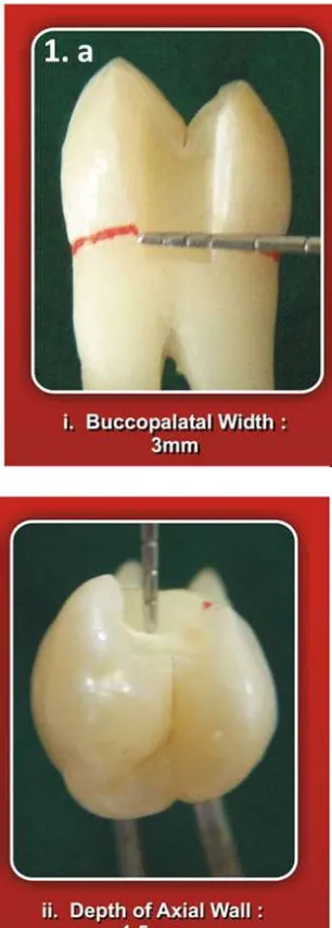
[image:2.595.342.529.51.246.2]Sixty freshly extracted human maxillary premolars free of caries, attrition, abrasion, erosion, restorations and craze lines were selected for the study. The teeth were cleaned and stored in distilled water for 1 week. Class II slot cavity preparations were made on the proximal (mesial / distal) surface of each sample using a FG-169L taper fissure carbide bur (S.S. White, Germany). All line angles were rounded. The gingival margin of the preparation was extended 1 mm below the cemento-enamel junction on the root surface. Cavity dimensions were 3 mm wide buccolingually, 4.5-5.5 mm in height and the axial wall 1.5 mm deep, all measured with a periodontal probe. (Fig 1.a) The enamel cavosurface margin was beveled (450, 0.5 mm) with a TF 11 diamond point (Mani). The teeth were randomly divided into three groups of 20 samples each, based on the restorative technique used. (Table 1)
[image:2.595.345.520.288.459.2]Figure 1.a. Cavity location and dimensions
[image:2.595.345.520.290.709.2]Figure 1.b: polyethylene fibers
Figure 1.c. Glass fibers
Figure 1.d. Restored tooth coated with nail varnish
[image:2.595.86.239.344.771.2]USA) were dispensed into separate wells.Liquid A was applied to the entire bonding area to obtain a continuous pink layer. A second brush tip was used to scrub Liquid B into the entire wetted surface of bonding area. The pink colour disappeared quickly indicating that the etching components had been activated. The bonding area was thoroughly air dried for 10 seconds and a second coat of Liquid B was applied to the entire bonding area. The adhesive layer was light cured for 10 seconds with OptiLite LD Max (Gnatus) LED curing unit. For samples of Group II, polyethylene fiber (Ribbond Triaxial, Ribbond Inc., Seattle, Washington, USA) was cut into pieces, each measuring 3 mm in length and 1 mm in width and wetted with few drops of bonding adhesive (Prime & Bond - Dentsply De Trey Gmbh, UK).(fig. 1.b)For samples of Group III, 3 mm long pieces of resin pre-impregnated glass fiber EverStick Ortho (StickTech Ltd, Turku, Finland), 0.75 mm in diameter each along with its silicone bedding were cut using sharp scissors. (fig 1.c). Less than 1 mm thick amount of resin composite Filtek P-60 Shade B2 (3M ESPE, St Paul, MN, USA) was first placed on the gingival floor.Then, 3 mm piece of respective fiber insert was placed onto the uncured composite increment and condensed through it to adapt it against the gingival floor and displace the composite to fill into the corners of the box.
It was light polymerized for 40 seconds from the occlusal surface using OptiLite LD Max (Gnatus) LED curing unit in soft start mode. Remaining cavity was restored using diagonal incremental technique. Restorations were then finished and polished with Shofu Super-Snap (ShofuInc, Kyoto, Japan) aluminum oxide discs of decreasing abrasiveness. The teeth were stored in distilled water at room temperature for two weeks. The restored teeth were thermocycled for 500 cycles at temperatures of 50C 20C and 550C 20C with a dwell time of 10 seconds in each water bath and a transfer time of 10 seconds between each bath.The samples were blotted dry and the root apices were sealed with sticky wax.The teeth were coated with two layers of nail varnish (Lakme) except for an area approximately 1 mm around the gingival margin of the restorations. (Fig 1.d)
Figure 2. Dye penetration scores
The teeth were then immersed in 2% methylene blue dye for 24 hours at room temperature, removed and thoroughly rinsed.They were then sectioned with a thin diamond disc (DFS, Germany) through the center of the restoration such that two sections were obtained from each tooth. The degree of dye penetration in each tooth was assessed under 20X magnification with a stereomicroscope (Olympus SZ40). Based on the ordinal ranking system (Ferrari et al., 1994), the
degree of dye leakage was determined as shown in Fig 2. Dye penetration at the restoration-tooth interface was scored for cervical margins only and data was tabulated. To determine statistically significant differences in leakage at cervical margin among three tested groups, non-parametric data were analyzed using Kruskal-Wallis one-way Analysis of Variance test and an intergroup comparison was performed by Mann Whitney-U test.
RESULTS
The mean microleakage scores indicate lowest mean leakage score of 0.40 0.6806for Group III followed by Group II (1.00
[image:3.595.42.281.525.685.2] 1.0260) and Group I (2.95 0.9445). (Table 2)Kruskal-Wallis one-way ANOVA indicated significant differences between groups (p < 0.0001). Intergroup comparison with Mann-Whitney U test showed that Group II and Group III showed significantly less dye leakage than Group I (p ≤ 0.001). Group III (mean rank – 19.42) showed slightly better results than Group II (mean rank – 23.55) with no statistical significance (Table 3)
Table 1. Description of groups
Group I Adper SE Plus adhesive + Filtek P 60 composite resin - Incremental technique with No fiber inserts
Group II Adper SE Plus adhesive + RibbondTriaxial Polyethylene fibers + Filtek P 60 composite resin - Incremental technique
Group III Adper SE Plus adhesive + EverStick Ortho Glass fibers
+ Filtek P 60 composite resin - Incremental technique
DISCUSSION
This study showed that significantly less microleakage was noted at the resin- dentin interface with the use of fiber inserts (group II and III) as compared to those with no inserts (Group I). The Leno Weave Ultra High Modulus (LWUHM) polyethylene fiber used in this study has a high modulus of elasticity and lower flexural modulus. Eskitasciogluet al13
reported the elastic modulus of 23.6 Gpa of a polyethylene fiber in combination with adhesive resin has a modifying effect on the interfacial stresses developed along the etched enamel-resin boundary. Glass fibers used here have also demonstrated their ability to withstand tensile stress and to stop crack propagation in composite material. The internal stress patterns of the restorative material can change by the application of a
glass fiber layer (Valittu, 1999). The Self-etch adhesive system
to composite resin matrix, hence they decrease the overall coefficient of thermal expansion of these restorations (Rossomando, 1995). Teeth were assessed for microleakage by dye penetration method using 2% methylene blue. This method is simple, economic, quick and is hence most employed (Alani, 1997). The results of this in vitro study indicate that none of the groups tested completely eliminated microleakage at cementum (dentin) margins. Both polyethylene fibers and glass fibers significantly decreased the gingival microleakage of class II composite resin restorations as compared to control group (p<0.05). If the total amount of composite material used to restore a Class II cavity could be reduced, the overall amount of polymerization shrinkage would be proportionately decreased owing to the presence of less organic matrix. Xu et al. (2003) stated that, when fiber inserts are placed in Class II composite restorations, they replace the part of the composite increment at this location, which results in a decrease in the overall volumetric polymerization contraction of the composite. Also, the fibers assist the initial increment of the composite in resisting pull-away from the margins toward the light source. Basavanna et al. (2012) reported that the reinforcing effect of glass fibers was more effective than polyethylene fibers due to difficulty in obtaining good adhesion between the polyethylene fibers and resin matrix. Hamza et al. (2004) used silane coupling agent and plasma treatment to increase the degree of adhesion of the polyethylene fibers to the resin and found no significant difference between the reinforcing effects of glass and polyethylene fibers. Both studies showed no statistically significant difference between the glass and polyethylene fibers (p>0.05). In the current study glass fibers used were pre-impregnated with polymethyl methacrylate and Bisphenol-A glycidyldimethacrylate (Bis-GMA), while polyethylene fibers were wetted with adhesive resin before insertion.
Less microleakage scores showed by glass fiber inserts as compared to polyethylene fiber inserts can be explained by the fact that glass fibers transmit light to the gingival increment of composite resin during curing thus increasing its hardness, physical properties and durability. Also, glass fibers were pre-impregnated with Bis-GMA (by manufacturer) which may produce better bond strength with composite resin as compared to polyethylene fibers which were wetted with adhesive resin (chairside). Fiber inserts modify other properties of composites too. Soderholm, (1984) showed that an inverse linear relationship exists between the volume fraction of filler in composite resins and its coefficient of thermal expansion. Thus, more the filler loading in composite resins, such as by inserting “Megafillers” like glass fibers or polyethylene fibers, more is the decrease in coefficient of thermal expansion of reinforced composite restorations.
Conclusion
Polyethylene fiber inserts and glass fiber inserts significantly reduced the gingival microleakage of Class II composite resin restorations but could not completely eliminate microleakage. Also, glass fiber inserts showed superior results as compared to polyethylene fiber inserts.
REFERENCES
Alani AH, Toh GC. 1997. Detection of microleakage around
dental restorations: a review. Oper Dent., 22: 173-185.
Attar N , Turgut MD, Güngör HC. 2004. The effect of flowable resin composites as gingival increments on themicroleakage of posterior resin composites. Operative Dentistry., 29(2): 162-167.
Baratieri LN, Ritter AV. 2001. Four-year clinical evaluation of posterior resin-based composite restorations placed using the total-etch technique. J EsthetRestor Dent.,13(1): 50-7.
Basavanna, RS., Garg, A., Kapur. R 2012. Evaluation of gingival microleakage of class II resin composite restorations with fiber inserts: An in vitro study. J Conserv Dent. Apr-Jun; 15(2): 166–169.
Brandt PD, de Wet FA, du Preez IC. 2006. Self-etching bonding systems: in vitro microleakage evaluation. SADJ., 61(6): 248-251.
Donly, K.J., Wild, T.W., Bowen, R.L. and Jensen, M.E. 1989. An in vitro Investigation of the Effects of Glass Inserts on the Effective Composite Resin Polymerization Shrinkage; J Dent Res August, 68(8):1234-1237.
Eskitascioglu G, Belli S, Kalkan M. 2002. Evaluation of two post core systems usingtwo different methods (fracture
strength test and a finite elemental stressanalysis). J
Endod., 28:629-633.
Feilzer A, de Gee A, Davidson CL. 1987. Setting stress in composite resin in relation to configuration of therestoratives.Journal of Dental Research., 66: 1636-1639.
Ferrari M, Yamomoto K, Vichi A & Finger WJ. 1994. Clinical and laboratory evaluation of adhesive restorative systems.
American Journal of Dentistry.,7: 217-219.
Gupta S., Khinda V. I., Grewal N. 2002. A Comparative study of Microleakage below Cementoenamel junction using Light Cure and Chemically Cured glass lonomer cement liners. J Indian SocPedoPrev Dent December., 20 (4) : 158-184.
Hamza TA, Rosentritt SF, Elhosary MM and Ibraheem RM. 2004. The effect of fiber reinforcement on the fracture toughness and flexural strength of provisional restorative
[image:4.595.40.547.73.125.2]resins. J Prosthetic Dent., 91(3); 258-264.
Table 2. Microleakage scores and statistical analysis on application of kruskal- wallis test
Score group
0 1 2 3 4 5 Mean score Standard deviation Mean rank Chi square value P value Result
Group I 01 02 04 10 02 01 2.95 0.9445 48.53
35.100 0.000
Significant as p<o.o5 Group II 10 05 04 01 00 00 1.00 1.0260 23.55
Group III 13 04 03 00 00 00 0.40 0.6806 19.42
Table 3. Results of Intergroup comparison using Mann- Whitney U test
[image:4.595.143.444.156.199.2]Kanca J III. 1999. Microleakage of five dentin bonding systems. Dental Materials; Vol. 5 (6), 415-416.
Oberholzer TG, Du Preez IC, Kidd M. 2005. Effect of LED curing on the microleakage, shear bondstrength and surface
hardness of a resin-based composite restoration.
Biomaterials; 26:3981-3986.
Pearson JD, Bouschlicher MR, Boyer DB. 1999. Polymerization shrinkage forces of condensable composites. J Dent Res., 78(special issue):448.
Perdigao J, Lambrechts P, Van Meerbeek B, Braem M et al. The interaction of adhesive systems with human dentin. Am J Dent 1996;9: 167-73.
Prati C, Chersoni S, Mongiorgi R, Pashley DH. 1998. Resin-infiltrated dentin layer. Formation of new bonding systems.
Op Dent., 23: 185-194.
Puckett A, Fitchie J, Hembree J, Smith J. 1992. Effect of incremental versus bulk techniques on the microleakage ofcomposite resin using a glass – ionomer liner. Operative Dentistry, 17:186-191.
Rossomando KJ, Wendt SL Jr. 1995. Thermocycling and dwell times in microleakage evaluation for bonded restorations.
Dent Mater., 11: 47-51.
Soderholm. K. J. M. 1984. Influence of Silane Treatment and Filler Fraction onThermal Expansion of Composite Resins;
J Dent Res November, 63(11):1321-1326.
Valittu PK. 1999. Flexural properties of acrylic polymers
reinforced with unidirectionaland woven glass fibers. J
Prosthet Dent., 81: 318-326.
Xu HH, Schumacher GE, Eichmiller FC, Peterson RC, Antonucci JM, Mueller HJ. 2003. Continuous fiber preform reinforcement of dental resin composite restorations.
Dental materials., 19(6); 523-530.