RESEARCH ARTICLE
CHARACTERIZATION OF PLANTAR PRESSURE AND FOOTPRINT IN ELITE AND SUBELITE
BADMINTON PLAYERS
*,1
Tong-Hsien Chow and
2Yih-Shyuan Chen
1
Department of Leisure Sport and Health Management, St. John's University, New Taipei 251, Taiwan
2Department of Digital Literature and Arts, St. John's University, New Taipei 251, Taiwan
ARTICLE INFO ABSTRACT
Context: The arch is an anatomical structure of the foot which influences movements of the lower limbs and causes changes in plantar pressure distributions. Plantar pressure measurement is the effective method for assessing plantar loading and can be applied to evaluating movement performance of the foot.
Objective: The purpose of this study is to explore the badminton players’ plantar loading
characteristics and pain profiles in static standing.
Methods: Experiments were undertaken on twenty elite badminton players (EB), twenty-four subelite badminton players (SB) and twenty-eight non-athletes (controls). ‘JC Mat’, the optical plantar pressure measurement, was applied to examining all subjects’ arch index (AI), plantar pressure
distributions (PPD), and footprint characteristics. Pain assessment and self-reported health status were undertaken for evaluating the badminton players’ common pain locations.
Results: Findings from the control group, the AI fell into the normal range. Thebadminton players’
arch type was classified as high-arched feet. PPD at the lateral longitudinal arch and the lateral heel of both feet, and the medial heel of the left foot was significantly higher in the badminton players, particularly the EB, than in the controls. PPD at the lateral metatarsal bone of both feet was significantly lower in the badminton players than in the controls. Compared with the SB, the EB had lower PPD in the lateral metatarsal bone of both feet. Footprint characteristics supported the results of the AI and PPD, and this reflected the corresponding pressure profiles.The badminton players’lateral ankle and knee joints and gastrocnemius were the most common pain locations.
Conclusion: Thebadminton players’ AI and PPD were generallyclassified as high-arched supinators, and their pain profiles paralleled the symptoms of foot supination. The findings reflected the possible link between badminton injuries and supinated feet, and the correlation is worth further studies.
Copyright©2016, Tong-Hsien Chow and Yih-Shyuan Chen. This is an open access article distributed under the Creative Commons Attribution License, which
permits unrestricted use, distribution, and reproduction in any medium, provided the original work is properly cited.
INTRODUCTION
Badminton is a high-intensity racket sport which requires rapid jumps and lunges, quick changes in direction, and instant arm movements with a wide variety of body postures. (Phomsoupha and Laffaye, 2015) Footwork is the fundamental skill in badminton competitions and enables athletes to reach the shuttlecock as quickly as possible while maintaining good balance and body control. (Kuntze et al., 2010) According to previous studies, the frequent execution of a lunge step in a badminton competition is generally considered to be the main risk factor for lower extremity injuries. (Hong et al., 2014) This
*Corresponding author: Tong-Hsien Chow,
Department of Leisure Sport and Health Management, St. John's University, New Taipei 251, Taiwan.
is mainly because badminton players exhibit higher vertical ground reaction forces which are approximately 2.1 to 2.5 times their body weight. (Kuntze et al., 2010; Hong et al., 2014; Hu et al., 2015) This, in turn, may cause their feet to experience a great amount of stress and lead to fatigue and painful conditions. (Hu et al., 2015) Badminton players’
dominant leg usually bears a great load, and their tendons are susceptible to suffer from overstrain. (Lee and Yoo, 2012) Ankle joints, knee joints, lower legs, feet and thighs are the
common injuries which occur in badminton players’ lower
extremities. Importantly, due to badmintonplayers’ unique and
repetitive movements, their Achilles tendon, plantar fascia, anterior talofibular ligament of the specific musculotendinous and ligamentous structures were verified to be at higher risks of serious injuries than athletes who were involved in other types of sports. (Jørgensen and Winge, 1990) Foot arch, an
ISSN: 0975-833X
International Journal of Current Research
Vol. 8, Issue, 08, pp.37522-37531, August, 2016
INTERNATIONAL JOURNAL OF CURRENT RESEARCH
Article History:
Received 16thMay, 2016
Received in revised form 25thJune, 2016
Accepted 26thJuly, 2016 Published online 31stAugust, 2016
Key words:
Elite badminton players, Subelite badminton players, Arch index,
Plantar pressure distributions, Supinated feet.
Citation: Tong-Hsien Chow and Yih-Shyuan Chen, 2016.“Characterization of plantar pressure and footprint in elite and Subelite badminton players”,
International Journal of Current Research, 8, (08), 37522-37531.
RESEARCH ARTICLE
CHARACTERIZATION OF PLANTAR PRESSURE AND FOOTPRINT IN ELITE AND SUBELITE
BADMINTON PLAYERS
*,1
Tong-Hsien Chow and
2Yih-Shyuan Chen
1
Department of Leisure Sport and Health Management, St. John's University, New Taipei 251, Taiwan
2Department of Digital Literature and Arts, St. John's University, New Taipei 251, Taiwan
ARTICLE INFO ABSTRACT
Context: The arch is an anatomical structure of the foot which influences movements of the lower limbs and causes changes in plantar pressure distributions. Plantar pressure measurement is the effective method for assessing plantar loading and can be applied to evaluating movement performance of the foot.
Objective: The purpose of this study is to explore the badminton players’ plantar loading
characteristics and pain profiles in static standing.
Methods: Experiments were undertaken on twenty elite badminton players (EB), twenty-four subelite badminton players (SB) and twenty-eight non-athletes (controls). ‘JC Mat’, the optical plantar pressure measurement, was applied to examining all subjects’ arch index (AI), plantar pressure
distributions (PPD), and footprint characteristics. Pain assessment and self-reported health status were undertaken for evaluating the badminton players’ common pain locations.
Results: Findings from the control group, the AI fell into the normal range. Thebadminton players’
arch type was classified as high-arched feet. PPD at the lateral longitudinal arch and the lateral heel of both feet, and the medial heel of the left foot was significantly higher in the badminton players, particularly the EB, than in the controls. PPD at the lateral metatarsal bone of both feet was significantly lower in the badminton players than in the controls. Compared with the SB, the EB had lower PPD in the lateral metatarsal bone of both feet. Footprint characteristics supported the results of the AI and PPD, and this reflected the corresponding pressure profiles.The badminton players’lateral ankle and knee joints and gastrocnemius were the most common pain locations.
Conclusion: Thebadminton players’ AI and PPD were generallyclassified as high-arched supinators, and their pain profiles paralleled the symptoms of foot supination. The findings reflected the possible link between badminton injuries and supinated feet, and the correlation is worth further studies.
Copyright©2016, Tong-Hsien Chow and Yih-Shyuan Chen. This is an open access article distributed under the Creative Commons Attribution License, which
permits unrestricted use, distribution, and reproduction in any medium, provided the original work is properly cited.
INTRODUCTION
Badminton is a high-intensity racket sport which requires rapid jumps and lunges, quick changes in direction, and instant arm movements with a wide variety of body postures. (Phomsoupha and Laffaye, 2015) Footwork is the fundamental skill in badminton competitions and enables athletes to reach the shuttlecock as quickly as possible while maintaining good balance and body control. (Kuntze et al., 2010) According to previous studies, the frequent execution of a lunge step in a badminton competition is generally considered to be the main risk factor for lower extremity injuries. (Hong et al., 2014) This
*Corresponding author: Tong-Hsien Chow,
Department of Leisure Sport and Health Management, St. John's University, New Taipei 251, Taiwan.
is mainly because badminton players exhibit higher vertical ground reaction forces which are approximately 2.1 to 2.5 times their body weight. (Kuntze et al., 2010; Hong et al., 2014; Hu et al., 2015) This, in turn, may cause their feet to experience a great amount of stress and lead to fatigue and painful conditions. (Hu et al., 2015) Badminton players’
dominant leg usually bears a great load, and their tendons are susceptible to suffer from overstrain. (Lee and Yoo, 2012) Ankle joints, knee joints, lower legs, feet and thighs are the
common injuries which occur in badminton players’ lower
extremities. Importantly, due to badmintonplayers’ unique and
repetitive movements, their Achilles tendon, plantar fascia, anterior talofibular ligament of the specific musculotendinous and ligamentous structures were verified to be at higher risks of serious injuries than athletes who were involved in other types of sports. (Jørgensen and Winge, 1990) Foot arch, an
ISSN: 0975-833X
International Journal of Current Research
Vol. 8, Issue, 08, pp.37522-37531, August, 2016
INTERNATIONAL JOURNAL OF CURRENT RESEARCH
Article History:
Received 16thMay, 2016
Received in revised form 25thJune, 2016
Accepted 26thJuly, 2016 Published online 31stAugust, 2016
Key words:
Elite badminton players, Subelite badminton players, Arch index,
Plantar pressure distributions, Supinated feet.
Citation: Tong-Hsien Chow and Yih-Shyuan Chen, 2016.“Characterization of plantar pressure and footprint in elite and Subelite badminton players”,
anatomical structure of the foot, is constituted of ligaments, muscles and bones, and could be regarded as a shock absorber in the human body. (Simkin et al., 1989) When running and jumping, the medial longitudinal arch (MLA) provides adequate elastic forces and twisting forces for absorbing the ground reaction force, and this is helpful for attenuating the shock from movement, mitigating injuries and deferring fatigue. (Kaye and Jahss, 1991) Arch height of the MLA is generally treated as the influential and key determinant of the function of the foot and lower limbs. (Razeghi and Batt, 2002) High-arched individuals were found to be associated with more laterally located bony injuries of the foot, ankle, knee, and lower extremity stress fractures. (Williams et al., 2001) Yet, low-arched individuals usually showed a greater incidence of medially soft tissue injuries, such as patellar tendinitis, plantar fasciitis and knee pain. (Dahle et al., 1991; McCrory et al., 1997) Based on previous studies, the arch index (AI) from footprints could be considered to be the reliable and valid method for characterizing the foot and MLA height. (McCrory
et al., 1997; Mickle et al., 2006; Cavanagh et al., 1987;
Wearing et al., 2004; Williams and McClay, 2000; Chu et al., 1995) The measurements of static arch height and arch height ratio of the foot may assist clinicians in estimating foot posture during dynamic activity in patients with lower-limb injuries. (Franettovich et al., 2007) Clinicians also commonly use static posture of the MLA to infer dynamic foot function in order to assess the potential for several foot-specific pathologies (Imhauser et al., 2004) and for lower extremity dysfunction which may increase injury risk. (Jonely et al., 2011) A recent prospective study suggests that the static characteristics of the flatfoot, high-arched foot and rearfoot range of motion are the risk factors for developing lower extremity overuse injuries in general. (Kaufman et al., 1999) Furthermore, the plantar pressure assessment of the footprints is one of the effective methods for evaluating the plantar loading characteristics during functional activities. The parameters gained from the plantar pressure assessment can be useful not only for understanding the variations in the plantar loading in different regions of the foot, but also for detecting the foot pathologies. (Hessert et al., 2005; Orlin and McPoil, 2000) High plantar pressure is thought to be the potential factor for sports-related injuries in the lower extremity. (Dowling et al., 2010) When plantar pressure in each region of the foot is distributed evenly, sports injuries could be reduced effectively. (Sneyers et al., 1995)
In a sense, badminton players’ static plantar pressure
characteristics are possibly significant for predicting their potential pain profiles. Nevertheless, few studies have been undertaken for exploring plantar pressure characteristics in badminton movements. (Hong et al., 2014; Hu et al., 2015; Fu, 2011) With a few exceptions, though, these studies tended to focus on dynamic footwork rather than static standing. Given the above context, the purpose of this study, therefore, was to investigate the differences among elite badminton players (EB), subelite badminton players (SB) and healthy non-athletes in terms of their static plantar pressure characteristics. The related parameters which were examined within this study are listed below: the arch index, three regional and six distinct sub-regional plantar pressure distributions (PPD), and footprint characteristics of both feet. Apart from this, data gained from
the pain assessments and badminton players’ self-reported health status within this study were used for evaluating the pain locations which occurred frequently in the body. It was our assumption that badminton players were classified into high arch type, and that their PPD were particularly concentrated in the rearfoot and the lateral foot regions. The plantar pressure characteristics and pain profiles may correlate with the features of the supinated foot.
MATERIALS AND METHODS
1. Subject Selection
The subjects participating in this study comprised three specific groups of college and university students in Taiwan. One of the groups, labelled asthe ‘elite badminton group’, was
composed of 20 right-handed first-division male badminton players. For the elite badminton group, the length of being the qualified first-division players is to have more than successive five-year experiences in badminton competitions. All subjects within the elite badminton group in this study were recruited from the sport university, school of kinesiology and three city sports centers in Taipei, Taiwan. Another group, the‘subelite
badminton group’, was constituted of 24 right-handed male badminton players who were the same age range (between 17 and 21 years old) as the elite badminton group. All subjects in the subelite badminton group were single-sport university athletes and participated only in team-based badminton training. They were playing badminton at least once a week at sports centers in Taipei. The other group, the control group within this study, included 28 healthy age-matched male university students without specialties in sports nor regular time for exercise (the average time for exercise weekly was less than 2 days or 6 hours).Each subject’s age, gender, height, body weight and body mass index (BMI) were recorded in the research process. Considering the effect of the body weight on
shape characteristics of the foot arch, each subject’s BMI
within this research was required to range between 18.5 and 24 and this particular range was defined by the World Health Organization (WHO) as healthy weight. A total of 72 subjects participated in this study, and their average age, height, weight
and BMI value were shown in Table 1. All subjects’ pain and
health assessments were based on the self-reported health status and measurements which were diagnosed by the professional physiotherapist at the rehabilitation department. The pain and health assessments were essential for this research to ensure that all subjects had no history of previous fracture and surgery, and that they were free from injuries in their ankle joints, knee joints, hip joints, spine, and bones and muscles of their lower limbs within a year as this study was underway. Prior to the experiments, all participants were informed of the purpose of the present study. They read and signed an informed consent form approved by the institutional review board. The entire process of the experiments within this study followed the guidelines of the local Ethical Committee and the recommendations of the Declaration of Helsinki.
2. Instruments and Equipment
as the main research tool for the present study. The measurement technology and principles of JC Mat were similar to the operation principles of Harris footprint measurement instrument. The key attributes of JC Mat were as follows: (1) the subtle characteristics of the foot were easily distinguished; (2) the plantar pressure distribution and footprints coincided with the weight calibration data (data not shown); (3) there were 25 sensors in each square centimeter for the plantar pressure measurement, and thus, 13600 sensors were on each side (32*17 cm) of JC Mat; (4) the pressure sensing was sensitive and the scope of the sensor was large. A smooth and delicate plantar pressure image was shown in the form of round dots; (5) the static pressure profiles from footprints and barefoot images were captured instantly; and (6) the built-in FPDS-Pro software was competent for analyzing the following parameters: the arch index, plantar pressure values, balance of the center of gravity, toe angles and footprints.
3. Methods and Procedures
It took approximately seven months to select the subjects and conduct the experiments for this study. Before the experiments were undertaken, all subjects were informed of the purpose and processes of this study and their consent to participate in this research was obtained. For the sake of consistency and trustworthiness of the experiments, time for each experiment was set between 2pm and 4pm. All subjects were required to measure their body weights and heights when the experiments were conducted. This was helpful for recording the basic and
accurate data of subjects’ physiological conditions in terms of weights and heights. The subjects’ weights and heights which were recorded during the experiments, associated with the given formula (body weight (kg)/height (m2)), served as the base for calculating the BMI values for this study. Apart from this, the subjects in the experimental processes were asked to follow the instructions listed below:
(1) Roll both trouser legs up to above the knees if necessary, in order to prevent the clothing from limiting movements of the extremities.
(2) Stand with bare feet on the sensing cushion with marks of the specific measuring range of JC Mat.
(3) Relax the body; then, control and balance the center of gravity by standing with feet shoulder-width apart and with body weight evenly distributed on both feet. (4) Stampede for 6-8 steps, and then, stand still with a
natural and comfortable posture and arms hanging straight down at sides.
(5) Face the guide of the experiment, and look the guide straight in the eye. Keep the body stationary and balanced until there were no obvious changes in the pressure values of both feet measured by JC Mat.
When the condition above was met, the subjects’ static
pressure profiles were acquired immediately.
4. Pain Assessment and Self-Reported Health Status of the Subjects
The process of the subjects’ pain assessment and self-reported health status was conducted though the assistance of a
professional physiotherapist at the rehabilitation department. This process functioned as the basis for the subject selection
criteria, the subjects’ physiological symptom assessment and
confirmation of the pain location. After the plantar pressure measurement, all subjects were asked to undergo the skeleton
arrangement and soft tissue pain assessment. The term ‘lower limb pain’ used in this study was defined as the
musculoskeletal pain which occurred during the past month and originated from the structures of the foot, ankle, knee, lower leg and thigh. This definition excluded intermittent cramps, dermatological conditions, digital calluses and night-time paresthesia from analysis. A standardised protocol for the questioning and examination techniques was used within this research for determining the precise nature of the complaint (e.g., metatarsalgia and plantar fasciitis). The procedures for evaluating the pain location which occurred frequently in the subjects were presented as follows:
(1) The professional physiotherapist evaluated and
documented the subjects’ self-reported health status and pain location which occurred frequently in the body. (2) The subjects were asked to stand with bare feet and roll
both trouser legs up to above the knees if necessary. (3)Inspection of subjects’ lower extremities by pressing
their foot (including phalanges, metatarsal bones, navicular bone, cuboid bone and calcaneus), ankle joints, knee joints, hip joints, tibias, fibulas and femur, and then, assessing the bones arrangement of their lower extremities.
The procedures for assessing the soft tissue pains were listed as follows:
(1)The professional physiotherapist pressed the subjects’
self-reported pain location and re-checked the corresponding location on the opposite side of the pain location.
(2) Based on their clinical experiences, the professional physiotherapist pressed and examined the specific
points in the subjects’ common pain locations,
including plantar metatarsal heads, plantar fascia, the inferior margin of navicular bones, the Achilles tendon, the medial and lateral sides of ankle joints, the medial and lateral fossas of knee joints, gastrocnemius, tibialis anterior and posterior, biceps and quadriceps femoris. This allowed the physiotherapist to definitely confirm
the subjects’ common pain locations.
5. Data Analysis
In order to examine the subjects’ plantar pressure distributions
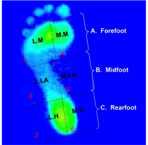
excluding the toes. Secondly, the software generated a set of four parallel lines which were perpendicular to the nearly vertical line and divide the footprint into three equal parts. In
this study, ‘regions A, B and C’ of the footprint were defined, respectively, as the ‘forefoot, midfoot and rearfoot’. ‘Sub
-regions 1, 2, 3, 4, 5 and 6’ were defined, respectively, as the
‘lateral metatarsal bone (L.M.), lateral longitudinal arch
(L.LA.), lateral heel (L.H.), medial metatarsal bone (M.M.), medial longitudinal arch (M.LA.) and medial heel (M.H.)’.
The arch index ratio method developed by Cavanagh and Rodgers assumed that the arch index (AI) was calculated as the ratio of the area of the middle third of the footprint divided by the entire footprint area excluding the toes, i.e. AI=B/ (A+B+C). Based on Cavanagh and Rodgers’ assertion, a
normal arched foot was defined by the ratio between 0.21 and 0.26, a high-arched foot was defined by the ratio lower than 0.21, and a flat-arched foot was defined by the ratio higher than 0.26.
6. Statistical Analysis
Descriptive statistics used for this study was to summarize all
subjects’ ages, heights, weights, BMI values and experience.
Numerical data gained in the research process was presented as mean ± SD. One-way ANOVA was used for distinguishing the differences among three groups in terms of their arch index, three regional and six sub-regional plantar pressure distributions. Post-ANOVA, paired t-testing with Scheffe correction were used for dealing with the between-group comparisons. All statistics were calculated with the Statistical Package for the Social Sciences for Windows (Version 17.0; SPSS Inc., Chicago, IL). Statistical significance was defined as
P < 0.05 (marked as *) between the EB and control groups, P < 0.05 (marked as †) between the SB and control groups, and
P < 0.05 (marked as #) between the EB and SB groups.
RESULTS
1. Arch Index
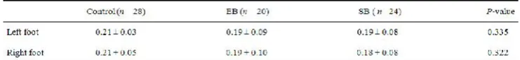
As Table 2 illustrates, no significant difference was found among the EB, SB and control groups in their arch indices of both feet. The results suggested that the arch index was similar among three groups.
2. Plantar Pressure Distributions of the Forefoot, Midfoot and Rearfoot Regions
The plantar pressure distributions were presented in the form of percentages of the relative load. The relative loads in the forefoot of both feet were lower in the EB and SB groups than in the control group (p < .01) (Table 3). The relative loads in the rearfoot of the left foot was higher in the EB group as compared with the control group (p < .05). Furthermore, no significant difference was found among the EB, SB and control groups in the midfoot region. Based on the findings from the EB group, the relative load was low in the forefoot of both feet and was particularly concentrated in the rearfoot of the left foot.
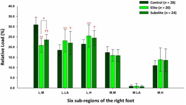
3. Plantar Pressure Distributions at the Six Sub-Regions
The calculation of the relative loads at the six distinct sub-regions was based on the data gained from the forefoot, midfoot and rearfoot. In the forefoot, the relative loads at the lateral metatarsal bone of both feet were lower in the EB (left: 18.47 ± 3.01%, right: 20.83 ± 3.47%) and SB (left: 22.01 ± 3.46%, right: 23.53 ± 2.62%) groups than in the control group (left: 29.22 ± 3.93%, right: 30.88 ± 3.67%) (p < .01). In the midfoot, the relative loads at the lateral longitudinal arch of both feet were higher in the EB (left: 24.10 ± 4.64%, right: 23.08 ± 5.44%) and SB (left: 23.68 ± 5.75%, right: 22.0 ± 5.63%) groups, the EB group in particular, than in the control group (left: 19.29 ± 3.56%, right: 18.46 ± 2.68%) (p < .01).
Passiflora edulis Xanthomonas axonopodis pv. passiflrae Oryza sativa Magnaporthe oryzae
Theobroma cacao Moniliophthora perniciosa Phaseolus vulgaris Colletotrichum lindemuthianum Brassica rapa pekinensis Xanthomonas campestris pv. campestris Zea mays Aspergillus flavus and A. parasiticus Solanum lycopersicum Botrytis cinerea
Solanum lycopersicum Botrytis cinerea Glycine max Phakospora pachyrhizi
Glycine max Macrophomina phaseolina and Phytophthora sojae Lolium perenne Magnaporthe oryzae
[image:4.595.146.457.614.708.2]Triticum spp Gaeumannomyces graminis var. tritici.
Table 1. Demographic characteristics and training experience in elite badminton (EB), subelite badminton (SB) athletes, and controls
[image:4.595.115.486.760.803.2]Data are represented as mean ± SD. The control group: male university students with neither specialties in sports nor regular time for exercise (the average time for exercise weekly was less than 2 days or 6 hours). The elite badminton group: right-handed first-division male badminton athletes. The subelite badminton group: single-sport university athletes and participated only in team-based badminton training (playing at least once a week).
Table 2. Arch index of the foot in elite badminton (EB), subelite badminton (SB) players, and controls
Table 3. Relative load of the forefoot, midfoot and rearfoot regions in elite badminton (EB), subelite badminton (SB) players, and controls
The percentage of relative load are represented as mean ± SD for each foot region during the static standing. P value for one-way ANOVA across groups. *P < 0.05, **P< 0.01, significant differences between the EB and control group. †P< 0.05, ††P < 0.01, significant differences between the SB and control
[image:5.595.82.519.262.458.2]group.
Table 4. Pain assessment and the self-reported health status in the badminton players
Pain assessment and self-reported health status of the elite badminton players (EB) and subelite badminton players (SB) was conducted though the assistance of a professional physiotherapist at the rehabilitation department.
[image:5.595.175.423.492.734.2]Figure 2. Plantar pressure distributions of six sub-regions of the left foot in static standing Data are expressed as mean ± SD. *P < 0.05, **P< 0.01, significant differences between the EB and control group. †P< 0.05, ††P < 0.01, significant differences between the SB and control group.#P < 0.05,# #P < 0.01, significant differences between EB and SB group
[image:6.595.143.462.288.468.2]In the rearfoot, the relative loads at the lateral heel of both feet were higher in the EB (left: 25.31 ± 4.25%, right: 25.43 ± 5.41%) and SB (left: 24.27 ± 4.03%, right: 24.44 ± 3.65%) groups, the EB group in particular, when compared with the control group (left: 21.47 ± 2.89%, right: 21.46 ± 2.21%) (p < .01). Again, the relative loads at the medial heel of the left foot were higher in the EB (14.19 ± 3.89%) and SB (13.46 ± 2.84%) groups, particularly the EB group, compared with the control group (10.95 ± 2.85%) (p < .01). The relative loads at the lateral metatarsal bone of both feet were lower in the EB group, the left foot in particular, than in the SB group (p < .01). The findings showed that the plantar pressure distributions at the lateral longitudinal arch and the lateral heel of both feet, and at the medial heel of the left foot were higher in the EB and SB groups, the EB group in particular; they, however,
were lower at the lateral metatarsal bones of both feet in two groups (Figure 2 and 3).
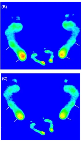
4. Footprint Characteristics
In Figure 4, it can be seen that footprints in the EB and SB groups displayed the lower pressure profiles in the forefoot of both feet. The higher pressure profiles were found to be concentrated in the midfoot and rearfoot of both feet, particularly the left foot.
5. Pain Assessment and Self-Reported Health Status of the Subjects
[image:7.595.161.443.51.534.2]As can be seen in Table 4 which illustrates the findings from the pain assessment and the self-reported health status of the
EB and SB groups, the six most common pain locations in bones are presented as follows: the lateral ankle joint (62.5-65%), the lateral knee joint (50-55%), the shoulder joint (37.5-55%), the elbows and wrists (25-45%), the calcaneus (33.3-40%) and the plantar metatarsal bone (25-29.2%). The six most common pain locations in soft tissues are listed below: the gastrocnemius (70.8-80%), the Achilles tendon (54.2-70%), the anterior cruciate ligament (45.8-60%), the quadriceps femoris (37.5-40%), the lower back (40-50%) and the plantar fascia (8.33-15%).
DISCUSSION
The purpose of this study was to examine the differences among the EB, SB and control groups by focusing particularly on the arch index, three regional and six distinct sub-regional plantar pressure distributions, footprint characteristics and pain profiles in the static standing posture. The results revealed that the static arch index of both feet was considerably close in the EB, SB and controls, respectively. No significant difference was found between the control group and the badminton groups (i.e. the EB and SB groups). It has been widely accepted that the arch index of the footprints could be considered to be the predictor of the arch height (McCrory et al., 1997; Mickle
et al., 2006; Cavanagh and Rodgers, 1987; Wearing et al.,
2004; Williams and McClay, 2000; Chu et al., 1995), and that the normal values of the arch index ranged between 0.21 and 0.26. (Cavanagh and Rodgers, 1987) On this basis, the foot arch of the control group within this study could be classified into the normal range, whereas the EB and SB groups generally fell into the high arch type. It is well known that a high-arched individual with an increased height of the MLA often experiences the supinated foot and decreased pronation during the stance phase. (Nigg et al., 1993) An over-supinated foot was defined as increased calcaneal inversion and may provide an advantage in reducing contact time when running. (Hasegawa et al., 2007) Hasegawa et al. indicated that runners with the greatest degree heel inversion at foot strike had the shortest contact time. (Hasegawa et al., 2007) A shorter contact time and a higher frequency of inversion at the foot contact may contribute to higher running economy. (Hasegawa
et al., 2007) Therefore, deformation of the foot arch appeared
to be crucial for transferring force and absorbing shock in high-impact sports, such as jump and sprint. (Chang et al., 2010) With regard to the results of plantar pressure distributions in the forefoot, midfoot and rearfoot, the relative loads in the forefoot of both feet were significantly smaller in the EB and SB groups than in the control group. The average of regional plantar pressure were mainly concentrated in the rearfoot of both feet of the EB and SB groups, particularly the left foot of the EB group. Furthermore, findings from the six distinct sub-regional plantar pressure distributions showed that the plantar pressure were mainly exerted on the lateral longitudinal arch and the lateral heel of both feet, and the medial heel of the left foot of the EB and SB groups, the EB group in particular. These findings seem to be consistent with the previous studies which verified that the plantar pressure is mainly concentrated in the heel and the lateral foot of the badminton players during forward lunges. (Fu, 2011) The studies by Hu et al. went further, arguing that the front-forward lunge showed lower plantar loads on the great toe region of the dominant leg of the
male right-handed badminton players compared with the left and right maximum forward lunges. (Hu et al, 2015) In Hong
et al.’s studies which investigated the in-shoe peak plantar pressure during left- and right-forward lunges, the left-forward lunge was found to have greater plantar pressure, higher loading rate and more vertical ground reaction forces at the total foot and heel regions compared with the right-forward and left/right-backward lunges. (Hong et al., 2014) Given the above context, these findings may support the results from this study that the relative load in the heel and the lateral foot, particularly in the left foot, was higher in right-handed badminton players. Moreover, findings showed that PPD at the lateral metatarsal bone of both feet was significantly lower in the badminton players (i.e. the EB and SB) than in the controls. These findings seem to be inconsistent with the studies by Fu et al. who argued that the plantar loadings were higher in the great toe, the first and second metatarsals of the forefoot than in other areas in technical revolve to jump. (Fu, 2011) They also asserted that the metatarsal heads, lateral heel and lateral foot could be the most contacting regions with the surface in different footwork. (Fu, 2011) The in-shoe pressure results from Hong et al.’s
research indicated that the great toe, first metatarsal head, medial heel, and lateral heel masks had relatively high peak pressure magnitude during lunge maneuvers. (Hong et al., 2014) Differences in the findings could be attributed to the differences in the static and dynamic states. Findings from this research, however, this could be initially confirmed that plantar pressure of the forefoot, rearfoot and lateral foot was crucial for badminton movements, to a certain degree.
Based on the findings from the badminton players, the common bone pains listed in order of frequency are as follows: the lateral ankle joint, the lateral knee joint, the shoulder joint, the elbows and wrists, the calcaneus and plantar metatarsal bone. The flowing listed the common pain locations in soft tissues in order of frequency: the gastrocnemius, the Achilles tendon, anterior cruciate ligament, the quadriceps femoris, the lower back and plantar fascia. The results seemed to support previous studies which verified that when landing after a jump while playing badminton, the body is upright while the ankle is maintained in a plantar-flexed position due to the activity of the gastrocnemius and soleus muscles. (Schepsis et al., 2002) Sports injuries, therefore, were caused by the ankle being forced upwards while in a plantar-flexed position when landing after a jump during badminton match play, leading to a compression injury of the Achilles tendon. (Lee and Yoo, 2012) The Achilles tendon is subjected to loads as high as 6-12 times the body weight while running and jumping. Such high repetitive loading is considered to be one of the main pathological stimuli to the Achilles tendon. (Paavola et al., 2002) Peers and Lysens suggested that frequent knee problems were probably related to the rapidly changing eccentric/concentric work of the quadriceps in the varying degrees of knee flexion and rotation, creating a high force load on the patellar tendon. (Peers and Lysens, 2005) Lee’s study
maintained that the mean peak vertical ground reaction force is approximately 2.44 times the body weight for left-forward lunges, and 2.2 times for right-forward lunges in badminton players. (Ryue et al., 2013) The differences in the plantar loads of the different lunge directions may be potential risks for
2015) Repetitive lunge steps may result in ruptures of the Achilles tendon. In addition, excessive shear force would be associated with the risk of anterior cruciate ligament injury. (Dowling et al., 2010) For all badminton players (i.e. the EB and SB groups) within this study, the regional plantar pressure were mainly concentrated in the lateral longitudinal arch and the lateral heel of both feet, and the medial heel of the left foot. Despite no significant difference between the badminton players and the control group in terms of the AI, the results of the AI showed that the badminton players could be classified as the high-arch type. Based on the results of the foot characteristics, the badminton players were generally classified as having supinated feet. Evidence indicated that high arches were prone to have high loads on the lateral foot, and this may resulted in problems with the lateral knees and ankles. (Simkin
et al., 1989; Williams et al., 2001) Molgaard et al. reported that
high arches had a high probability of ankle injuries, heel pains and stress fractures. (Mølgaard et al., 2010) This is because high arches move with a stiffer lower extremity and higher loading rates during running. (Williams et al., 2014) High-arched individuals tend to suffer from over supination, and this results not only in a decrease in pronation throughout the stance phase, but also in an increase in supination in the forefoot and rearfoot during exercise. (Nigg et al., 1993) Therefore, the plantar pressure and the integration of pressure over time are usually higher in the metatarsal and calcaneal regions. This could lead to a high risk of injuries on the lateral sides of their knees and ankle joints. (Williams et al., 2001)
Conclusion
It could be summarized from the findings by saying that
badminton players’ arch index and plantar pressure
characteristics were generally classified as high-arched supinators. The results from the pain assessment and the self-reported health status confirmed that the lateral ankle/knee joint and the gastrocnemius were the most common musculoskeletal pains in badminton players. These findings were consistent with the symptoms of foot supination. Although some studies have stressed the link between badminton injuries and footwork, little research has been conducted for exploring the relationships between badminton injuries and foot supination. Therefore, the correlation between badminton injuries and the development of the foot supination is worth further studies.
Acknowledgements
The authors have declared that no conflict of interest exist. This study was supported by the Ministry of Science and Technology, Taipei, Taiwan under Grant MOST 104-2622-H-129-001-CC3.
REFERENCES
Cavanagh PR, Rodgers MM. The arch index: a useful measure from footprints. J Biomech. 1987; 20(5):547-551. [PubMed: 3611129]
Chang YW, Hung W, Wu HW, Chiu YC, Hsu HC. Measurements of Foot Arch in Standing, Level Walking, Vertical Jump and Sprint Start. Int J Sports Med. 2010; 2(2):35-42.
Chu WC, Lee SH, Chu W, Wang TJ, Lee MC. The use of arch index to characterize arch height: a digital image processing approach. IEEE Trans Biomed Eng. 1995; 42(11):1088-1093. [PubMed: 7498912]
Dahle LK, Mueller MJ, Delitto A, Diamond JE. Visual assessment of foot type and relationship of foot type to lower extremity injury. J Orthop Sports Phys Ther. 1991; 14(2):70-74. [PubMed: 18796826]
Dowling AV, Corazza S, Chaudhari AM, Andriacchi TP. Shoe-surface friction influences movement strategies during a sidestep cutting task implications for anterior cruciate ligament injury risk. Am J Sports Med. 2010; 38(3):478-485. [PubMed: 20194954]
Franettovich MM, McPoil TG, Russell T, Skardoon G, Vicenzino B. The ability to predict dynamic foot posture from static measurements. J Am Podiatr Med Assoc. 2007; 97(2):115-120. [PubMed: 17369317]
Fu WJ. The Role of Footwear on Plantar Pressure Performance during Badminton Movements. Appl Mech Mater. 2011; 55-57:1675-1678.
Hasegawa H, Yamauchi T, Kraemer WJ. Foot strike patterns of runners at the 15-km point during an elite-level half marathon. J Strength Cond Res. 2007; 21(3):888-893. [PubMed: 17685722]
Hessert MJ, Vyas M, Leach J, Hu K, Lipsitz LA, Novak V. Foot pressure distribution during walking in young and old adults. BMC Geriatr. 2005; 5:8. [PubMed: 15943881] Hong Y, Wang SJ, Lam WK, Cheung JT. Kinetics of
badminton lunges in four directions. J Appl Biomech. 2014; 30(1):113-118. [PubMed: 23878207]
Hu X, Li JX, Hong Y, Wang L. Characteristics of Plantar Loads in Maximum Forward Lunge Tasks in Badminton. PLoS One. 2015; 10(9):e0137558. [PubMed: 26367741] Imhauser CW, Siegler S, Abidi NA, Frankel DZ. The effect of
posterior tibialis tendon dysfunction on the plantar pressure characteristics and the kinematics of the arch and hindfoot. Clin Biomech. 2004; 19(2):161-169. [PubMed: 14967579] Jonely H, Brismée JM, Sizer PS Jr, James CR. Relationships
between clinical measures of static foot posture and plantar pressure during static standing and walking. Clin Biomech. 2011; 26(8):873-879. [PubMed: 21632159]
Jørgensen U, Winge S. Injuries in badminton. Sports Med. 1990; 10(1):59-64. [PubMed: 2197700]
Kaufman KR, Brodine SK, Shaffer RA, Johnson CW, Cullison TR. The effect of foot structure and range of motion on musculoskeletal overuse injuries. Am J Sports Med. 1999; 27(5):585-593. [PubMed: 10496574]
Kaye RA, Jahss MH. Tibialis posterior: a review of anatomy and biomechanics in relation to support of the medial longitudinal arch. Foot Ankle. 1991; 11(4):244-247. [PubMed: 1855713]
Kuntze G, Mansfield N, Sellers W. A biomechanical analysis of common lunge tasks in badminton. J Sports Sci. 2010; 28(2):183-191. [PubMed: 20391092]
Lee JH, Yoo WG. Treatment of chronic Achilles tendon pain by Kinesio taping in an amateur badminton player. Phys Ther Sport. 2012; 13(2):115-119. [PubMed: 22498152] McCrory JL, Young MJ, Boulton AJM, Diamond JE. Arch
index as a predictor of arch height. The Foot. 1997; 7:79-81.
obese young children: are they flat or fat? Obesity. 2006; 14(11):1949-1953. [PubMed: 17135610]
Mølgaard C, Lundbye-Christensen S, Simonsen O. High prevalence of foot problems in the Danish population: a survey of causes and associations. Foot. 2010; 20(1):7-11. [PubMed: 20382520]
Nigg BM, Cole GK, Nachbauer W. Effects of arch height of the foot on angular motion of the lower extremities in running. J Biomech. 1993; 26(8):909-916. [PubMed: 8349716]
Orlin MN, McPoil TG. Plantar pressure assessment. Phys Ther. 2000; 80(4):399-409. [PubMed: 10758524]
Paavola M, Kannus P, Järvinen TA, Khan K, Józsa L, Järvinen M. Achilles tendinopathy. J Bone Joint Surg Am. 2002; 84-A(11):2062-2076. [PubMed: 12429771]
Peers KH, Lysens RJ. Patellar tendinopathy in athletes: current diagnostic and therapeutic recommendations. Sports Med. 2005; 35(1):71-87. [PubMed: 15651914]
Phomsoupha M, Laffaye G. The science of badminton: game characteristics, anthropometry, physiology, visual fitness and biomechanics. Sports Med. 2015; 45(4):473-495. [PubMed: 25549780]
Razeghi M, Batt ME. Foot type classification: a critical review of current methods. Gait Posture. 2002; 15(3):282-291. [PubMed: 11983503]
Ryue J, Lam WK, Cheung J, Lee KK. Effect of shoe heel modifications on shock attenuation and joint loading during extreme lunge movement in elite badminton players. Footwear Sci. 2013; 5(sup1):S72-S73.
Schepsis AA, Jones H, Haas AL. Achilles tendon disorders in athletes. Am J Sports Med. 2002; 30(2):287-305. [PubMed: 11912103]
Simkin A, Leichter I, Giladi M, Stein M, Milgrom C. Combined effect of foot arch structure and an orthotic device on stress fractures. Foot Ankle. 1989; 10(1):25-29. [PubMed: 2788605]
Sneyers CJ, Lysens R, Feys H, Andries R. Influence of malalignment of feet on the plantar pressure pattern in running. Foot Ankle Int. 1995; 16(10):624-632. [PubMed: 8574374]
Wearing SC, Hills AP, Byrne NM, Hennig EM, McDonald M. The arch index: a measure of flat or fat feet? Foot Ankle Int. 2004; 25(8):575-581. [PubMed: 15363380]
Williams DS 3rd, McClay IS, Hamill J. Arch structure and injury patterns in runners. Clin Biomech. 2001; 16(4):341-347. [PubMed: 11358622]
Williams DS 3rd, Tierney RN, Butler RJ. Increased medial longitudinal arch mobility, lower extremity kinematics, and ground reaction forces in high-arched runners. J Athl Train. 2014; 49(3):290-296. [PubMed: 24840580]
Williams DS, McClay IS. Measurements used to characterize the foot and the medial longitudinal arch: reliability and validity. Phys Ther. 2000; 80(9):864-871. [PubMed: 1096093]