.
, Issue 03, 2016 5
Vol
www.wjpr.net 1949
CORNEAL ENDOTHELIUM OUTCOME AFTER TORSIONAL
PHACOEMULSIFICATION SURGERY
Hassan Shamselden Yousef*
Sohag El-Balyina, Lecturer of Ophthalmology, AL-Azhar University.
ABSTRACT
Purpose: Corneal Endothelium Outcome After Torsional
Phacoemulsification surgery. Desigen: prospective randomized study.
Methods: This is a prospective study that was performed on 20
patients to estimate postoperative corneal endothelial outcome after
cataract surgery performed with torsional phacoemulsification in senile
cataracts. The current study has been conducted at Al-Azhar University
Hospital Asyuit. Results: This is a prospective study that was
performed on 20 patients to estimate postoperative corneal endothelial
outcome after cataract surgery performed with torsional
phacoemulsification in senile cataracts. Preoperative data including
age, sex and grading of nucleus are Presented in Table (3). The Age group ranges from 55-65
years, including 12 males & 8 Females, all have Nuclear cataract grade II-III. In our trial we
also conducted a more detailed study about the corneal endothelial cells as regard,
Endothelial cell Count (Endothelial density) (CD), Average cell size (AVE), Maximum cell
size (MAX), Minimum cell size (MIN), Coefficient of variation in cell size (CV), Standard
deviation in cell density (SD), Pachymetry (PACHY), Best corrected visual acuity (BCVA),
as well as the Percentage of the hexagonal cells (6A). Conclusion: Our results indicate that
the torsional phacoemulsification is a safe method of removing uncomplicated senile cataract
with less endothelial cell loss. Torsional phacoemulsification has the advantage of reducing
UST as well as effective energy used. Phaco duration was the most significant intraoperative
factor affecting the corneal endothelium. So Specular microscopy is a useful tool in
preoperative assessment of cataract patients especially in cases undergoing
phacoemulsification. This study is limited by a small sample size in hard cataracts.
Nevertheless, the findings have meaningful clinical relevance to support the efficiency and
safety of torsional phacoemulsification in medium density cataracts.
Volume 5, Issue 3, 1949-1962. Research Article ISSN 2277– 7105
*Corresponding Author
Hassan Shamselden Yousef
Sohag El-Balyina, Lecturer
of Ophthalmology,
AL-Azhar University. Article Received on 20 Jan 2016,
Revised on 10 Feb 2016, Accepted on 1 Mar 2016
.
, Issue 03, 2016 5
Vol
www.wjpr.net 1950
KEYWORDS: Corneal Endothelium, Torsional Phacoemulsification, Specular microscopy.
INTRODUCTION
Phacoemulsification has transformed cataract surgery into an operation following which
visual rehabilitation is almost immediate and postoperative restrictions are few. The principal
advantage is a smaller incision size which decrease the amount of postoperative pain and
inflammation, and provides a more rapid anatomical healing and refractive stabilization with
less astigmatism induced by the procedure.[1] Endothelial injury may occur during cataract
surgery due to a number of factors, such as corneal distortion, aspiration of nuclear
fragments, intraocular lens contact, and release of free radicals.[2]
The corneal endothelial cell layer cannot regenerate after injury. Repair process involve
enlargement of the residual cells, a mitotic nucleus division, migration, and the rosette
phenomenon, which leads to a reduction in cell density, a proportional increase in mean cell
size, and disruption of the normal hexagonal cell pattern. The normal corneal endothelial cell
density is approximately 2500 cells per mm².Corneal decompensation occurring when cell
density falls to 700 cells per mm² or more.[3]One of the goals of ocular surgery is to minimize
iatrogenic effects on the delicate structures of the eye.[4]
Assessing postoperative changes in cornealcurvature as well asendothelial cell dysfunction
can indicate the quality of surgery performed.[5,6] Torsional phacoemulsification using an
angled tip required shorter cumulative tip travel and less procedure time imply increased
nuclear followability, and increased phacoemulsification efficiency and safety.[7] Corneal
endothelial cells are very sensitive to trauma, which affect cell density as well as cell
morphology. Endothelial cell loss during surgery affects the functional capacity of the cornea
to maintain transparency with subsequent visual deterioration.[8] When the endothelium is
stabilized after a period of rearrangement, the coefficient of variation (CV) and the
hexagonality shift toward the preoperative status. Specular microscopy should be performed
3 months postoperatively when cell loss and reorganization have stabilized.[9]
In general, changes in the CV and the percentage of hexagonal cells are thought to be the
early changes that precede a decrease in the endothelial cell density. Pleomorphism and
polymegathism indicates that the cornea is under stress, and endothelial cell density decrease
.
, Issue 03, 2016 5
Vol
www.wjpr.net 1951
PATIENTS AND METHODS
This is a prospective study that was performed on 20 patients to estimate postoperative
corneal endothelial outcome after cataract surgery performed with torsional
phacoemulsification in senile cataracts.
Written informed consent was obtained from all participants or a legally responsible person
after approval by the Institutional Ethic Committee.
Case selection
INCLUSION CRITERIA
Patient with senile nuclear cataract (n II- III).
Preoperative endothelial cell count (not less than 1500 cells/mm2).
Age group ranges from 55-65 years.
EXCLUSION CRITERIA
Patients with any other types of cataract.
Patients with any type of glaucoma.
Patients with previous history of ocular surgeries.
Patients with previous history of ocular trauma.
Patients with corneal dystrophies.
Patients with any medical disease that can affect the eye.
Patients with an endothelial cell count of < 1500 cells/mm2 before surgery.
Pre-operative evaluation
Full medical history.
Best corrected visual acuity (BCVA).
Slit-lamp biomicroscopy. Nuclear hardness was evaluated according to the color of
nucleus and retroillumination using the Lens Opacities Classification System III
.
, Issue 03, 2016 5
Vol
[image:4.595.171.426.71.223.2]www.wjpr.net 1952
Figure 1: Lens Opacities Classification System III.[11]
1. Nuclear opalescence and color is visualized through oblique illumination and compared
with the standard nuclear images (grades 1-6).
2. Cortical cataract is visualized through retroillumination and compared with standard
grades 1-5 cortical opacities.
3. Posterior subcapsular opacities are graded for posterior focused retroillumination and
compared with standard grades of 1-5 posterior subcapsular opacities
4. Intraocular pressure measurement using applanation tonometry.
5. Fundus examination using indirect ophthalmoscope
6. Specular microscopy to detect endothelial cell count.
7. Biometry for IOL power calculation.
Preoperative Preparations
Preoperative medications
- Includes topical administration of an antibiotic (Ofloxacin 0.3% eye drops) five times
daily for two days before surgery.
- Patients were dilated using cyclopentolate hydrochloride 1% given twice (30 minutes
apart) and tropicamide 0.5 % once prior to surgery to achieve proper papillary dilatation
during this operation. Phenylephrine hydrochloride 10% was also used twice (10 minutes
apart) 30.
- Surgeries were performed under local peribulbar anesthesia using Xylocaine 2% and
Bupivacaine 0.5%. Supine position, plastic sterile drape, eye speculum. A 2mm long,
2mm wide clear corneal tunnel incision was performed superiorly between 10 and 12
o’clock using the bevel up crescent knife (8065-940002, Alcon surgical, Fortworth,
.
, Issue 03, 2016 5
Vol
www.wjpr.net 1953
- This is followed by entry into the anterior chamber using a 2.8mm keratome (2.8mm
angled slit knife 8065-993261, Alcon surgical, Fortworth, Texas, USA).
- Then 2 MVR incisions using MVR a 19 gauge (1.6 mm) blade, were done at 3 and 9
o’clock position (V- Lance 8065-911901, Alcon Surgiacl, Fortworth, Texas, USA).
- Then Viscoelastic (sodium hyaluronate 10mg/ml) was injected through the side port into
the anterior chamber (Ophthalin. Fermetech medical limited research, Scotland,
distributed by CIBA Vision).
- A central opening in the anterior capsule is performed using a cystitome (25 gauge bent
insulin needle tip) to raise a small flap of the anterior capsule.
- A Continous Curvilinear Capsulorhexis using rhexis forceps ranging in size from 5 to 6
mm in diameter.
- Viscoelastic (sodium hyaluronate 10mg/ml) was again injected into the anterior chamber
from the main incision to reform the anterior chamber.
Foldable three piece hydrophobic acrylic IOL (Acrysof MA60BM, Alcon Surgical,
Fortworth, Texas, USA)
Tabel-2.
Characteristic Acrysof
Model MA60BM
Optic material UV- absorbing acrylate /methacylate copolymer Index of refraction 1.55
Optic configuration biconvex Optic diameter (mm) 6mm
Optic edge Square edge Total diameter (mm) 13mm Haptic material PMMA Haptic configuration Modified C
Postoperative treatment
Topical steroid and antibiotic eye drops 6 times daily for 1 week then tapered gradually
over a month.
.
, Issue 03, 2016 5
Vol
www.wjpr.net 1954
Postoperative follow-up
Complete ocular examination was done on the first day, 1 week and one month
postoperatively including the following.
1. Slit lamp biomicroscopy for:
- Corneal edema
- Anterior chamber flare and cells
- State of the IOL
2. Refraction
3. Best corrected visual acuity (BCVA)
4. Specular microscopy and at 1 week, then 1 month, then 3 month follow up.
Machine
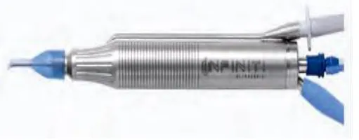
All surgeries were performed using the INFINITI® Vision System using a high
[image:6.595.171.428.369.465.2]performance Infiniti™ U/S handpiece: piezoelectric, slim, Lightweight, autoclavable.
Figure 2: OZil® Torsional Handpiece (Alcon Handpieces, 2011).
[image:6.595.172.428.510.610.2].
, Issue 03, 2016 5
Vol
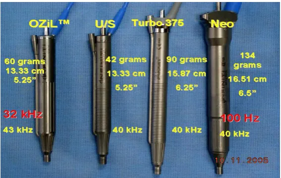
[image:7.595.156.440.70.250.2]www.wjpr.net 1955
Figure 4: INFINITI® Vision System Handpieces (Alcon Handpieces, 2011).
RESULTS
This is a prospective study that was performed on 20 patients to estimate postoperative
corneal endothelial outcome after cataract surgery performed with torsional
phacoemulsification in senile cataracts.
Preoperative data including age, sex and grading of nucleus are Presented in Table 4. The
Age group ranges from 55-65 years, including 12 males & 8 Females, all have Nuclear
cataract grade II-III.
Tabel-3.
20 No. of Patients
12 Males
8 Females
55 - 65 Age range
II - III Nuclear Grading
In our trial we also conducted a more detailed study about the corneal endothelial cells as
regard, Endothelial cell Count (Endothelial density) (CD), Average cell size (AVE),
Maximum cell size (MAX), Minimum cell size (MIN), Coefficient of variation in cell size
(CV), Standard deviation in cell density (SD), Pachymetry (PACHY), Best corrected visual
acuity (BCVA), as well as the Percentage of the hexagonal cells (6A).
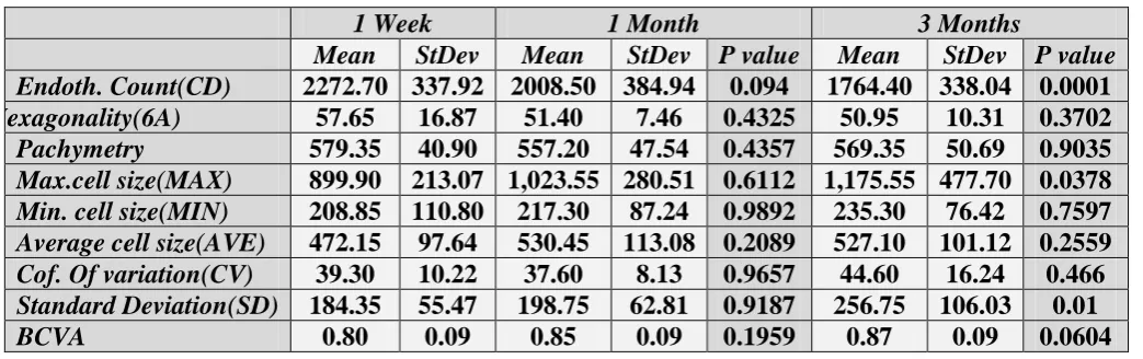
During the follow up, There was significant Endothelial Cell Loss, significant increase in the
Average cell size between Pre-Operative & 1 week post-operative, Pre-Operative & 1Month
.
, Issue 03, 2016 5
Vol
www.wjpr.net 1956
Significant increase in the Maximum cell size and the Standard Deviation between
Pre-Operative & 1Month post-operative, pre-operative & 3 months post-operatively.
Significant increase in the Coefficient of variation between Pre-operative & 3 months
post-operatively.
Significant Improvement in the visual acuity between Pre-Operative & 1 week
operative, Pre-Operative & 1Month operative, pre-operative & 3 months
post-operatively.
Otherwise, there was no significant difference concerning other parameters, as shown in table
[image:8.595.39.561.479.643.2](5) & (6) & (7).
Table 4: Showing range of endothelial cell count in the study cases pre operative and post
operative by 1 week, 1month, and 3 months and also the mean and the standard deviation
of each item.
Torsional phaco Range Mean ± SD
CD
Pre 2155 - 3558 2631.80 ± 353.68
Post 1 week 1712 2881 2272.70 ±- 337.92
Post 1 month 1356 - 2865 2008.50 ± 384.94
Post 3 month 1204 - 2560 1764.40 ± 338.04
Table 5: Comparison between endothelial cell parameters throughout the study.
1 Week 1 Month 3 Months
.
, Issue 03, 2016 5
Vol
[image:9.595.40.562.88.254.2]www.wjpr.net 1957
Table 6: Comparison between endothelial cell parameters throughout the study.
1 Week 1 Month 3 Months
[image:9.595.40.562.283.445.2]Mean StDev Mean StDev P value Mean StDev P value Endoth. Count(CD) 2272.70 337.92 2008.50 384.94 0.094 1764.40 338.04 0.0001 Hexagonality(6A) 57.65 16.87 51.40 7.46 0.4325 50.95 10.31 0.3702 Pachymetry 579.35 40.90 557.20 47.54 0.4357 569.35 50.69 0.9035 Max.cell size(MAX) 899.90 213.07 1,023.55 280.51 0.6112 1,175.55 477.70 0.0378 Min. cell size(MIN) 208.85 110.80 217.30 87.24 0.9892 235.30 76.42 0.7597 Average cell size(AVE) 472.15 97.64 530.45 113.08 0.2089 527.10 101.12 0.2559 Cof. Of variation(CV) 39.30 10.22 37.60 8.13 0.9657 44.60 16.24 0.466 Standard Deviation(SD) 184.35 55.47 198.75 62.81 0.9187 256.75 106.03 0.01 BCVA 0.80 0.09 0.85 0.09 0.1959 0.87 0.09 0.0604
Table 7: Comparison between endothelial cell parameters throughout the study.
1 Week 1 Month 3 Months
Mean StDev Mean StDev P value Mean StDev P value Endoth. Count(CD) 2272.70 337.92 2008.50 384.94 0.094 1764.40 338.04 0.0001 Hexagonality(6A) 57.65 16.87 51.40 7.46 0.4325 50.95 10.31 0.3702 Pachymetry 579.35 40.90 557.20 47.54 0.4357 569.35 50.69 0.9035 Max.cell size(MAX) 899.90 213.07 1,023.55 280.51 0.6112 1,175.55 477.70 0.0378 Min. cell size(MIN) 208.85 110.80 217.30 87.24 0.9892 235.30 76.42 0.7597 Average cell size(AVE) 472.15 97.64 530.45 113.08 0.2089 527.10 101.12 0.2559 Cof. Of variation(CV) 39.30 10.22 37.60 8.13 0.9657 44.60 16.24 0.466 Standard Deviation(SD) 184.35 55.47 198.75 62.81 0.9187 256.75 106.03 0.01 BCVA 0.80 0.09 0.85 0.09 0.1959 0.87 0.09 0.0604
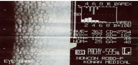
Taking one patient as example:- A 50 years old patient with senile nuclear cataract (n II-
III ), Preoperative endothelial cell count 2754 (not less than 1500 cells/mm2), no past
history of ocular surgeries or ocular trauma, without any type of glaucoma or corneal
dystrophies.
Fig (5-a): Left preoperative specular microscope of 50 years old patient showing that,
Endothelial cell Count (Endothelial density) (CD) is 2754, Average cell size (AVE) is 363,
Maximum cell size (MAX) is 777, Minimum cell size (MIN) is 129, Standard deviation in cell
density (SD) is 162, Coefficient of variation in cell size (CV) is 44, Pachymetry (PACHY) is
595, the Percentage of the hexagonal cells (6A) is 48, as well as the Number of cell (NUM) is
[image:9.595.185.411.538.644.2].
, Issue 03, 2016 5
Vol
www.wjpr.net 1958
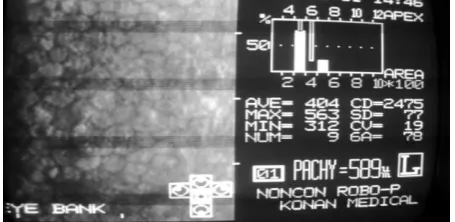
Fig (5-b):- Left postoperative 1 week specular microscope of 50 years old patient who had
Phaco showing that, Endothelial cell Count (Endothelial density) (CD) is 2475, Average cell
size (AVE) is 404, Maximum cell size (MAX) is 563, Minimum cell size (MIN) is 312,
Standard deviation in cell density (SD) is 77, Coefficient of variation in cell size (CV) is 19,
Pachymetry (PACHY) is 589, the Percentage of the hexagonal cells (6A) is 78, as well as the
Number of cell (NUM) is 9.
Fig (5-c):- Left postoperative 1 month specular microscope of 50 years old patient who had
Phaco showing that, Endothelial cell Count (Endothelial density) (CD) is 2057, Average cell
size (AVE) is 486, Maximum cell size (MAX) is 883, Minimum cell size (MIN) is 185,
Standard deviation in cell density (SD) is 175, Coefficient of variation in cell size (CV) is 36,
Pachymetry (PACHY) is 638, the Percentage of the hexagonal cells (6A) is 30, as well as the
Number of cell (NUM) is 23.
Fig (5-d):- Left postoperative 3 month specular microscope of 50 years old patient who had
Phaco showing that, Endothelial cell Count (Endothelial density) (CD) is 2070, Average cell
[image:10.595.184.412.72.183.2] [image:10.595.189.409.321.435.2] [image:10.595.181.415.573.707.2].
, Issue 03, 2016 5
Vol
www.wjpr.net 1959
Standard deviation in cell density (SD) is 180, Coefficient of variation in cell size (CV) is 37,
Pachymetry (PACHY) is 584, the Percentage of the hexagonal cells (6A) is 14, as well as the
Number of cell (NUM) is 14.
DISCUSSION
For the lens to be removed efficiently and safely, the risk of ultrasound induced endothelial
cell loss should be minimized. Reducing phacoemulsification energy and time are the main
objectives of future improvement.[12]
The purpose of this prospective, randomized study is to study the effect of torsional
phacoemulsification as regards: Endothelial cell Count (Endothelial density) (CD), Average
cell size (AVE), Maximum cell size (MAX), Minimum cell size (MIN), Coefficient of
variation in cell size (CV), Standard deviation in cell density (SD), Pachymetry (PACHY),
Best corrected visual acuity (BCVA), as well as the Percentage of the hexagonal cells (6A).
Kim et al (2010)[13]reported that using torsional phacoemulsification showed less endothelial
cell loss and central corneal thickening at postoperative day seven in moderate cataracts.
They found that in moderate cataract group percentage endothelial cell loss at 1 week
postoperative was 5.12% in torsional group, which turned out to be not significantly different
by one month after operation 3.19%, while in hard cataract group percentage endothelial cell
loss was 23.52%.
Kim et al (2010)[13]concluded that torsional phacoemulsification showed superior efficiency
for moderate cataracts. Based on these studies, we used torsional phaco only to minimize
intraoperative factors affecting the corneal endothelium.
Walkow et al (2000)[6] found a significant correlation between phaco time and central
endothelial cell loss, but not between phaco energy and cell loss. O’Brien et al (2004)[14]
found a significant association between phaco time, mean US power and endothelial cell loss.
As regarding the BCVA at 30 days postoperative there was no statistically significant
difference. Similar results were encountered in Kim et al (2010),[13] Vasavada et al (2010)[15] and Reushel et al (2010)[16]
Our results demonstrate that torsional phacoemulsification produces a safe and efficient
mode of phacoemulsification with reduced mean ultrasound time and Cumulative Dissipiated
.
, Issue 03, 2016 5
Vol
www.wjpr.net 1960
Our results also matched with Liu et al [17] they reported that The torsional mode may provide
more effective lens removal with less endothelial cell loss. At 7 days and 30 days, the mean
central corneal endothelial cell count was 2272.7 ± 337.92 cells/mm2 and 2008.50 ± 384.94
cells/mm2 (P < 0.001). Also matched with what Fakhry et al (2011)[18] found (P < 0.001).
Gonen T et al (2012)[19] concluded that the percentage of mean endothelial cell loss was between 35.4% and 39.1%.
Gogate and associates (2010)[20] conducted a study to compare endothelial cell loss in cataract surgery by phacoemulsification surgery over 6 weeks, the study evaluated 100
patients. The mean endothelial cell loss in percent after 1 week postoperative was 13.2 % and
after 6 weeks postoperatively it was 15.5 %, also they found that best corrected visual acuity
at 6 weeks was better than 6/18 in 98.5 % of eyes which is matched with our study.
CONCLUSION
Cataract extraction constitutes the largest surgical workload in ophthalmic units throughout
the world. The torsional mode provides an effective and safe method for cataract removal
with lower energy usage.
Our results indicate that the torsional phacoemulsification is a safe method of removing
uncomplicated senile cataract with less endothelial cell loss. Torsional phacoemulsification
has the advantage of reducing UST as well as effective energy used.
Phaco duration was the most significant intraoperative factor affecting the corneal
endothelium. So Specular microscopy is a useful tool in preoperative assessment of cataract
patients especially in cases undergoing phacoemulsification.
This study is limited by a small sample size in hard cataracts. Nevertheless, the findings have
meaningful clinical relevance to support the efficiency and safety of torsional
phacoemulsification in medium density cataracts.
RECOMMENDATION
1- Proper preoperative assessment of the corneal endothelium using specular microscopy in
.
, Issue 03, 2016 5
Vol
www.wjpr.net 1961
2- Proper choice of the surgical procedure for cataract extraction in patients having
compromised corneal endothelium to avoid more endothelial damage. For example; using
phacoemulsification technique with the least possible Phaco duration.
REFERENCES
1. Neumann AC., McCarty GR., Sanders DR., et al: Small incision to control astigmatism
during cataract surgery. J. Cataract Refractive Surgery, 1989 Jan; 15(1): 78-84.
2. Cameron MD., Poyer JF. and Aust SD.: Identification of free radicals produced during
phacoemulsification. J Cataract Refract Surg, 2001 mar; 27(3): 463-470.
3. Bourne RRA., Minassian DC., Dart JKG., et al: Effect of cataract surgery on the corneal
endothelium; modern phacoemulsification compared with extracapsular cataract surgery.
Ophthalmology, 2004; 111(4): 679-685.
4. Vasavada V., Raj SM. and Vasavada AR. Intraoperative performance and postoperative
outcomes of microcoaxial phacoemulsification; observational study. J Cataract Refract
Surg, 2007; 33: 1019-1024.
5. Davison JA., Cionni RJ., Snowdon JR., et al: Simultaneous surgeon and side view
videoanalysis comparing in situ fracture and stop-and-chop phacoemulsification. J
Cataract Refract Surgery, 2005 Feb; 31(2): 274-279.
6. Walkow T., Anders N. and Klebe S. Endothelial cell loss after phacoemulsification:
relation to preoperative and intraoperative parameters. J Cataract Refract Surg, 2000;
26: 727-732.
7. Davison JA. Cumulative tip travel and implied followability of longitudinal and torsional
phacoemulsification. J Cataract Refract Surgery, 2008 Jun; 34: 986-90.
8. Kohlhaas M., Klemn M., Kammann J., et al: Endothelial cell loss secondary to two
different phacoemulsification techniques. Ophthalmic Surgery and Lasers, 1998; (29):
890-895.
9. Mishima S.: Corneal thickness. Surv. Ophthalmol, 1968; 13: 57-96. Quoted from
Mishima S.: Clinical investigations of the corneal endothelium. XXXVIII Edward Jackson
Memorial Lecture. Am. J. Ophthalmol, 1982 Jan; 93(1): 1-29.
10.MacRae SM., Matsuta M., Shellans S., et al: The effects of hard and soft contact lenses
on the corneal endothelium. Am J Ophthalmol, 1986; (102): 50–7.
11.Chylack LT., Wolfe JK., Singer DM., et al: Lens Opacities Classification System III.
.
, Issue 03, 2016 5
Vol
www.wjpr.net 1962
12.Waring GO.: Posterior collagenous layer of the cornea. Ultrastructural classification of
abnormal collagenous tissue posterior to Descemet’s membrane in 30 cases. Arch
Ophthalmol, 1982; 100: 122–134.
13.Kim DH., Wee WR., Lee JH., et al: The comparison between torsional and conventional
mode phacoemulsification in moderate and hard cataracts. Korean J Ophthalmol, 2010;
24(6): 336-340.
14.O’Brien PD., Fitzpatrick P., Kilmartin DJ., et al: Risk factors for endothelial cell loss
after phacoemulsification by a junior resident. J Cataract Refract Surg, 2004; (30):
839-843.
15.Vasavada AR., Raj SM. and Vasavada VL.: Comparison of torsional and microburst
longitudinal phacoemulsification: A prospective, randomized, masked clinical trial.
Ophthalmic Surgery, Lasers and Imaging, 2010; 41(1): 109-114.
16.Reuschel A., Bogatsch H., Barth T., et al: Comparison of endothelial changes and
power settings between torsional and longitudinal phacoemulsification. J Cataract
Refract Surg, 2010 Nov; 36: 1855-1861.
17.Liu Y., Zeng M., Liu X., et al: Torsional mode versus conventional ultrasound mode
phacoemulsification; randomized comparative clinical study. J Cataract Refract Surg,
2007 FEB; 33(2): 287-292.
18. Fakhry MA. and El Shazly MI.: Torsional ultrasound mode versus combined torsional
and conventional ultrasound mode phacoemulsification for eyes with hard cataract. Clin
Ophthalmology, 2011; 5: 973- 8.
19.Gonen T., Sever O., Horozoglu F., et al: Endothelial cell loss: Biaxial small-incision
torsional phacoemulsification versus biaxial small-incision longitudinal
phacoemulsification. J Cataract Refract Surg, 2012 Nov; 38(11): 1918-24.
20.Gogate P., Ambardekar P., Kulkarni S., et al: Comparison of endothelial cell loss after
cataract surgery: Phacoemulsification versus manual small- incision cataract surgery six
week results of a randomized control trial. J Cararact Refract Surg, 2010 Feb; 36(2):
![Figure 1: Lens Opacities Classification System III.[11]](https://thumb-us.123doks.com/thumbv2/123dok_us/816191.590177/4.595.171.426.71.223/figure-lens-opacities-classification-system-iii.webp)