Acta Cryst.(2001). E57, o1087±o1088 DOI: 10.1107/S1600536801017536 Bond and Davies C6H7N
o1087
organic papers
Acta Crystallographica Section E Structure Reports Online
ISSN 1600-5368
3-Picoline
Andrew D. Bond* and John E. Davies
Department of Chemistry, University of Cambridge, Lensfield Road, Cambridge CB2 1EW, England
Correspondence e-mail: [email protected]
Key indicators Single-crystal X-ray study
T= 120 K
Mean(C±C) = 0.003 AÊ
Rfactor = 0.040
wRfactor = 0.106 Data-to-parameter ratio = 8.1
For details of how these key indicators were automatically derived from the article, see http://journals.iucr.org/e.
#2001 International Union of Crystallography Printed in Great Britain ± all rights reserved
The crystal structure of 3-picoline (3-methylpyridine, C6H7N)
has been determined at 120 (2) K following in situ crystal growth from the liquid. The molecules pack in a herring-bone-type arrangement in the non-centrosymmetric space group
Pna21.
Comment
The picolines (methylpyridines) comprise a series of empirical formula C6H7N, with weak intermolecular interactions and
low melting points. The crystal structure of 4-picoline (4-methylpyridine; m.p. 276 K) has been determined previously from a crystal grown using an elaborate modi®ed Bridgman technique (Ohms et al., 1985). We report here the crystal structure of 3-picoline (m.p. 255 K), determined at 120 (2) K from a crystal grownin situin a 0.3 mm glass capillary. This work forms part of a study devoted to improving techniques for determining the crystal structures of substances that are liquids at room temperature (see, for example, Bond & Davies, 2001).
Molecules of (I) (Fig. 1) pack in a herring-bone-type arrangement in the non-centrosymmetric space groupPna21
(Fig. 2). There are no apparent directional CÐH N inter-actions: the closest contacts to N1 are made by H4 and H5, with geometric parameters H4 N1i= 2.77 (3) AÊ and C4Ð
H4 N1i = 124 (2), and H5 N1i = 2.90 (2) AÊ and C5Ð
H5 N1i = 120 (2) [symmetry code: (i) 1.5ÿx, 0.5+y,
0.5+z].
Experimental
The sample (99%) was obtained from the Aldrich Company and was used without further puri®cation. The crystal was grown in a 0.3 mm glass capillary tube at 240 K (a temperature only slightly less than the melting point of the solid in the capillary tube) using a technique described previously (Davies & Bond, 2001). The crystal was cooled subsequently to 120 (2) K for data collection. The length of the cyl-indrical crystal was not estimated, but it exceeded the 0.35 mm collimator diameter.
Crystal data
C6H7N Mr= 93.13
Orthorhombic,Pna21 a= 9.3516 (9) AÊ b= 9.7925 (10) AÊ c= 5.7651 (3) AÊ V= 527.94 (8) AÊ3 Z= 4
Dx= 1.172 Mg mÿ3
MoKradiation Cell parameters from 3581
re¯ections
= 1.0±27.5
= 0.07 mmÿ1 T= 120 (2) K Cylinder, colourless 0.15 mm (radius)
Data collection
Nonius KappaCCD diffractometer Thin-slice!and'scans Absorption correction: none 2718 measured re¯ections 665 independent re¯ections 643 re¯ections withI> 2(I)
Rint= 0.040 max= 27.5 h=ÿ12!8 k=ÿ9!12 l=ÿ6!7
Re®nement
Re®nement onF2 R[F2> 2(F2)] = 0.040 wR(F2) = 0.106 S= 1.08 665 re¯ections 82 parameters
H atoms treated by a mixture of independent and constrained re®nement
w= 1/[2(F
o2) + (0.0633P)2 + 0.0757P]
whereP= (Fo2+ 2Fc2)/3 (/)max= 0.002
max= 0.17 e AÊÿ3
min=ÿ0.16 e AÊÿ3
H atoms on the pyridyl ring were located in difference Fourier maps and were allowed to re®ne with independent isotropic displacement parameters. Methyl H atoms were placed geometrically and re®ned with one common isotropic displa-cement parameter, with the methyl group allowed to rotate about its local threefold axis. Friedel pairs (478) were
aver-aged prior to merging of data inPna21; the reported value of Rintcorresponds to subsequent merging of equivalent re¯ec-tions in this space group.
Data collection:COLLECT(Nonius, 1998); cell re®nement:HKL
and SCALEPACK (Otwinowski & Minor, 1997); data reduction:
HKL, DENZO and SCALEPACK (Otwinowski & Minor, 1997); program(s) used to solve structure: SIR92 (Altomare et al., 1994); program(s) used to re®ne structure:SHELXL97 (Sheldrick, 1997); software used to prepare material for publication:SHELXL97.
We thank the EPSRC for ®nancial assistance towards the purchase of the Nonius CCD diffractometer.
References
Altomare, A., Cascarano, G., Giacovazzo, C., Guagliardi, A., Burla, M. C., Polidori, G. & Camalli, M. (1994).J. Appl. Cryst.27, 435±436.
Bond, A. D. & Davies, J. E. (2001).Acta Cryst.E57, o1039±o1040. Davies, J. E. & Bond, A. D. (2001).Acta Cryst.E57, o947±o949. Nonius (1998).COLLECT. Nonius BV, Delft, The Netherlands.
Ohms, U., Guth, H., Treutmann, W., DannoÈhl, H., Schweig, A. & Heger, G. (1985).J. Chem. Phys.83, 273±279.
Otwinowski, Z. & Minor, W. (1997). Methods in Enzymology, Vol. 276, Macromolecular Crystallography, Part A, edited by C. W. Carter and R. M. Sweet, pp. 307±326. London: Academic Press.
Sheldrick, G. M. (1993).XP. University of GoÈttingen, Germany. Sheldrick, G. M. (1997).SHELXL97. University of GoÈttingen, Germany. Watkin, D. J., Prout, C. K. & Pearce, L. J. (1996).CAMERON. Chemical
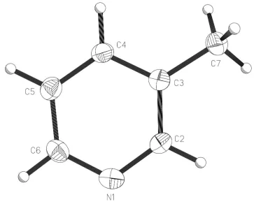
Crystallography Laboratory, University of Oxford, England. Figure 1
The molecular structure and atom-labelling scheme for (I) showing displacement ellipsoids at 50% probability for non-H atoms (XP; Sheldrick, 1993).
Figure 2
supporting information
sup-1
Acta Cryst. (2001). E57, o1087–o1088supporting information
Acta Cryst. (2001). E57, o1087–o1088 [doi:10.1107/S1600536801017536]
3-Picoline
Andrew D. Bond and John E. Davies
S1. Comment
The picolines (methylpyridines) comprise a series of empirical formula C6H7N, with weak intermolecular interactions and
low melting points. The crystal structure of 4-picoline (4-methylpyridine; m.p. 276 K) has been determined previously
from a crystal grown using an elaborate modified Bridgman technique (Ohms et al., 1985). We report here the crystal
structure of 3-picoline (m.p. 255 K), determined at 120 (2) K from a crystal grown in situ in a 0.3 mm glass capillary.
This work forms part of a study devoted to improving techniques for determining the crystal structures of substances that
are liquids at room temperature (see, for example, Bond & Davies, 2001).
Molecules of (I) (Fig. 1) pack in a herring-bone-type arrangement in the non-centrosymmetric space group Pna21 (Fig.
2). There are no apparent directional C—H···N interactions: the closest contacts to N1 are made by H4 and H5, with
geometric parameters H4···N1i = 2.77 (3) Å and C4—H4···N1i = 124 (2)°, and H5···N1i = 2.90 (2) Å and C5—H5···N1i =
120 (2) Å [symmetry code: (i) 1.5 - x, 0.5 + y, 0.5 + z].
S2. Experimental
The sample (99%) was obtained from the Aldrich Company and was used without further purification. The crystal was
grown in a 0.3 mm glass capillary tube at 240 K (a temperature only slightly less than the melting point of the solid in the
capillary tube) using a technique described previously (Davies & Bond, 2001). The crystal was cooled subsequently to
120 (2) K for data collection. The length of the cylindrical crystal was not estimated, but it exceeded the 0.35 mm
collimator diameter.
S3. Refinement
H atoms were placed geometrically and allowed to refine with independent isotropic displacement parameters (one
common parameter for the methyl-H atoms). Friedel pairs (478) were merged prior to merging in Pna21; the reported
Figure 1
The molecular structure and atom-labelling scheme for (I) showing displacement ellipsoids at 50% probability for non-H
supporting information
[image:5.610.124.485.70.432.2]sup-3
Acta Cryst. (2001). E57, o1087–o1088Figure 2
Projection of (I) onto (001) showing the herring-bone packing arrangement (CAMERON; Watkin et al., 1996).
3-methylpyridine
Crystal data C6H7N Mr = 93.13
Orthorhombic, Pna21 a = 9.3516 (9) Å b = 9.7925 (10) Å c = 5.7651 (3) Å V = 527.94 (8) Å3 Z = 4
F(000) = 200
Dx = 1.172 Mg m−3 Melting point: 254.9 K Mo Kα radiation, λ = 0.7107 Å Cell parameters from 3581 reflections θ = 1.0–27.5°
µ = 0.07 mm−1 T = 120 K
Cylinder, colourless 0.15 mm (radius)
Data collection Nonius KappaCCD
diffractometer
Radiation source: fine-focus sealed tube Thin–slice ω and φ scans
2718 measured reflections 665 independent reflections
643 reflections with I > 2σ(I) Rint = 0.040
θmax = 27.5°, θmin = 4.1° h = −12→8
Refinement on F2 Least-squares matrix: full R[F2 > 2σ(F2)] = 0.040 wR(F2) = 0.106 S = 1.08 665 reflections 82 parameters 1 restraint
Primary atom site location: structure-invariant direct methods
Secondary atom site location: difference Fourier map
Hydrogen site location: inferred from neighbouring sites
H atoms treated by a mixture of independent and constrained refinement
w = 1/[σ2(F
o2) + (0.0633P)2 + 0.0757P] where P = (Fo2 + 2Fc2)/3
(Δ/σ)max = 0.002 Δρmax = 0.17 e Å−3 Δρmin = −0.16 e Å−3
Special details
Experimental. Grown in situ in a 0.3 mm Lindemann capillary tube at 240 K. Freidel pairs (478) were averaged for the refinement.
Geometry. All e.s.d.'s (except the e.s.d. in the dihedral angle between two l.s. planes) are estimated using the full covariance matrix. The cell e.s.d.'s are taken into account individually in the estimation of e.s.d.'s in distances, angles and torsion angles; correlations between e.s.d.'s in cell parameters are only used when they are defined by crystal symmetry. An approximate (isotropic) treatment of cell e.s.d.'s is used for estimating e.s.d.'s involving l.s. planes.
Refinement. Refinement of F2 against ALL reflections. The weighted R-factor wR and goodness of fit S are based on F2, conventional R-factors R are based on F, with F set to zero for negative F2. The threshold expression of F2 > σ(F2) is used only for calculating R-factors(gt) etc. and is not relevant to the choice of reflections for refinement. R-factors based on F2 are statistically about twice as large as those based on F, and R- factors based on ALL data will be even larger.
Fractional atomic coordinates and isotropic or equivalent isotropic displacement parameters (Å2)
x y z Uiso*/Ueq
N1 0.81677 (17) −0.01946 (16) 1.0686 (3) 0.0269 (4) C2 0.70900 (19) 0.02815 (16) 0.9380 (3) 0.0231 (4) H2 0.691 (3) −0.016 (2) 0.789 (5) 0.028 (6)* C3 0.62234 (17) 0.13911 (16) 0.9967 (3) 0.0211 (4) C4 0.65209 (18) 0.20425 (18) 1.2061 (3) 0.0239 (4) H4 0.597 (3) 0.286 (3) 1.247 (6) 0.042 (7)* C5 0.7631 (2) 0.15703 (17) 1.3440 (4) 0.0263 (4) H5 0.783 (2) 0.204 (2) 1.495 (5) 0.030 (6)* C6 0.8417 (2) 0.04500 (19) 1.2694 (4) 0.0263 (5) H6 0.919 (3) 0.011 (2) 1.358 (5) 0.043 (7)* C7 0.5034 (2) 0.18534 (18) 0.8400 (4) 0.0286 (4) H7A 0.4235 0.2191 0.9339 0.071 (6)* H7B 0.4710 0.1084 0.7449 0.071 (6)* H7C 0.5381 0.2587 0.7389 0.071 (6)*
Atomic displacement parameters (Å2)
U11 U22 U33 U12 U13 U23
supporting information
sup-5
Acta Cryst. (2001). E57, o1087–o1088C5 0.0268 (9) 0.0249 (8) 0.0273 (9) −0.0030 (7) −0.0009 (8) −0.0029 (10) C6 0.0205 (8) 0.0266 (8) 0.0319 (11) −0.0005 (6) −0.0022 (7) 0.0026 (8) C7 0.0254 (8) 0.0300 (8) 0.0304 (9) 0.0016 (7) −0.0057 (8) −0.0022 (9)
Geometric parameters (Å, º)
N1—C6 1.339 (3) C3—C7 1.503 (3) N1—C2 1.342 (2) C4—C5 1.387 (3) C2—C3 1.397 (2) C5—C6 1.389 (3) C3—C4 1.393 (2)