COMPARATIVE STUDY OF MAST CELL STABILIZING EFFECT OF
EXTRACT OF SELECTED PLANTS WITH PROVEN H
1ANTAGONIST
ACTIVITY
Krishna Prasad1, Rashmi Rajappa2, Jessen George2, Shastry C.S1, Magesh S. Bojan2*
1
NGSM Institute of Pharmaceutical Sciences, Nitte University, Paneer, Deralakatte, Mangalore - 575018
2
Department of Water and Health, Faculty of Life Sciences, JSS University, SS Nagar, Mysore, Karnataka, India-570015
ABSTRACT
Mast cells play a key role in allergy and inflammation which have profound patho-physiological role in asthma. In the present study a comparative effect of mast cell stabilizing activity of different plant extracts Momordica dioica fruit pulp (family - Cucurbitaceae),
Cuminum cyminum seeds (family - Apiaceae), Piper nigrum seeds (family - Piperaceae), Boerhaavia diffusa roots (family - Nyctaginaceae.), Withania somnifera roots (family - Solanaceae),
Mangifera indica leaves (Anacardiaceae), Plantago ovata
(Plantaginaceae) was carried out. For this purpose plant extracts were subjected to the preliminary phytochemical analysis and mast cell stabilizing action by assessing the prevention of degranulation of mast cells by known degranulating compound 48/80. The result showed a higher stabilization potential in ethanolic extract of Piper nigrum seeds which showed 55.17±1.56, 64.00±1.18 and 70.50±0.96 percentage inhibition of mast cell degranulation. Minimal activity was observed in the ethanolic extract of Plantago ovata with percentage inhibition of 21.17±1.54, 22.17±2.21, 24.33±2.20 at three dose levels. Other plant extracts showed intermediate activity. Further investigation of the 5-Lipooxygenase activity, leukotriene antagonist activity and anti-inflammatory activity along with the findings of the present work would give us more clear understanding of the mechanism by which the above extracts exert beneficial effect in the management of asthma.
KEY WORDS: Mast cell, Inflammation, degranulation, compound 48/80.
Volume 4, Issue 11, 981-993. Research Article ISSN 2277– 7105
*Correspondence for
Author
Magesh S. Bojan
Department of Water and
Health, Faculty of Life
Sciences, JSS University,
SS Nagar, Mysore,
Karnataka, India-570015 Article Received on 20 Aug 2015,
INTRODUCTION
Asthma is a chronic airway inflammatory disorder with inflammation due to complex interactions between inflammatory cells, mediators, and airway cells. This is characterized by airway hyper reactivity to a variety of non-specific stimuli, leading to a variable degree of airway obstruction, some of which may become irreversible over many years.[1] The mechanism of the inflammatory response resulting in asthma is complex and involves numerous cell types, including mast cells.[2] Mast cell activation causes process of degranulation that result in releasing of mediators, such as histamine and an array of inflammatory cytokines,[3, 4] on activation, mast cells immediately released the preformed and the de novo synthesized mediators such as histamine, proteases, leukotrienes, prostaglandins, and cytokines.[5] As a consequence, the acute reactions such as vasodilation, increased vascular permeability, and bronchoconstriction were induced. In addition, allergic responses also trigger the influx and activation of a variety of inflammatory cells including eosinophills and lymphocytes.[6] Therefore, mast cell stabilization is a key factor in controlling the occurrence of asthma.
Various systems of medicine such as Ayurveda, Unani, Siddha and Chinese employed plant based drugs for the treatment of diseases which dates back to 5000 B.C. Because of the low incidence of adverse reactions and cost effectiveness, many countries have now shifted their focus to plant based medicines.[7] It is the need of the hour to ensure the perseverance of the knowledge base of traditional medicines and employ them in the management of ailments.[8] The formidable challenges facing the traditional medicine are developing standards and guidelines maximizing the economic potential to ensure safe and efficacious provision of medicines WHO (2002). It is reported in various literature that the different parts of plants
MATERIALS AND METHODS Chemicals
The Compound 48/80 was purchased from Sigma Aldrich, Bangalore, India and preserved in deep freeze until use. The standard protective drug disodium chromoglycate was procured from Yarrow chemicals and stored as per storage requirements. All the other chemicals were of analytical grade.
Plant material and preparation of extract
Momordica dioica fruit pulp (family - Cucurbitaceae), Cuminum cyminum seeds (family-Apiaceae), Piper nigrum seeds (family - Piperaceae), Boerhaavia diffusa roots (family - Nyctaginaceae.), Withania somnifera roots (family - Solanaceae), Mangifera indica leaves (Anacardiaceae), Plantago ovata (Plantaginaceae) seeds were collected from different regions of Mangalore, Karnataka, India. The plants were collected in the month of May and June and were authenticated by Professor Krishnakumar G, Head of Applied Botany, Mangalore University, Konaje, Mangalore, India. The reference voucher number MU/AB/July, 2014 was assigned to the plant samples. The extraction procedure was carried out by the method of Handa et al., (2008).[17] Approximately 500 g of plant materials were extracted separately in a soxhlet apparatus using 95 % ethanol. The solvent from the total extract was removed by means of rotary flash evaporator, later made to a syrupy consistency by evaporating the excess of solvent on a water bath. These extracts were then suspended in 0.6% Sodium CMC to get concentrations of 100 μg/ml, 200 μg/ml, 400μg/ml for screening.
Table I. Percentage yield of of different plants extracts
Plant Colour Consistency Percentage
yield (%)
Boerhaavia diffusa Dark brown Semi-solid 1.32
Cuminum cyminum Dark green Semi-solid, pasty 10.50
Piper nigrum Dark green Semi-solid 3.02
Magnifera indica Dark green Viscous, Semi
solid 20.82
Momordica dioica Dark brown Semi-solid 2.34
Withania somnifera Amber coloured,
Brown Resinuous 11.62
Plantago ovate Black Semi solid 1.08
Qualitative Phytochemical Estimations
Test for Alkaloids-Dragendroff’s test: To 1 ml of Dragendroff’s reagent was added to different plant extracts. Formation of orange or orange red precipitate indicates the presence of alkaloids.
Hager’s test: To 1 ml of different plant extract, few drops of Hager’s reagent were added.
Formation of yellow precipitate indicates the presence of alkaloids.
Wagner’s test: To 1 ml of different plant extract Wagner’s reagent was added. Yellow or
brown precipitation indicates the presence of alkaloids.
Mayer’s test: 1 ml of Mayer’s reagent was added to different plant extract. Formation of pale
yellow precipitate indicates the presence of alkaloids.
Test for Carbohydrates-Molischs’s test: Plant extracts were mixed with few drops of molish reagent and concentrated sulphuric acid was added through the side of the test tube. Formation of red-violet ring at junction indicates the presence of carbohydrates.
Benedict’s test: To 0.5 ml of different plant extract, few drops of Benedict’s solution was
added and heated. Formation of brick red color indicates the presence of carbohydrates.
Fehling’s test: 1 ml of Fehling’s solution A and Fehling’s solution B were mixed with 2 ml
of different plant extracts and heated. Formation of red color indicates the presence of reducing sugar.
Test for Flavonoids- Shinoda test: To 0.5 ml of different plant extract, few magnesium turnings and few drops of dilute hydrochloric acid were added and heated. Formation of pink or reddish brown indicates the presence of flavonoids.
Test for anthraquinone glycosides-Borntrager’s test: Different plant extracts were boiled with ferric chloride solution for about 5 min. The mixture was cooled and shaken with equal volume of benzene. The benzene layer was separated and treated with half of its volume of ammonia solution. Formation of rose pink or cherry red colour in the ammonical layer indicates the presence of anthraquinone glycosides.
concentrated sulphuric acid was added along the sides of the test tube. Formation of green color indicates the presence of steroids.
Salkowski reaction: Different extract were treated with few drops of concentrated sulphuric acid. Formation of red and yellow color indicates the presence of steroids and triterpenoids respectively.
Test for resins: 1 ml of different plant extract was dissolved in acetone and the solution was poured in distilled water. Turbidity indicates the presence of resins.
Test for saponins: 5 ml of different plant extract was taken in test tubes; a drop of sodium bicarbonate was added. The test tube was shaken well and left for 3 minutes. Formation of stable froth indicates the presence of saponins.
Test for tannins: Add 2 ml of lead acetate solution to different plant extracts. Formation of white cloudy precipitate indicates the presence of tannins.
Experimental design
Male albino rats of Wistar strain (180-220 g) were obtained from central animal house KSHEMA, Deralakatte, Mangalore. The animal experiments were executed after obtaining Institutional Animal Ethical clearance in accordance with the guidelines of the committee for the purpose of control and super vision of experiments on animals (CPCSEA) formed by the government of India (approval number; KSHEMA/IAEC/17/2014). Rats were housed in polypropylene cages under an ambient temperature of 25 ± 2°C, Relative humidity 50 ± 60% with dark and light cycle 12h/12h. Animal were maintained on a standard pellet diet and water ad libitum. The animals were allowed for acclimatization for a period of seven days before initiation of the experiments.
In vitro mesenteric mast cell degranulation
The plant extracts were suspended in 0.6% Sodium CMC to get concentrations of 100 μg/ml, 200 μg/ml and 400 μg/ml for evaluation of mast cell stabilization effect. The overnight fasted
rats were sacrificed by cervical dislocation. The abdomen was cut open to expose the intestine and the small pieces of the mesentery were cut and placed in a petri dish containing Ringer Locke solution (NaCl 154 mM , KCl 5.6 mM, CaCl2 2.2 mM, NaHCO3 6.0 mM and
Petri dish I: Vehicle control (Ringer Locke solution only) Petri dish II: Positive control (0.8μg/ml of C.48/80 only)
Petri dish III: Standard (disodium chromogylate-DSCG, 1 mg/ml) Petri dish IV: 100μg/ml of plant extract.
Petri dish V: 200μg/ml of plant extract. Petri dish VI: 400μg/ml of plant extract.
Each petri dish was incubated for 15 min at 37º C and later compound 48/80 at 0.8μg/ml was added to each petri dish except vehicle control and again incubated for 10 min at 37º C. After incubation, all pieces were immersed in 4% formaldehyde solution containing 0.1 % toluidine blue for 20-30 min and then were treated with acetone and then xylene for 5 min and mounted on slides. The stained mesentery pieces were focused through a digital light microscope at 100x magnification. Minimum 100 mast cells were counted and percentage of intact and fragmented mast cells was determined. Each cell was considered either fragmented or not fragmented, and percentage protection from degranulation of mast cells was calculated using the following formula (Norton et al., 1954).[19]
Percentage of intact mast cells = Total no of mast cells - total no of degranulated cells X 100 Total no of mast cells
RESULTS
Qualitative phytochemical estimations
The plant extracts were examined for preliminary phytochemical screening through different standards showing the presence of alkaloids, carbohydrates, steroids, flavonoids, saponins, steroids, tannins, and triterpenoids in the phytochemical screening, on the basis of number of secondary metabolites (Table II).
Table II. Phytochemical analysis of ethanolic extract of different plants
Sl. No Test EBD EMD ECC EPN EMI EWS EPO
1.
Test for alkaloids a) Dragendorff’s test b) Hager’s test c) Wagner’s test d) Mayer’s test
+ + + + + + - + - + + + + + + + + + + + + + + + - - - - 2.
Test for carbohydrates a) Benedict’s test
b) Fehling’s test c) Molisch’s test
(a) Shinoda’s test + + + + + - -
4.
Test for glycosides (a) Anthracene test (Modified
borntraggers test) +
+
+ + + + -
5.
Triterpenoids and steroids
(a) Liebermann – Burchard test
(b) Salkowski reaction
+
-
-
-
+
+
+
+
+
+
+
+
-
-
6. Test for resins - - + + + + -
7 Test for saponins + + + + - - +
9. Test for tannins + + + + + + -
+: present, -: absent
Momordica dioica fruit pulp (EMD), Cuminum cyminum seeds (ECC), Piper nigrum seeds (EPN), Boerhaavia diffusa roots(EBD.), Withania somnifera roots (EWS), Mangifera indica
leaves (EMI), Plantago ovata (EPO)
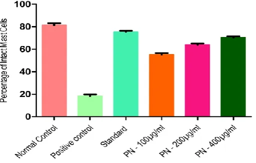
In vitro mesenteric mast cell degranulation
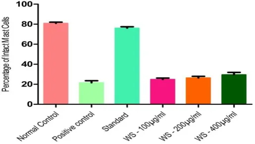
The result showed a higher stabilization potential was exhibited by ethanolic extract of Piper nigrum seeds which showed 55.17±1.56, 64.00±1.18 and 70.50±0.96 percentage inhibition of mast cell degranulation (Figure 1). Minimal activity was observed in the ethanolic extract of
Plantago ovata with percentage inhibition of 21.17±1.54, 22.17±2.21, and 24.33±2.20 at three dose levels (Figure 2). Other plant extracts showed intermediate activity (Figure 3-7).
[image:7.595.192.449.501.664.2]Figure 2. Effect of Ethanolic extract of Plantago ovata seeds on Mast cell stabilization activity on rat mesentery tissue
Figure 3. Effect of ethanolic extract of Boerhaavia diffusa roots on Mast cell Stabilization activity on rat mesentery tissue
[image:8.595.193.447.80.227.2] [image:8.595.190.449.316.470.2] [image:8.595.189.447.560.704.2]Figure 5. Effect of Ethanolic extract of Mangifera indica leaves on Mast cell Stabilization activity on rat mesentery tissue
Figure 6. Effect of Ethanolic extract of Cuminum cyminum seeds on Mast cell Stabilization activity on rat mesentery tissue
[image:9.595.192.448.81.231.2] [image:9.595.190.449.322.472.2] [image:9.595.190.448.561.706.2]DISSCUSSION
The present work has been taken up with an objective of screening a large number of plants which have been reported to possess anti-histaminic, anti-allergic and immunomodulatory activity. Several approaches have been tried for the prevention and cure of asthma, which affects nearly 25% of the population. The environmental pollution and rapid industrialization have contributed to a dramatic increase in the incidence of asthma in the recent times.[20] It has been clearly established that the airway inflammation is the main cause of asthma, which leads to bronchoconstriction and mucus secretion.[21, 22] With this understanding, various approaches for the management of asthma have been investigated. Steroidal anti-inflammatory drugs, bronchodilators, β-adrenergic stimulants, leukotriene antagonists, phosphor diesterase inhibitors, anti -IgE antibodies, monoclonal antibodies, 5- lipoxygenase inhibitors and mast cell stabilizers have been used in the prevention and cure of asthma.[23]
In the present study, we have concentrated on the screening of several plants for their ability to prevent the degranulation of mast cell and release of mediators of inflammation such as histamine, tryptase, chymase, heparin, leukotrienes, interleukins, nitric oxide etc. Our findings show that the efficacy of the plant extracts in asthma may be partially due to the mast cell stabilizing effect. Based on the literature survey, we have selected seven plants which are employed in the treatment of allergy and respiratory disorders in the traditional or folklore medicine. The selected plants are Momordica dioica fruit pulp (family - Cucurbitaceae), Cuminum cyminum seeds (family - Apiaceae), Piper nigrum seeds (family - Piperaceae), Boerhaavia diffusa roots (family - Nyctaginaceae.), Withania somnifera roots (family - Solanaceae), Mangifera indica leaves (Anacardiaceae), Plantago ovata seeds (Plantaginaceae).
eosinophils, macrophages, monocytes and neutrophils generate more ROS in the asthmic condition.[26] In addition to the cells, the mediators like lipid mediators, chemokines, adhesion molecules, and eosinophil granule proteins are potential stimuli of ROS production, so released in the process of inflammation also add to the oxidative stress in the individuals.[27] The ROS can perturb the airways and can worsen the pathophysiological changes which are associated with the asthma. ROS also cause the release of histamine from the mast cells and cause mucus secretion from airway epithelial cells.[28] ROS are also involved in production of a number of inflammatory mediators, most notably eicosanoids, by activating phospholipase A2 (PLA2).[29] The plants have their own strategy to detoxify the free radicals which may be done by direct enzymatic breakdown of oxidant radicals using SOD, catalase, ascorbate peroxidase, peroxidase, glutathione reductase and monodehydro ascorbate reductase which convert oxygen radicals to reduced products.[30]
On the basis of the present finding it is concluded that extract of Piper nigrum seeds showed the highest percentage of inhibition compared to the other extracts and can be useful for the disorders associated with the mediators of inflammation released from the mast cells. However, further studies on other experimental models are needed to support the hypothesis. A detailed study needs to be conducted to evaluate the phytoconstituent responsible to produce above result and their clinical efficacy in the treatment of asthmatic patients.
Conflict of interest
We authors declare no conflict of interest.
REFERENCES
1. Anupama, A., Suralkar, Gayatri, S., Vaidya, Abhijeet, R., Borate, Asha, S., Jadhav, and Kuldeep, K., Gaikwad, Antiallergic, Antianaphylactic and Mast Cell Stabilizing Activity of Pterocarpus marsupium Roxb. International Journal of Research in Pharmaceutical and Biomedical Sciences, 2012; 3: 1691-97.
2. Jignesh, I., Patel, Shrikalp, S., Deshpande,. Mast cell stabilising activity of various subfractions of leaves of Vitex Negundo. International Journal of Green Pharmacy, 2011; 5: 273-75.
3. Kambayashi, T., and Koretzky, G.A., Proximal signaling events in FC, RI mediated mast cell activation. J. Allergy and Clinical Immunol, 2007; 119: 544-52.
5. Hofeseth, L.J., Ying, L., 2006. Identifying and defusing weapons of mass inflammation in carcinogenesis. Biochim Biophys Acta 1765: 74-84.
6. Tejas Patel, Chimkode Rajshekar, and Rakesh Parmar,. Mast cell stabilizing activity of Myrica nagi bark. Journal of Pharmacognosy and Phytotherapy, 2011;3: 114-7.
7. Evans, W.C., Treas and Evans, 1996. Pharmacognosy. 15th ed. London.
8. Payyappallimana, U., Role of Traditional Medicine in Primary Health Care. Yokohama Journal of Social Sciences, 2010;14: 723- 43.
9. Anonymous, 2002. Regional Strategy for Traditional Medicine in the Western Pacific. Manilla: World Health Organization Western Pacific Region.
10.Maharudra, S., Rakh, Amol, N., khedkar, Nilesh, N., Aghav, sanjay, R., Chaudhari,. Antiallergic and analgesic activity of Momordica dioica Roxb. Willd fruit seed. Asian Pacific Journal of Tropical Biomedicine, 2012; 2: 192–96.
11.Boskabady, M.H., Kiani, S., Azizi, H., Relaxant effect of Cuminum cyminum on guinea pig tracheal chains and its possible mechanism(s). Indian Journal of Pharmacology, 2005; 37: 111-15.
12.Savithramma, N., Sulochana, C., Rao, K.N., Ethnobotanical survey of plants used to treat asthma in Andhra Pradesh, India. Journal of Ethnopharmacology, 2007; 113: 54-61. 13.Goyal, B.M., Bansal, P., Gupta, V., Kumar, S., Singh, R., Maithani, M., Pharmacological
Potential of Boerhaavia diffusa: An Overview. International Journal of Pharmaceutical Sciences and Drug Research, 2010; 2: 17-22.
14.Qamar, Uddin, L., Samiulla, Singh, V.K., Jamil, S.S., Phytochemical and Pharmacological Profile of Withania somnifera Dunal: A Review. Journal of Applied Pharmaceutical Science, 2012; 02: 170-75.
15.Dagmar, Garcia,Rivera; Ivones, Hernandez, Nelson, merino, Yilian Luque, Alina Alvarez, Yanet Martin et al. Mangifera indica L. extract (Vimang) and mangiferin reduce the airway inflammation and Th2 cytokines in murine model of allergic asthma. Journal of Pharmacy and Pharmacology, 2011; 63: 1336–45.
16.Rodriguez-Cabezas, M.E., Galvez, J., Camuesco, D., Lorente, M.D., Concha, A., Martinez-Augustin, ORedondo, L., Zarzuelo, A., Intestinal anti-inflammatory activity of dietary fiber (Plantago ovata seeds) in HLA-B27 transgenic rats. Clin Nutr., 2003; 22: 463-71.
Trieste, Chapter 1, An Overview of Extraction Techniques for Medicinal and Aromatic Plants., 2003 21-30.
18.Kokate, C.K., 1988. Practical Pharmacognosy, 2nd ed. New Delhi: Vallabhprakasham. 19.Norton, S., Quantitative Determination of Mast Cell Fragmentation by Compound 48/80.
British Journal of Pharmacology, 1954; 9: 494–97.
20.Alexey, V., Polonikov, Vladimir, P., Ivanov, Maria, A., Solodilova,. Genetic variation of genes for xenobiotic-metabolizing enzymes and risk of bronchial asthma: the importance of gene–gene and gene–environment interactions for disease susceptibility. Journal of Human Genetics, 2009; 54: 440-49.
21.Van den Berge, M., Ten Hacken, N.H.T, Van Der Wiel, E., Postma, D.S., Treatment of the bronchial tree from beginning to end: targeting small airway inflammation in asthma. Allergy, 2013; 68: 16-26.
22.O'Byrne, P.M., Airway inflammation and asthma. Alimentary Pharmacology & Therapeutics, 1996;10: 18–24.
23.Klaus, J., Erb Domnic, H., Martyres, Peter Seither, 2010. Antiasthmatic Agents. Ullmann's Encyclopedia of Industrial Chemistry.
24.Hyo-Hyun Park, Soyoung Lee, Hee-Young Son, Seung-Bin Park, Mi-Sun Kim, Eun-Ju Choi,. Flavonoids inhibit histamine release and expression of proinflammatory cytokines in mast cells. Archives of Pharmacol Research, 2008; 31: 1303-11.
25.Sangilimuthu, A., Sathish Kumar, R., Teepica Priya Darsini, D., Anitha, J., Ravi Subban,. A Review on Phytoconstituents Against Asthma. Int. J. Pharm. Sci. Rev. Res., 2015; 30: 7-16.
26.Nirmal, S.A, Patel, A.P., Bhawar, S.B., Pattan, S.R., Antihistaminic and antiallergic actions of extracts of Solanum nigrum berries: Possible role in the treatment of asthma. Journal of Ethnopharmacology, 2012; 14: 91–97.
27.Ryszard Dworski,. Oxidant stress in asthma. Thorax, 2000; 55: 51–53.
28.Paul, A.J., Henricks, Frans, P., Nijkamp,. Reactive Oxygen Species as Mediators in Asthma. Pulmonary Pharmacswcology & Therapeutics, 2001; 14: 409–21.
29.Ewa Pniewska, Rafal Pawliczak, 2013. The Involvement of Phospholipases A2 in Asthma and Chronic Obstructive Pulmonary Disease. Mediators of Inflammation Article ID 793505: 12.