ORIGINAL ARTICLE
Using venous blood gas analysis in the assessment
of COPD exacerbations: a prospective cohort study
Tricia M McKeever,
1Glenn Hearson,
2Gemma Housley,
3Catherine Reynolds,
2William Kinnear,
4Tim W Harrison,
2Anne-Maree Kelly,
5Dominick E Shaw
2,3▸ Additional material is published online only. To view, please visit the journal online (http://dx.doi.org/10.1136/ thoraxjnl-2015-207573).
For numbered affiliations see end of article.
Correspondence to Dr Dominick E Shaw, Respiratory Research Unit, Division of Respiratory Medicine, University of Nottingham, Clinical Sciences Building, Nottingham City Hospital, Edwards Lane, Nottingham NG5 1PB, UK; dominic.shaw@nottingham.ac. uk
Received 15 July 2015 Revised 6 October 2015 Accepted 9 October 2015 Published Online First 1 December 2015
▸ http://dx.doi.org/10.1136/ thoraxjnl-2015-208205
To cite:McKeever TM, Hearson G, Housley G,et al. Thorax2016;71:210–215.
ABSTRACT
Introduction Identifying acute hypercapnic respiratory failure is crucial in the initial management of acute exacerbations of COPD. Guidelines recommend obtaining arterial blood samples but these are more difficult to obtain than venous. We assessed whether blood gas values derived from venous blood could replace arterial at initial assessment.
Methods Patients requiring hospital treatment for an exacerbation of COPD had paired arterial and venous samples taken. Bland–Altman analyses were performed to assess agreement between arterial and venous pH, CO2and HCO3. The relationship between SpO2and SaO2was assessed. The number of attempts and pain scores for each sample were measured.
Results 234 patients were studied. There was good agreement between arterial and venous measures of pH and HCO3 (mean difference 0.03 and−0.04, limits of agreement−0.05 to 0.11 and−2.90 to 2.82, respectively), and between SaO2and SpO2(in patients with an SpO2of >80%). Arterial sampling required more attempts and was more painful than venous (mean pain score 4 (IQR 2–5) and 1 (IQR 0–2), respectively, p<0.001).
Conclusions Arterial sampling is more difficult and more painful than venous sampling. There is good agreement between pH and HCO3 values derived from venous and arterial blood, and between pulse oximetry and arterial blood gas oxygen saturations. These agreements could allow the initial assessment of COPD exacerbations to be based on venous blood gas analysis and pulse oximetry, simplifying the care pathway and improving the patient experience.
INTRODUCTION
Exacerbations of COPD are the second most common cause of emergency hospital admission in the UK, with an estimated 94 000 per year.1COPD exacerbations have a very high risk of mortality; 50% of people with a severe exacerbation will die within 4 years of an admission.1
The recognition that high flow oxygen therapy can induce hypercapnia in susceptible patients during exacerbations of COPD,2 and that respira-tory acidosis is associated with a worse outcome3 4
led to a rise in arterial blood gas (ABG) sampling to measure pH, PaCO2, PaO2 and HCO3. The
current National Institute for Health and Care Excellence (NICE) COPD guidelines recommend obtaining an ABG in all patients admitted to hos-pital with a COPD exacerbation.5Arterial sampling
is more technically difficult and reportedly more painful6 than venous blood gas (VBG) sampling.
Administration of local anaesthetic prior to arterial sampling is recommended but seldom used, as shown in a recent survey of junior doctors where 91% never or rarely used local anaesthesia when performing arterial puncture.7 Using less invasive measures of pCO2and SaO2 could greatly benefit
patients by both decreasing pain and streamlining the care pathway.
Recent meta-analysis data suggest good agree-ment between venous and arterial measureagree-ments of pH, HCO3 and base excess.8–11 In diabetes care arterial sampling has been replaced with venous for the monitoring of diabetic ketoacidosis.12 The use of venous samples to guide treatment in COPD exacerbations has been limited, perhaps because the relationship between arterial and venous measures of CO2is less strong, although a PvCO2of >6 kPa
has been shown to have 100% (95% CI 97% to 100%) sensitivity in identifying patients with clinic-ally relevant hypercapnia.13
We set out to assess the relationship between arterial and venous measures of PCO2, pH and
HCO3, and between arterial and pulse oximetry oxygen saturations during exacerbations of COPD, in order to establish whether VBG analysis combined with pulse oximetry could replace ABG analysis in the initial assessment of COPD exacerbations.
Open Access Scan to access more
free content
Key messages
What is the key question?
▸ Can venous blood gas analysis replace arterial blood gas sampling in the initial assessment of patients with COPD exacerbations?
What is the bottom line?
▸ Over two-thirds of arterial blood gas samples could be replaced by the simpler and safer use of venous blood gas analysis.
Why read on?
METHODS
Patients admitted to Nottingham University Hospitals Trust with a doctor-diagnosed exacerbation of COPD were considered for inclusion. Patients were included in the study unless they refused. If they were unable to give informed consent an approved alternative decision maker was approached. Recruited patients had an ABG and pulse oximetry performed by a junior doctor as per routine care, and a parallel paired VBG sample. Care was guided by the ABG results as per current clinical guidelines.
Arterial samples were collected via a heparinised needle and syringe system, and venous samples were aspirated into a separ-ate heparinised blood gas syringe via a butterfly needle or needle. Samples were processed as soon as possible on the same ward-based blood gas analyser. Analysers were calibrated regu-larly in accordance with the department of medical physics standard operating procedure. Research nurses collected data on demographics, body mass index, smoking status, pain scores (visual analogue scores 0=no pain, 10=worst pain imaginable) for arterial and venous sampling and the number of attempts taken to acquire each sample.
The main outcomes were the agreements between ABG and VBG parameters, and between ABG and pulse oximetry mea-sures of oxygen saturation. Secondary outcome meamea-sures included pain scores, and the number of attempts taken to obtain blood.
Statistical analysis
Agreements between venous and arterial samples for CO2, pH
and HCO3, and between SaO2 and SpO2were assessed using
the Bland–Altman method. Previous studies found 40% of patients attending the Emergency Department with an exacerba-tion of COPD had arterial hypercarbia, and it was estimated that 200 patients would allow us to calculate the sensitivity of the venous CO2 screening threshold for detection of arterial
hypercarbia with CIs of <5%.9 13Using this sample size of 200,
our 95% CI around the limits of agreement are estimated at 0.24× the SD of the mean difference for which pH is ±0.001.14
Missing data were not imputed. Receiver operating character-istic curves were used to estimate how venous CO2, pH, HCO3
predicted arterial values and how SpO2 predicted SaO2. Area
under the curve, sensitivity and specificity were calculated to predict an arterial pH≥7.35 and bicarbonate≥21, and for the pulse oximetry result to predict an SaO2of≥92%.
RESULTS
Over the time course of the study (from 3 February 2013 to 10 January 2014), there were 1376 admissions with a coded diag-nosis of COPD exacerbation. Of these, 234 participants were recruited and had at least one paired sample of blood gases. Twelve patients declined study participation. The mean age of the population was 71 years (SD 10.8) and 50% of the popula-tion was male. Characteristics of the populapopula-tion are shown in
table 1.
[image:2.595.309.551.59.245.2]There was good agreement between arterial and venous pH, and between arterial and venous HCO3 (figures 1 and 2 and
Table 1 Characteristics of study population
n N
Age (years) mean (SD) 71.0 (10.8) 234
Sex N (%)
Male 118 (50.4)
Female 116 (49.6)
BMI (kg/m2) mean (SD) 26.2 (7.9) 223
Smoking status N (%) 230
Never 14 (6.1)
Ex 146 (63.5)
Current 70 (30.4)
Clinical measures mean (SD)
Heart rate (bpm) 98.7 (20.8) 234
Respiratory rate 23.6 (5.9) 234
Systolic blood pressure (mm Hg) 130.7 (23.7) 234 Diastolic blood pressure (mm Hg) 71.3 (13.1) 234
[image:2.595.162.548.500.748.2]BMI, body mass index.
Figure 2 Bland–Altman plot for arterial and venous blood bicarbonate levels.
Table 2 Agreement between arterial and venous pCO2, pH and HCO3−
ABG (mean) (SD) VBG (mean) (SD) Mean difference (ABG–VBG) (95% CI) 95% limits of agreement N
pH 7.40 (0.09) 7.37 (0.08) 0.03 (0.02 to 0.04) −0.05 to 0.11 234
HCO3 (mEq/L) 29.7 (6.3) 29.7 (6.4) −0.04 (−0.22 to 0.15) −2.90 to 2.82 232
pCO2(kPa) 6.89 (2.40) 7.63 (2.41) −0.75 (CI−0.89 to−0.61) −2.91 to 1.41 225
[image:3.595.51.556.373.746.2]ABG, arterial blood gas; VBG, venous blood gas.
Table 3 Agreement between SaO2and SpO2
SaO2(mean) (SD) SpO2(mean) (SD) Mean difference (SaO2–SpO2) (95% CI) 95% limits of agreement N
Oxygen percentage saturation* 91.2 (6.0) 91.0 (4.0) −0.17 (CI−0.89 to 0.56) −11.12 to 10.78 224
*In patients with SpO2≥80%.
tables 2 and 3), however, the CO2 values varied significantly
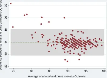
(figure 3). There was also a good agreement between SaO2and
SpO2 in those patients with an SpO2of ≥80% (figure 4 and
tables 2and3). These relationships were not significantly differ-ent in the 20 patidiffer-ents with an admission systolic blood pressure of <100 mm Hg (see online supplementary material).
Venous blood cut points for managing acute exacerbations of COPD
Given the relationships observed, we calculated the sensitivity and specificity of a VBG pH and HCO3 to correctly identify an arterial pH of≥7.35, and an arterial HCO3 of≥21, as well as an SpO2to identify an SaO2of≥92% (table 4). A venous pH of
7.34, a venous HCO3 of 21.45 and an SpO2 of 91.5 would
have correctly classified 87% (95% CI 82% to 91%), 97% (95% CI 93% to 98%) and 71% (95% CI 65% to 77%) of patients, respectively. In terms of specificity, 96% of patients with an ABG pH of≥7.35 also had a VBG pH of≥7.35 (tables 5and6).
We used current oxygen guidelines15 to calculate how many
ABG samples may have been avoided if the venous cut points were used instead. Of our 234 patients, 72 (31%) had a venous pH <7.35. Of the 162 with a venous pH≥7.35, only two had an arterial pH of <7.35.
Consequently, we estimate approximately two-thirds of ABGs can safely be avoided in the initial assessment of COPD exacer-bations. This figure does not factor in the repeat attempts needed to obtain arterial blood. Using these data, we suggest a new algorithm for the management of COPD exacerbations based on the current guidelines (figure 5).
Timing of samples
We assessed the mean difference in time between the paired arterial and venous samples to see if a delay between samples had an effect on the relationship between ABG and VBG parameters. The mean time difference was −4.18 min, SD 16.92, range−58.98 to 78.64 min (arterial–venous). Given the range, we repeated our analysis using the 168 paired samples that were performed within 15 min of each other. This did not affect the relationships (see online supplementary table).
Pain score
[image:4.595.202.554.49.306.2]The median pain score was significantly higher for ABG sam-pling as compared with VBG ( p<0.001). In addition, there was
Figure 4 Bland–Altman plot for SaO2and SpO2levels. (This graph only
[image:4.595.43.286.573.750.2]includes patients with a pulse oximetry value of >80%.)
Table 4 Pain score and number of venesection attempts
n N
Pain score (median and IQR)
Arterial pain score 4 (2–5) 187
Venous pain score 1 (0–2) 205
Number of attempts (N and %) Arterial number of attempts
1 162 (69.2)
2 55 (23.5)
3 10 (4.3)
≥4 7 (3.0) 234
Venous number of attempts
1 211 (90.2)
2 18 (7.7)
[image:4.595.310.551.648.725.2]≥3 5 (2.1) 234
Table 5 Predictive performance of venous blood gas parameters
Venous blood cut-off AUC
Sensitivity %
Specificity %
Correctly classified
%* N
Arterial pH
≥7.35
7.34 0.92 88.9 95.6 87 234
HCO3 (mEq/L)≥21
21.45 0.98 96 100 97 232
a significantly greater number of attempts taken to obtain an ABG sample (69.2% achieved at first attempt) compared with VBG, where 90.2% were obtained at the first attempt ( p<0.001) (table 4).
DISCUSSION
Exacerbations of COPD are a major cause of morbidity and mortality worldwide,16 and our local figures reflect this. In
2010, there were 1343 admissions into Nottingham University Hospitals Trust. The management of COPD exacerbations depends upon quickly identifying acute hypercapnic respiratory failure. In this study, we set out to establish if ABG analysis obtained for the initial assessment of COPD exacerbations
could be replaced by VBG analysis and pulse oximetry when assessing for acute hypercapnic respiratory failure.
We examined the agreement between ABG and VBG para-meters and between ABG and pulse oximetry measurements of oxygen saturation in COPD exacerbations and found acceptable agreement for pH, HCO3 and for SaO2at an SpO2>80%. We
found that 96% of patients with an ABG pH of <7.35 also had a VBG pH of <7.35 and that only two patients were
misclassi-fied as having a normal venous pH but a low arterial pH. A meta-analysis offive studies examining the utility of periph-eral VBG analyses in exacerbations of COPD in the emergency department found that there was agreement between arterial and venous pH and HCO3.9The weighted average difference for pCO2 was 0.79 kPa (n=440), whereas those for pH and
HCO3 were 0.028 and 1.34 mmol (n=239), respectively. The relatively weaker relationship seen between SpO2and SaO2 at
lower levels is unsurprising as commercial pulse oximeters are more accurate at higher oxygen saturations and significantly less accurate below 80%.17 18 The proposed algorithm reflects this; any patient with an SpO2of <80% needs an immediate ABG.
Our study has limitations. There was a small time gap between sample acquisition and processing, although results did not change significantly when this was factored in. We also had
Table 6 Predictive performance of pulse oximetry
SpO2
cut-off AUC
Sensitivity %
Specificity %
Correctly classified* N
SaO2 91.5% 0.75 78 72 71 233
[image:5.595.92.505.344.723.2]*Correctly classified refers to percentage of patients correctly classified both above and below given parameter.
a pragmatic approach to sample collection which depended upon our junior doctors and specialist nurses; consequently, it was difficult to fully exclude mixed arterial/venous stabs which may explain why the sensitivities and specificities to predict an arterial pH of <7.35 were not 100%. We stress in our algorithm that if there is a risk or actual clinical deterioration an arterial analysis should be performed.
Arterial sampling was more painful than venous and required more attempts. While the pain of arterial sampling can be reduced by using local anaesthetic, it is not widely used.7
Although capillary sampling is used in speciality wards, wide-spread adoption is difficult because of the extra training, resources and time needed. As patients with exacerbations of COPD almost always have venipuncture to obtain samples for full blood count and blood chemistry analysis, VBG analysis can be performed on the same sample.
Our results suggest that the close relationship between venous and arterial acid base parameters, and between oxygen satura-tions obtained from pulse oximetry and arterial blood, could allow the initial assessment of acute COPD exacerbations to be based on a combined measurement of a VBG pH and SpO2.
This would mean a change in practice as the current oxygen guidelines published by the British Thoracic Society15 state that any patient requiring supplemental oxygen to achieve a target SpO2of 92%–94% should have an arterial blood or arteriolised
capillary blood gas performed.
We suggest that arterial sampling is reserved for patients with a venous pH of <7.35. The approach of using venous blood
first has obvious benefits. Only one blood draw would be required resulting in less pain and a lower risk of bruising and associated side effects. Less training would be required to ini-tially assess acid/base status, and fewer attempts to draw blood would be needed, needing less equipment and simplifying the care pathway for COPD exacerbations. We conservatively esti-mate that >66% of ABG attempts would be avoided and replaced by VBG sampling.
CONCLUSION
There is a good agreement between pH and HCO3 values derived from venous and arterial blood, and between pulse oximetry and ABG oxygen saturations. These agreements could allow the initial assessment of COPD exacerbations to be based on VBG analysis and pulse oximetry rather than ABG analysis, simplifying the care pathway.
Author affiliations 1
Division of Epidemiology, University of Nottingham, Nottingham, UK 2Respiratory Research Unit, Division of Respiratory Medicine, University of Nottingham, Nottingham, UK
3Medical Informatics, East Midlands Academic Health Sciences Network, Nottingham, UK
4Respiratory Medicine, Nottingham University Hospital Trust, Nottingham, UK 5
Emergency Medicine, Joseph Epstein Centre for Emergency Medicine Research, Western Health, St Albans, Victoria, Australia
Correction notice This article has been corrected since it was published Online First. Data in the abstract and in Table 2 has been corrected. Figure 3 has been updated due to data corrections.
Twitter Follow Glenn Hearson at @NottinghamRRU
Acknowledgements The authors would like to acknowledge the expertise and help of Rachel King and Adeline Sheehan for recruiting the patients running the study. They would like to thank all the ward staff and junior doctors working in the Emergency Department, Emergency Admissions Unit, Medical High Dependency Unit
and Respiratory Wards at Queens Medical Centre and Nottingham City Hospital for their help with the study, and the hospital at night nurse coordinator team. The authors also wish to acknowledge Dr Dale Gardiner for his expert blood gas input. Contributors TMM performed the bulk of the data analyses and commented on the manuscript. GH helped with data acquisition and cleaning and commented on the manuscript. GH helped with data acquisition and follow-up data analyses and commented on the manuscript. CR and WK helped with data acquisition and commented on the manuscript. TWH helped with study design, data interpretation and commented on the manuscript. A-MK helped with study design, data interpretation and commented on the manuscript. DES had the original idea for the study, designed the study and obtained funding. He wrote thefirst draft. Funding This study was funded by the National Institute for Health Research’s (NIHR) research for patient benefit programme, grant number PB-PG-0211-24049. The views expressed are those of the authors and not necessarily those of the NHS, the NIHR or the Department of Health.
Competing interests None declared.
Ethics approval Nottingham Research and Ethics Committee East Midlands Nottingham 1 12/EM/0323.
Provenance and peer reviewNot commissioned; externally peer reviewed. Data sharing statement Data on mortality are available; these were not included at the request of reviewer 3 but will be presented in abstract form.
Open Access This is an Open Access article distributed in accordance with the terms of the Creative Commons Attribution (CC BY 4.0) license, which permits others to distribute, remix, adapt and build upon this work, for commercial use, provided the original work is properly cited. See: http://creativecommons.org/ licenses/by/4.0/
REFERENCES
1 APPG. All party parliamentary group on respiratory health: report on enquiry into respiratory deaths. 2014.
2 Aubier M, Murciano D, Milic-Emili J,et al. Effects of the administration of O2on ventilation and blood gases in patients with chronic obstructive pulmonary disease during acute respiratory failure.Am Rev Respir Dis1980;122:747–54.
3 Hudson LD. Survival data in patients with acute and chronic lung disease requiring mechanical ventilation.Am Rev Respir Dis1989;140(Pt 2):S19–24.
4 Jeffrey AA, Warren PM, Flenley DC. Acute hypercapnic respiratory failure in patients with chronic obstructive lung disease: risk factors and use of guidelines for management.Thorax1992;47:34–40.
5 NICE.Chronic obstructive pulmonary disease: management of chronic obstructive pulmonary disease in adults in primary and secondary care. London, UK: Royal College of Physicians, 2010.
6 Dar K, Williams T, Aitken R,et al. Arterial versus capillary sampling for analysing blood gas pressures.BMJ1995;310:24–5.
7 Mangera Z, Gunasekera C, Kinley J,et al. P113 the use of local anaesthesia in improving the patient experience of arterial blood gases: students and trainers are still not getting the message.Thorax2014;69(Suppl 2):A127.
8 Kelly AM, Kyle E, McAlpine R. Venous pCO(2) and pH can be used to screen for significant hypercarbia in emergency patients with acute respiratory disease.
J Emerg Med2002;22:15–9.
9 Lim BL, Kelly AM. A meta-analysis on the utility of peripheral venous blood gas analyses in exacerbations of chronic obstructive pulmonary disease in the emergency department.Eur J Emerg Med2010;17:246–8.
10 Kelly AM. Review article: can venous blood gas analysis replace arterial in emergency medical care.Emerg Med Australas2010;22:493–8.
11 Kelly AM, Klim S. Agreement between arterial and transcutaneous PCO2in patients undergoing non-invasive ventilation.Respir Med2011;105:226–9.
12 Kelly AM, McAlpine R, Kyle E. Venous pH can safely replace arterial pH in the initial evaluation of patients in the emergency department.Emerg Med J2001;18:340–2. 13 Kelly AM. Can VBG analysis replace ABG analysis in emergency care?Emerg Med J
2014.
14 Bland JM, Altman DG. Statistical methods for assessing agreement between two methods of clinical measurement.Lancet1986;327:307–10.
15 O’Driscoll BR, Howard LS, Davison AG. BTS guideline for emergency oxygen use in adult patients.Thorax2008;63(Suppl 6):vi1–68.
16 Rabe KF, Hurd S, Anzueto A,et al. Global strategy for the diagnosis, management, and prevention of chronic obstructive pulmonary disease: GOLD executive summary.
Am J Respir Crit Care Med2007;176:532–55. 17 Jubran A. Pulse oximetry.Crit Care1999;3:R11–7.