Phase Characterization of Re-Based Diffusion Barrier Layer on Nb Substrate
Eni Sugiarti
1;2;*, Youngmin Wang
1, Naoyuki Hashimoto
1, Somei Ohnuki
1and Toshio Narita
3 1Laboratory of Advanced Materials, Faculty of Engineering, Hokkaido University, Sapporo 060-8628, Japan2Research Center for Physics, Indonesian Institute of Sciences (LIPI), Indonesia 3
Advanced Barrier-Coating Laboratory, Faculty of Engineering, Hokkaido University, Sapporo 060-8628, Japan
An electron microscopy phase characterization was carried out for a Re-based diffusion barrier layer, which was deposited on the Nb substrate used as an ultra high temperature material. The coating process produced three layers; an outer Cr(Re) layer, an intermediate Cr-Nb-Re layer, and an inner Nb(Re) layer. The Cr-Nb-Re layer is considered to act as a diffusion barrier layer between the substrate and the outer Cr(Re) reservoir layer. The Cr(Re) and Nb(Re) layers are in single phase with a similar bcc structures, but they are different in structure from the intermediate layer, which is composed of a dual phase of Re63Cr20Nb17with a cubic structure and Nb42Re33Cr25with a hexagonal structure
determined by transmission electron microscopy (TEM) in this study. [doi:10.2320/matertrans.MB201022]
(Received September 2, 2010; Accepted November 24, 2010; Published January 19, 2011)
Keywords: phase characterization, crystal structure, diffusion barrier layer, niobium, transmission electron microscopy
1. Introduction
Niobium has attracted attention as the basis for new materials for high temperature applications in place of Ni-based superalloys. In order to increase the properties of Nb or Nb-based alloys for applications that require the highest creep resistance, Re as one of the refractory metals has been selected for coating element.1,2) Fundamental understanding of the crystal structure of the Cr-Nb-Re phase in the Re-based coating layer is important to control the coating process and to understand the mechanical properties of the coated layer. Moreover, the formation process of coatings with an Re-based diffusion barrier on Nb-Re-based alloys to improve the low oxidation resistance of these alloys was investigated by various methods in previous studies.3–5)However, the crystal structure of the Cr-Nb-Re phases formed by the coating process has not been determined.
An obstacle in resolving this issue is that no Cr-Nb-Re ternary phase diagram has been established. It is well known that Re forms intermetallic compound phases by alloying with Nb or Cr: Re-Cr -phase, Re-Nb -phase, and Re-Nb
-phase.6–8) Nevertheless, estimates of the Cr-Nb-Re phase based only on the composition would be unreliable. Further, analysis by X-ray diffraction (XRD), which is the most general and conventional method for determining crystal structure of unknown phases, is also complicated in applying to the coating layer, because the coating layer has a multi-layer structure and usually has a thickness of about severalmm.
The objective of this study is to identify the crystal structure of the various phases in the Cr-Nb-Re coating system by means of TEM. The paper will describe details of the selected area electron diffraction (SAED) patterns involving crystal structure and lattice constant analysis of the Cr-Nb-Re coating system. In the absence of other exper-imental data, this study will offer guidelines for developing Re-based diffusion barrier coatings on Nb or Nb-based alloys.
2. Experimental
A 99.99% Niobium ingot was cut into coupons of about 1 mm thickness. The specimen and it was polished to a 150-grit finish, then degreased in a methanol/benzene solution under ultrasonic agitation. A coating was developed involv-ing electroplatinvolv-ing of Re film from aqueous solution at temperature of 323 K, followed by heat-treated in vacuum at 1723 K for 10.8 ks to form a homogeneous Re film and pack cementation with Cr at 1673 K for 36 ks in vacuum. Details of the formation process of the coating were presented else-where.3)
The cross-section of the coated layer structure was first investigated by SEM. After the initial characterization, a cross-sectional TEM (XTEM) specimen, containing a dif-fusion barrier layer, was prepared by using an argon ion slicer (IS, JEOL EIS 9100) to ensure the minimum structural damage. The sample had to be cut in a size to2:80:45
0:1mm prior to the IS, causing spallation of the outer layer due to the weak bonding of the coated layer during cutting and polishing. Additionally, a focused ion beam (FIB, JFIB 2300) technique was used to obtain XTEM specimens at specific sites, which is not possible by the IS method. With these two methods, it is possible to prepare XTEM specimens which yield good results. The TEM observations were performed using a JEOL JEM 2010F at 200 kV equipped with an energy dispersive X-ray spectroscope (EDS).
3. Results and Discussion
3.1 Coating-layer structure and composition
The cross-sectional structure of an as-coated specimen is shown in Fig. 1(a). It was found from a SEM image in Fig. 1(a) and a EDS result in Fig. 1(b) that the coating comprises three-layers: the first layer of Cr(Re), the second layer of Cr-Nb-Re, the third layer of Nb(Re), and then there is the Nb substrate. The EDS result (Fig. 1(b)), which was performed along the line in (a), indicates that the outer Cr(Re) layer contains (100–78 at%)Cr and (22–1 at%)Re, the intermediate Cr-Nb-Re layer contains (60–22 at%)Re, (45– *Corresponding author, E-mail: [email protected]. Graduate
Student, Hokkaido University
24 at%)Cr and (18–12 at%)Nb, and the inner Nb(Re) layer contains (98–60 at%)Nb, (33–20 at%)Re and (6–3 at%)Cr. It was confirmed that the intermediate Cr-Nb-Re layer formed homogeneously (Fig. 1(a)) and the concentrations of Cr, Nb and Re varied sharply at the interfaces (Fig. 1(b)).
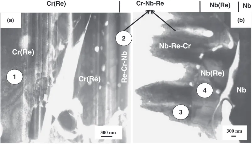
XTEM images of the coating layer are shown in Fig. 2(a) and (b) obtained from FIB and IS specimens, respectively. Table 1 shows the results of the EDS analysis obtained from the circled area in Fig. 2. In agreement with the results in Fig. 1, the layer-structure, from the surface, consists of an outer Cr(Re) layer with a thickness of about 15mm, an intermediate Cr-Nb-Re layer of about 6mm, and an inner Nb(Re) layer of about 6mm. The roughness of the Cr-Nb-Re surface in Fig. 2(b) is a local artifact and probably caused by the IS process. In addition, there are many defects, cracks,
4 3 2 1
10 µm
31.7 µm
4 3 2 1
Cr
Re
Nb
(a) (b)
Fig. 1 (a) Cross-sectional SEM image and (b) concentration profiles for three layers after Re plating followed by Cr-pack cementation on Nb substrate at 1673 K for 36 ks. (1) outer Cr(Re), (2) intermediate Cr-Nb-Re, (3) inner Nb(Re), and (4) Nb substrate.
Re-Cr-Nb
Cr(Re)
Cr(Re)
Cr-Nb-Re Nb(Re)
Cr(Re)
Nb-Re-Cr
Nb(Re)
Nb
300 nm 300 nm
(a) (b)
Nb
1
3 2
4
Fig. 2 Bright field XTEM images of the Cr-Nb-Re system (a) two layers at the top which are Cr75Re24and Re63Cr20Nb17and (b) three
layers at the bottom which are Nb42Re33Cr25, Nb70Re27and Nb substrate. There are cracks in the Cr(Re) layer and on the Nb(Re)-Nb
[image:2.595.88.505.70.265.2]interface. Cavities are formed mainly in the Cr-Nb-Re layer.
Table 1 Composition determined from the circled area of the coated layers (EDS analysis in TEM).
Layer Element (at%)
Nb Re Cr
Cr(Re)
(area 1 in Fig. 2(a)) 1 24 75 Re-Cr-Nb
(area 2 in Fig. 2(a)) 17 63 20 Nb-Re-Cr
(area 3 in Fig. 2(b)) 42 33 25 Nb(Re)
[image:2.595.97.498.319.548.2] [image:2.595.305.549.668.785.2]and pores in the interface between Cr(Re), Cr-Nb-Re and Nb(Re) layers, influencing the durability of the coating system.
The Re-riched intermediate Cr-Nb-Re layer is considered to act as a diffusion barrier between the substrate and an additional outer reservoir layer for both inward and outward diffusion, and the phases here are directly related to the stability and durability of the coating. Therefore, under-standing the phases, especially the Cr-Nb-Re phase, in the coating system is critical for the design of the Re-based diffusion barrier layer on the Nb or Nb-based alloy substrate. However, the phase of the Cr-Nb-Re compounds cannot be determined only from the EDS results because of the reason as mentioned earlier. The phase identification using electron diffraction method was carried out in this study.
3.2 Phase identification of the coating layer
It is possible to determine the crystal structures of the outer-layer Cr(Re) and inner layer Nb(Re) phases, because the phases were almost composed of only the two elements and the binary phase diagrams for these elements can be applied. The binary phase diagrams suggest the phases to be solid solutions with similar crystal structures, a body-center-cubic (bcc) structure, and verification of this was performed using SAED as shown in Fig. 3. The lattice constants determined from the patterns are shown in Table 2, and the results are in accord with the lattice constants of Cr70Re30and Nb60Re40phases in the Powder X-ray database.6)Because the atomic radiuses of Cr, Re, and Nb in the crystals are different: Cr (0.125 nm)<Re (0.137 nm)<Nb (0.143 nm), the lattice constant of the Cr(Re) phase is slightly larger than that of pure Cr, the lattice constant of the Nb(Re) phase is slightly smaller than that of Nb. With the data in Ref. 6) and the atomic radiuses, the differences in the lattice constants compared with pure Cr and Nb phases can be calculated to represent a 2.5% expansion for the Cr(Re) phase and a 1.2% shrinkage for the Nb(Re) phase, in good agreement with the experimental results.
For the phase of the Cr-Nb-Re layer, as previously mentioned, it is difficult to determine the crystal structure only by the composition, because of the absence of a ternary phase diagram based on experiments. Similar to the-phase formed in Ni-based alloys, an intermetallic compound phase was suggested to be formed on the Nb substrate in this study.
According to the Cr-Re, Cr-Nb, and Nb-Re binary diagrams, Cr-Re phase, Cr2Nb phase, Cr2Nb phase, Nb-Re
phase, and Nb-Rephase are possibly formed as a Cr-Nb-Re phase.8)In the case of limited experimental phase equilibrium data, electron diffraction is an option for determining the crystal structure of a new phase.9)From this reasoning, the crystal structure of the Cr-Nb-Re phase was determined by using SAED. In the area as shown in Fig. 2, the Cr-Nb-Re layer is formed by a dual phase, suggested as the EDS results indicate different compositions in the layer from the top and bottom sides. Therefore, to improve the accuracy and reliability of the dual phase identification for the Cr-Nb-Re layer, several SAED patterns were obtained from the same crystal grain in each phase by tilting the specimen (shown by the B value in the Fig. 4 and 5).
All of the SAED patterns correspond well to cubic and hexagonal structures rather than cubic (-Cr2Nb phase) or tetragonal (Cr-Re or Nb-Rephase) structures. To determine the crystal structure of the first phase of the Cr-Nb-Re layer, Ref. 7) shows that cubic NbRe2and Nb37Re63-phases have a lattice contant of a¼0:9670nm and a¼0:9675nm, respectively. Therefore, the Re63Cr20Nb17 phase can be identified as phase due to the lattice constant calculated from the SAED patterns in Fig. 4 witha¼0:953nm which is smaller than the value in Ref. 7) and probably influenced by the Cr constituent in the compound here. The second phase of the Cr-Nb-Re layer can be determined by comparing the lattice constants and the structure of the -Cr2Nb phase appears as the likely candidate. The -Cr2Nb phase is isostructural to a MgZn2type laves phase with 12 atoms per unit cell, and the lattice constants are a¼0:4976nm and c¼0:8059nm.6)According to the SAED patterns in Fig. 5, the lattice constant calculations of the Nb42Re33Cr25 phase B=[I11]
110 01I
121
Cr75Re24 (a)
110 121
(b)
B=[I11] Nb70Re27
110 121
B=[I11] (c)
Nb-base –
01I–
01I–
– –
–
[image:3.595.86.512.73.214.2]Fig. 3 SAED patterns showing a bcc structure of (a) Cr75Re24, (b) Nb70Re27, and (c) Nb substrate layers.
Table 2 Unit cell parameters determined from the coated layers.
Layer Crystal structure Unit Cell Parameter (nm)
a c
Cr75Re24 bcc 0.297 —
Re63Cr20Nb17 Cubic 0.953
Nb42Re33Cr25 Hexagonal 0.512 0.823
Nb70Re27 bcc 0.322 —
[image:3.595.304.550.280.367.2]are a¼0:512nm and c¼0:823nm, which are somewhat larger than those in-Cr2Nb phases. It should be noted that a
-Cr2Nb phase is a high temperature equilibrium phase formed only at temperatures above 1873 K, but an isostruc-tural Nb42Re33Cr25phase forms at a relatively lower temper-ature of 1673 K due to the the Re constituent in this compound. It must be mentioned that only the cubicphase and a hexagonal structure could be determined in the XTEM specimens here. We were unable to find any of the phase that has been reported to act as a diffusion barrier in Ni-based alloys.
4. Conclusions
This investigation reports a cross-sectional TEM analysis performed to identify the crystalline structure and composi-tion of an Re-based diffusion barrier layer on an Nb substrate,
which consists of an outer Cr75Re24layer with a thickness of about 15mm, an intermediate Cr-Nb-Re layer of about 6mm, and an inner Nb70Re27 layer of about 6mm. A crystal structure of Cr75Re24 and Nb70Re27 layers were determined by combining the phase descriptions of the three sub-binary systems and it was verified by lattice constant calculations from the SAED patterns, showing the same crystal structure, a bcc structure. For the phase of the Cr-Nb-Re layer, there are a number of phases which could be formed in accord with the sub-binary diagrams, Cr-Rephase, Cr2Nbphase, Cr2Nb
phase, Nb-Rephase, and Nb-Rephase. Several SAED patterns were confirmed from the same crystal grain of the Cr-Nb-Re layer and suggested a dual phase with cubic
phase and a hexagonal structure. Overall, the results of the calculations of the Cr-Nb-Re lattice constants suggest a Re63Cr20Nb17 with cubic phase of a¼0:953nm and a Nb42Re33Cr25with hexagonal structure ofa¼0:512nm and
020 I10 200
B=[001] B=[011]
01I 21I 200
B=[023]
200
132 I32
Fig. 4 SAED patterns showing a cubic crystal structure of a Re63Cr20Nb17phase.
B=[4223]
01I0 3I22
32I2
B=[01I0]
0002 2110
2112
10I0
I100 01I0
B=[0001]
01II 2II0 202I
B=[01I2]
10I0
1I01 0I11
B=[1213]
01II
B=[7253]
I2I2 I103
[image:4.595.85.516.73.214.2] [image:4.595.86.512.251.544.2]c¼0:823nm. In the absence of experimental data, the results here provide a guideline and will be useful for improving the performance of an Re-based diffusion barrier layers on Nb and Nb-based substrates, something that is necessary before these coated layer systems can be consid-ered for service or commercial alloys.
Acknowledgments
The author wish to thank all colleagues involved in the project for their help and input especially to all of staff in the advanced barrier-coating laboratory for providing the coating samples. One of the authors will express her gratitude to Inpex Foundation with grants for scholarships. This work was supported by Japan Aerospace Exploration Agency (JAXA) of Japan.
REFERENCES
1) R. C. Reed: The Superalloy:Fundamentals and Applications, (Cam-bridge University Press, 2006).
2) T. Jin, W. Wang, X. Sun and Z. Hu: Mater. Sci. Forum638–642(2010) 2257–2262.
3) K. Saito, S. Hayashi, T. Narita, I. Iwanaga and R. Tanaka: Mater. Sci. Forum522–523(2006) 309–316.
4) Y. Matsumura, M. Fukumoto, S. Hayashi, A. Kasama, I. Iwanaga, R. Tanaka and T. Narita: Oxid. Metals61(2004) 105–124.
5) M. Fukumoto, Y. Matsumura, S. Hayashi, K. Sakamoto, A. Kasama, R. Tanaka and T. Narita: Oxid. Metals60(2003) 335–346.
6) J. M. Joubert: Progr. Mater. Sci.53(2008) 528–583.
7) J. M. Joubert and M. Phejar: Progr. Mater. Sci.54(2009) 945–980. 8) T. B. Massalski: Binary Alloy Phase Diagram, 2nd ed., (ASM
International, 1990).